The organs of the bird respiratory system possess some outstanding anatomical features from that of mammals. Here, I will show you the anatomy of every single organ from the bird respiratory system with a labeled diagram. In most of the organ anatomy, I have taken examples from the chicken respiratory system. But, I will also show you some primary differences in the organs of the respiratory system from different avian species at the end of this article.
So, if you want to learn the bird’s anatomy (like chicken, duck) respiratory organs, this article is for you.
Bird respiratory system organs anatomy (unique features)
You will love to learn the interesting anatomical facts of the bird respiratory system organs. The following unique features distinguish the bird respiratory organs from that of mammals. These features are not enough to learn the respiratory organ anatomy. If you are a veterinary student, you might read the article till the end to achieve a good piece of knowledge on bird’s respiratory system organs.
- The nostril of the bird is a slit-like opening in the upper beak.
- You will find narrow nasal cavities in a bird with three compartments – vestibule, respiratory, and olfactory.
- There are both larynx and syrinx present in the respiratory system. The larynx comprises four different cartilages. On the other hand, the bird’s syrinx is a unique anatomical feature responsible for voice production.
- The trachea of the bird is supported by a series of completed cartilaginous rings (signate rings).
- There are right and left flat, triangular-shaped lungs present in the bird. They are not lobed as like the mammal’s lungs. The dorsal border of bird’s lung has five deep impressions of ribs.
- The bronchi of the bird lung ends at the entrance of the air sacs.
- There are nine air sacs present in the bird respiratory system. These are unique structures of birds that serve to keep the body light.
So, if you are interested to learn the details anatomical facts of these organs from a bird, you may continue this article.
Organs of bird respiratory system and diagram
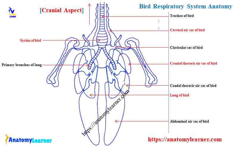
You know the bird respiratory system consists of the following organs. First, you might try to identify these organs from the actual sample with the help of the below-mentioned bird respiratory organs labelled diagram.
- Nostril and nasal cavity of the bird
- Larynx and laryngeal cartilage of bird
- The trachea of a bird
- Syrinx and its associated structures in bird
- A lung of bird and
- Different types of air sacs of a bird
Here, I will represent the chicken respiratory system organs with the labeled diagram.
The nasal cavity of the bird respiratory system
The nasal cavity of the bird locates to the left and right of the median nasal septum. You will find a considerable variation in the position of the nostril in different species of bird. In chicken, the nostril locates at the base of the beak. But, in duck, it is more caudal to beak.
Okay, let’s know what nostril is. The nostril is an elongated narrow slit in the upper beak of a bird. Sometimes, you will find a rigid plate (operculum) that project from the dorsal border of the nostril of the chicken. But, in pigeon, you will find a fleshy operculum on the dorsal edge of the nostril.
“The nasal septum of duck and goose perforates by the small opening at the level of the nostril.”
The nasal cavity of the bird is roughly cone-shaped, with the apex pointed rostrally. You will find three freely communication compartments in the nasal cavity of a bird.
Vestibular compartment or vestibulae rostrally
A respiratory compartment in the middle and
An olfactory compartment caudally
Before going to the detailed description of the compartments of the bird nasal cavity, you might have a good piece of knowledge of the different types of nasal conchae.
Nasal conchae in birds
You will find three nasal conchae in most of the birds. In contrast to mammals, these nasal conchae are arranged in rostrocaudal rather than dorsoventral sequence.
The three nasal conchae of birds are –
- Rostral nasal conchae or concha nasalis rostralis,
- Middle nasal conchae or concha nasalis media, and
- Caudal nasal conchae or concha nasalis caudalis.
The rostral nasal conchae of a bird locates at ventrally or cranially of the nasal cavity. Cranially, it is a pointed cone, wide at dorsoventrally, and have a smooth, flat medial surface. The caudal end of this nasal conchae of the bird is entirely closed by a flat plate of cartilage at the right angle.
In addition, the middle nasal conchae is the largest in a bird. In the transverse section, you will find a scroll-like appearance with one and one-half turns.
Again, the caudal nasal conchae of a bird is a hemisphere that projects from the lateral wall of the nasal cavity. It has an interior connection with the infraorbital sinus by a short and narrow canal.
“The infraorbital sinus is a relatively large, triangular space in the lateral region of the upper jaw. It bounds by the eyeball on its dorsocaudal aspect.”
Compartments of bird nasal cavity
The vestibular compartment of the bird nasal cavity consists of rostral conchae and lies with the non-glandular mucosa (stratified squamous epithelium). You will find a constricted area between the vestibular compartment and the middle compartment of the bird nasal cavity.
The nasal vestibule of the bird respiratory system receives the secretion of the nasal gland. It serves to humidify the nasal cavity of the bird. The nasal gland of most of the bird has lateral and media lobes, each with its ducts.
You will find only the medial lobe in the nasal vestibule of a chicken. The duct empties by a slit-like opening on the nasal septum near the rostral nasal conchae.
“There is no nasal gland present in the nasal cavity of a pigeon.”
The respiratory compartment of the bird nasal cavity consists of the middle nasal conchae and connect with the oral cavity through the conchal opening. Histologically, the respiratory region of bird nasal cavity lines with the pseudostratified ciliated epithelium.
In addition, the olfactory compartment of the nasal cavity consists of caudal nasal conchae and lies with the epithelium. You will find a typically yellowish olfactory region in chickens and also in some waterbirds. The olfactory part is a small circumscribed area on the caudal nasal conchae and the caudal nasal septum of the bird.
A nasal cavity in other birds
The nostril of the turkey resembles that of the chicken. But, in duck and goose, the nostril locates more caudal to the beak and is a large elongated structure rather than the narrow slit. You will not find any operculum in duck or goose’s beak.
The shape of the nasal cavity may vary with the modification of beak in different species of birds. You will find a narrow, elongated opening at the rostroventral part of the nasal septum.
The rostral nasal conchae of a turkey is similar to that of a chicken. But in duck, it reduces into a simple, elongated, and narrow shelf. Again, the middle nasal conchae of a duck is relatively longer than that of a chicken.
In the duck, the caudal nasal conchae is a narrow, elongated triangle with a long axis. The infraorbital sinus is relatively more prominent than that of a chicken and reaching to the bill. This sinus ends caudally at about the level of the lateral angle of the eye.
Larynx anatomy from bird respiratory system
The larynx of a bird is a conspicuous mound in the ventral oropharynx, caudal to the tongue. It is heart-shaped, and the point being directed rostrally into a transverse mucosal fold at the base of the tongue. Each side of the laryngeal mound consists of four rows of caudally directed papillae pharyngeal.
The inlet of the bird larynx is a narrow slit in rest condition. Again, this inlet continues caudally by the laryngeal fissure or dorsal furrow of the larynx.
There are two groups of laryngeal glands present in the larynx of a bird respiratory system. The caudal group (cricoarytenoid glands) lies in the caudal region of the laryngeal mound. On the other hand, the lateral group of glands locates at the lateral wall of the laryngeal mound of a bird.
“Together, these two groups of laryngeal glands (lateral and caudal) are known as the cricoarytenoid salivary glands in birds”.
The cartilage of bird larynx
You will find four different cartilage in the bird larynx. These are the median cricoid, procricoid, and paired arytenoid cartilages.
- Cricoid cartilage (cartilago cricoidea) of bird
- Procricoid cartilage (cartilago procricoidea), and
- Arytenoid cartilage (cartilago arytenoidea) of bird (paired)
The cricoid cartilage is unpaired and consists of the median body, left-wing, and right-wing. The cricoid cartilage of a male bird is longer than that of a female bird.
The median body of the cricoid cartilage is a large, gutter-like plate and concave dorsally. It is partly ossified, but you may also find a cartilaginous part. The left and right wings of the cricoid cartilage join with the body’s lateral aspect by a thin line of flexible cartilage.
The rostral border of the wings is thickened and have contact with the caudal edge of the body of the arytenoid cartilage of the bird.
In addition, the procricoid cartilage is median, dorsal and small in a bird. It is comma-shaped with a cranial body and a caudal tail. This cartilage has different facets on its lateral and dorsal aspect that connects with the right and left arytenoid cartilage bodies. The tail of the procricoid cartilage of bid has right and left facets that join with the right and left wings of the cricoid cartilage.
Again, you will find paired arytenoid cartilages that possess a body, rostral process, and a caudal process. The body of the arytenoid cartilage joins with the procricoid caudomedially. It also has a connection with the wings of the cricoid cartilage.
You will not find any thyroid and epiglottis laryngeal cartilage in any bird species.
Laryngeal ligaments of bird larynx
I will provide a little information on the laryngeal ligaments and muscles of the bird larynx. You will find five pairs of intrinsic ligaments, one unpaired intrinsic, and one pair of an extrinsic ligaments in the bird’s larynx.
The five pairs of intrinsic ligaments of the bird larynx are –
- Lateral cricoid ligament of the larynx
- The peripheral cricoarytenoid ligament of the larynx
- The caudal cricoarytenoid ligament of the larynx
- A capsular ligament of procricoarytenoid joint and
- The capsular ligament of the procricocricoid joint
The lateral cricoid ligament of the bird larynx runs from the most caudal and lateral part of the cartilaginous tip of the cricoid cartilage. Again, the peripheral cricoarytenoid ligament is a thin sheet of connective tissue. It joins the cranial process and body of the arytenoid and the cricoid.
You will find a stronger caudal cricoarytenoid ligament in the bird larynx. That joins the body of the arytenoid cartilage to the dorsal surface of the cricoid wings.
The unpaired intrinsic ligament of the bird larynx is the arytenoarytenoid ligament. It is a strong ligament that joins with the ventral surface of the caudal processes of each arytenoid. This ligament also has a connection with the dorsal surface of the body of procricoid cartilage.
In addition, the paired extrinsic ligament of the bird larynx is arytenoglossal. It is a cord of elastic tissue that runs from the cranial tips of the cranial process of arytenoid cartilage to the paraglossal bone of the tongue. You will find a much longer and stronger arytenoglossal ligament in a male bird than in a female.
Laryngeal muscles of bird larynx
I will enlist only the muscles of a laryngeal mound of a bird. You will find two paired intrinsic muscles, two pairs of extrinsic muscles, and one unpaired extrinsic muscle in the larynx of a bird.
The superficial intrinsic muscles of the bird larynx
A deep intrinsic muscle of the bird larynx (horseshoe-shaped muscle)
A rostral extrinsic muscle of bid larynx (basibranchial laryngeal muscle)
The caudolateral extrinsic muscle like sternotracheolaryngeous lateralis muscle
A caudomedial extrinsic muscle (sternotracheolaryngeous medialis muscle)
The larynx of other bird species
You will find a similar larynx structure in the turkey as a chicken. But in ducks, the laryngeal mound is relatively elongated. Rostrally, it blends relatively smooth with the pharyngeal floor. Again, in goose, it is longer than that of a duck.
The cricoid cartilage is much shorter rostrocaudally in a turkey than that of a chicken. The procricoid cartilage of the turkey is flattened dorsoventrally.
Trachea from bird respiratory system
The trachea of the bird respiratory system posses some unique anatomical features than that of mammals. It begins at the caudal end of the cricoid cartilage of the bird. In chicken, the upper part of the trachea remains in the midline of the cervical region. It courses then continue, together with the oesophagus on the right side of the neck. Again, the trachea of the chicken regains the median position upon entering the thoracic inlet.
But in other bird species, the trachea may be longer and coils than that of a chicken. It will lie in between the skin and muscles or within the sternum itself.
Cartilage rings of bird trachea
You will find a series of complete cartilaginous rings that tends to become ossify (except in pigeon). These tracheal rings resemble a signet ring, with the expanded portion alternately forming the left and right half of each subsequent ring.
You will find a considerable variation in the number of cartilage rings in different species of birds. The number of a ring may vary from hundred eight to hundred twenty-five in other species of bird.
The first ring follows immediately after the cricoid cartilage, and the last one precedes the tympanum of the syrinx. Each ring of the birds trachea is a complete circle. The first and last are simple rings; the rest are signet rings.
The width of the cartilaginous rings increases progressively throughout the cranial third of the trachea. Again, the width significantly decreases throughout the caudal third of the birds’ trachea.
The rings of the cranial third of the birds’ trachea are almost transversely oval. In addition, the rest of the tracheal rings are circular, but you may find some variation in different bird species.
Muscles of bird trachea
You will find a band-like tracheal muscle along the length of the caudal part of the bird’s trachea. The muscles of the birds trachea are as follow –
- Tracheolateralis muscles of bird trachea
- Cleidohyoideus muscle of bird trachea
- Sternotrachealis muscle of bird trachea and
- Cleidotrachealis muscle of bird trachea.
The tracheolateralis muscle arises from the syrinx and passes along the lateral trachea to the cricoid cartilage. Again, the cleidohyoideus muscle of the bird runs cranially and joins with the cricoid cartilage and hyobranchial apparatus.
At the craniolateral process of the bird sternum, a sternotrachealis muscle originated and inserted laterally at the caudal end of the trachea. In addition, the cleidotrachealis muscle arises from the clavicle of the bird and inserts on the trachea.
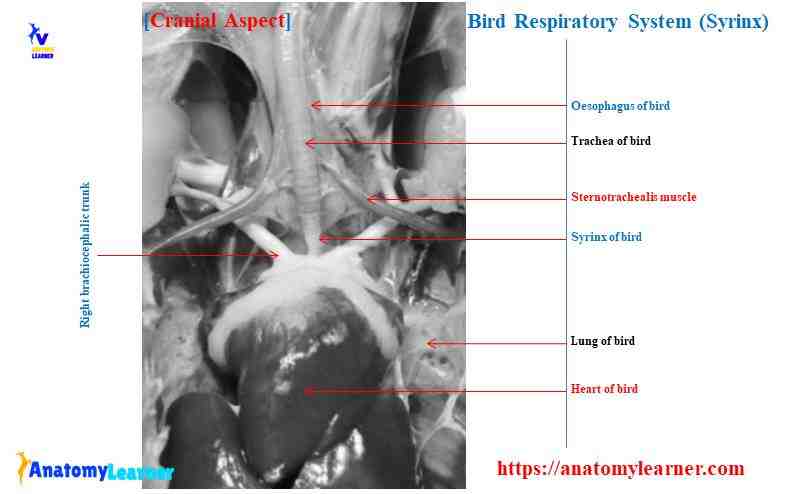
Syrinx of bird respiratory system
The syrinx is a unique anatomical feature in the bird respiratory system. It is the voice organ and lies at the bifurcation of the trachea into the right and left bronchi. You may quickly identify the syrinx of a bird by external appearance. Externally the syrinx of the bird is a marked lateral narrow area in the trachea.
You will find four cartilaginous components in the syrinx of a bird. The four cartilaginous components of bird syrinx (chicken) are –
- The cranial cartilage or tympanum
- A wedge-shaped pessulus
- The intermediate cartilage of syrinx, and
- Caudal cartilage or bronchial half rings
The cranial cartilage of the syrinx consists of four trachea rings in the male bird and three in a female bird. These are slightly greater in diameter than the preceding tracheal rings.
The plessulus are the wedge-shaped cartilage of the syrinx. Its sharp edge splits the lumen of the trachea into the two primary bronchi.
The intermediate cartilage of the syrinx is four in number on each side. This cartilage joins with the plessulus on its ventral end. The dorsal end of these intermediate cartilage remains free.
You will find three rings in the caudal cartilage of the syrinx. The first ring joins with the plessulus on both ends. Again, the second one is attaché on the ventral end of the trachea. The third cartilage is free at both ends.
Structures that are responsible for producing voice in bird
You will find two vibrating membranes in the construction of a syrinx. There are lateral and medial tympanic membranes present in a bird’s syrinx; these are responsible for producing the voice.
The lateral tympanic membrane lies on the lateral aspect of the bird syrinx. It runs from the caudal edge of the last intermediate cartilage to the cranial edge of the first caudal cartilage.
The medial tympanic membrane forms the medial modified medial wall of the cranial end of a primary bronchus. This medial tympanic membrane has a connection with the caudal border of the pessulus.
You will find the syringeal muscles in most songbirds, but in chicken or duck, these muscles are absent.
Syrinx of other bird species
The syrinx of a duck is asymmetrical in the male. It forms the large, dilated box on the left slide of the trachea (known as bulla tympaniformis). So, the structure of this tympaniformis is different from that of a chicken.
You will find three cartilaginous components in the syrinx of a duck. The tympaniformis of duck consists of six cranial cartilage, four bronchial half-rings, and pessulus.
Lung anatomy of bird (chicken)
Do you know the lung of a bird differs from that of a mammal? The lungs (right and left) of a bird (chicken) are flattened nearly rectangular structure lies on either vertebral column. The medial border of the bird lung is thick, and the lateral wall is thin.
You will not find any lobes in the right or left lung of a bird. But, you will find five distinctive ribs impressions on the lung’s dorsal surface that separates it into different segments (tori intercostales). These impressions are formed by the second to six vertebral ribs of the bird.
The size may vary from bird to bird due to variation in blood remaining in the lungs. You will find the following structures (a division of bronchi) in the lung of a bird.
- Two primary bronchi (bronchi primarii),
- The secondary bronchi (bronchi secundarii),
- A parabronchus, and
- The air capillaries (penumocapillaries) of bird lung
If you want to know more about these divisions of bronchi of bird lung, continue this article.
Bronchi of bird lung
The syrinx of the bird gives rise to the right and left primary bronchi. It enters and runs through the right and left lungs and ends at the right and left abdominal air sacs entrance.
The wall of the primary bronchi of the bird contains incomplete C-shaped cartilage rings. In the primary bronchi, the lumen surrounds by the respiratory epithelium. You will find the smooth muscle in the primary bronchi, which is mostly circular.
Each primary bronchi give rise to the four groups of secondary bronchi in the bird respiratory system. According to the direction in which they pass, they are grouped into –
- Four medioventral secondary bronchi
- Eight mediodorsal bronchi,
- Eight laterodorsal secondary bronchi, and
- Twenty-five to thirty laterodorsal secondary bronchi
The four mediodorsal secondary bronchi arise in the spiral line along the primary bronchus dorsomedial wall immediately after entering the lungs. These are the most prominent secondary bronchi in the bird lung.
The eight mediodorsal secondary bronchi arise dorsally and dorsomedially in the spiral line. These are the second largest secondary bronchi in the lung of a bird.
The eight laterodorsal secondary bronchi arise from the ventral aspect of the primary bronchus of the bird. You will find a similar diameter in the first and second laterodorsal secondary bronchi of a bird.
The twenty-five laterodorsal secondary bronchi are small in calibre like parabronchi. They arise from the lateral aspect of the primary bronchus.
Parabronchi and air capillaries of bird lung
The secondary bronchi of the bird lung raise the numerous parabronchi. They are invariably anastomose with one another. These parabronchi are the functional unit of a bird lung. The parabronchi arrange in the parallel array of the elongated tubule.
Again, the individual parabronchi are separated by the interparabronchial septa. Due to the arrangement of the septa, the parabronchi appears hexagonal in the transverse section. The parabronchi of the avian lung have some distinguished features –
- They anastomose with one another,
- Their wall contains chambers called atria,
- They have a gas exchange unit, and
- Their diameter is uniform within the species.
The wall of every parabronchus pierces by many opening that leads into a roughly polygonal chamber (atria). These lead into smaller, intermediate cavities – the infundibula. Again, these infundibula give rise to an anastomosing three-dimensional network of tubular air capillaries.
The diameter of the ari capillaries may vary with the different bird species. In addition, the air capillaries intermesh with the dense network of blood capillaries, permitting gas exchange to take place across the blood air barrier.
You will find a considerably thinner blood air barrier in a bird than that of a mammal. The blood air barrier of a bird consists of three elements –
- The endothelium cells of the blood capillaries,
- A fused basal membrane of blood and air capillaries, and
- The epithelium of air capillaries.
The surface of gas exchange is ten times higher in birds than that of a mammal.
Air sacs of bird respiratory system
The air sacs are the unique features in the bird respiratory system. These are the thin-walled deformable cavities attached to the avian lungs. The air sacs provide mechanical ventilation to the avian lungs.
The air sacs of birds may vary with the species. In chicken, you will find typically eight air sacs –
- A clavicular air sac in chicken (single)
- Median cervical air sac (single, but you may find paired cervical air sacs in some birds),
- Cranial thoracic air sacs in bird (paired),
- The caudal thoracic air sacs (paired), and
- Paired abdominal air sacs in a bird.
The air sacs of birds communicate with the secondary bronchi of the lungs (except the abdominal). But the abdominal air sacs communicate directly to the primary bronchi of the lungs.
Cervical air sacs of bird
The cervical air sac consists of a median chamber and a diverticular. It extends to from the twelve cervical vertebrae to the third thoracic vertebrae. The cervical air sac is in close contact with the ventral muscle of the vertebral column and clavicular air sac ventrally.
You will find the oesophagus that is trapped in between the cervical and clavicular air sacs. The caudal half of the median chamber interposes between the lung of the bird. Again, the cranial half gives rise to three arches dorsally that curves over the intervertebral spaces.
The diverticula of the cervical air sac arise from the three arches of the median chamber. These comprise extraspinal and intraspinal pairs of tubes.
Clavicular air sacs of bird
The clavicular air sac of a bird is extensive and complex. It lies at the base of the neck between the level of the shoulder joint and thoracic inlet. This air sac has a comprehensive connection with the coracoid bone and its muscles.
The clavicular air sac consists of a median chamber and paired lateral compartments. Again, in the median chamber, you will find the followings –
- A round craniomedial diverticulum
- The large left and right craniolateral diverticulum,
- The thin leaf-like left and right stenocardia diverticulum, and
- Left and right bronchial diverticulum.
The lateral chamber of the clavicular air sac arises from the median chamber by a long, wide canal compressed between two of the coracoid muscles. You will find the following three diverticula in the lateral compartment of the clavicular air sac –
The pectoral diverticulum,
A humeral diverticulum, and
The axillary diverticulum.
Cranial and caudal thoracic air sacs of bird
The cranial thoracic air sacs are paired and more or less symmetrical and simple. Each sac lies between the saccopleural membrane and the saccoperitoneal membrane. Dorsally it attaches to the ventral median process of the fifth and sixth thoracic vertebrae of the bird.
You will find three surfaces in each sac – sternocostal surface, pulmonary surface, and ventromedial surface. The sternocostal surface of the cranial thoracic air sac is attached to the most mobile part of the thoracic cage. It is reaching in the lateral border of the sternum.
In addition, the pulmonary surface of the cranial thoracic air sac relates to the concave ventral surface of the lung. The ventromedial surface encloses the heart, liver, caudal end of the oesophagus, and the proventriculus.
On the other hand, the caudal thoracic air sacs are paired, small, flattened, ear-shaped sacs smaller than that of cranial thoracic. Each sac is entirely excluded from direct contact with the visceral organs.
You will not find any diverticulum in the cranial and caudal air sacs of the bird.
Abdominal air sacs of bird
The abdominal air sacs project around the abdominal viscera of the bird. Their volume far exceeds that of the other air sacs. The right abdominal air sac is more significant than that of the left one.
Each abdominal air sac consists of a body and diverticula. The body of the abdominal air sacs extends from the sixth thoracic vertebrae to the last sacral vertebrae.
At the cranial end, the abdominal air sacs are capped by the liver. Dorsally, the lateral surface of both abdominal air sacs joins to the body wall. Again, the ventral border of both air sacs is free and mobile. The medial surface of both air sacs has no attachments.
The diverticulum of the abdominal air sacs comprises a pelvic and femoral group. You will find three perineal diverticula (cranial, middle, and caudal) in the pelvic group. There are also three pairs of diverticulum present in the femoral group.
The abdominal air sacs play a vital role in the mechanical ventilation of the avian lung.
Bird respiratory system organs diagram
I have already shown you the important labelled diagram from the bird respiratory system. Here, I tried to show you the detailed anatomical facts of most organs from a bird (chicken). If you want to get a more detailed diagram and high-quality image, make sure you join with anatomy learner on social media.
Frequently asked questions on bird respiratory organs
Again, in this part of the article, I will try to solve your queries. If you have any questions on bird respiratory organs, feel free to ask me.
How does a bird’s respiratory system work?
The ribs move craniolaterally during inspiration, pushing the sternum ventrally and cranially. The movement of the ribs and sternum also draw the abdominal wall ventrally and laterally. Thus the diameter of the celom increase during inspiration transversely, dorsoventrally, and craniocaudally.
The overall enlargement of the body cavity leads to a general fall of pressure within the celom and air sacs. As a result, ais moves through the lung to the air sacs.
The air sac is reaching to the caudal air sac and travel directly through the primary bronchi. Again, the air enters into the cranial air sac and passes through the parabronchi.
During the expiration, the air expels from the cranial air sac through the primary bronchi and trachea. The fresh air of the caudal air sac passes through the parabronchi, where it participates in gas exchange.
Why do birds have the most efficient respiratory system?
Do birds have a one-way respiratory system?
How does a bird respiratory system differ from a mammal respiratory system?
Why are the lungs of birds more efficient than human lungs?
Why is it important for the bird to have a unique respiratory system?
Do all birds have 9 air sacs?
No, the number of the air sacs may vary with different species of bird. Ideally, you will find the nine air sacs in bird – single clavicular and paired cervical, cranial thoracic, caudal thoracic, and abdominal. In chicken, this number of air sacs may also vary. Some of the chicken species possess nine air sacs (including paired cervical), some have eight.
In turkey, you will find a significant variation in the number and structure of air sacs. The paired cervical air sacs fuse with the lateral clavicular air sac and forms a cervico-clavicular air sac. Again, the paired caudal thoracic air sacs are suppressed in turkey. In duck, the number of air sacs is similar to the number of chickens, but you will find some variation in their structure.
Conclusion
I think you got a basic idea of the anatomy of every single organ from the bird respiratory system. The syrinx and different air sacs were the most interesting and unique anatomical features in the bird respiratory system. You might try to identify all the organs of respiration from a bird (don’t forget to take help from the bird respiratory labelled diagram). The structure of the birds trachea is somewhat different from that of mammals. You have also found a distinguished variation in the anatomy of bird lungs than that of a mammal. Please make a short note where you might enlist the most interesting anatomical facts of the different respiratory organs.