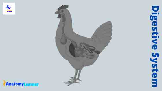
The chicken digestive system has some outstanding anatomical features than that of a mammal. But the basic pattern of the digestive tract is similar to some of the mammals. The upper part of the avian digestive tract shows adaptation for the flight. This article will show you the essential anatomical facts of different organs from the chicken digestive system with a labeled diagram.
In most bird species, you will find almost the same anatomical features in the digestive system organs like chicken. I will also provide a little information on the exceptional features in the digestive system of other different species of a bird compared to a chicken.
Chicken digestive system anatomy
First, I would like to enlist the unique anatomical facts from the chicken digestive system organs. Fine, these features might help you to understand why they are different from that of a mammal.
There are lots of exceptional anatomical features in their digestive organs, but here, I will enlist only the most important features –
- The presence of a short, narrow beak in the mouth cavity of a chicken,
- You will not find any clear boundary in between the mouth and pharyngeal cavities of a chicken,
- The absence of cheeks, teeth, and lips in the mouth cavity of a chicken,
- The esophagus posses a saccular diverticulum (crop) immediately cranial to the thoracic inlet,
- You will find a glandular proventriculus and muscular gizzard in the stomach of a chicken,
- There is a Meckel’s diverticulum present in the middle of the jejunoileal,
- The presence of caeca and the caecal tonsil in the large intestine of a chicken,
That nice, you will find three different sections on the cloaca of a chicken.
“For your kind information, this single article is not enough for learning the details anatomy of the digestive organ. You might learn the anatomical facts of each organ from the chicken digestive system separately.”
I tried to show you all the anatomical facts of different organs from a chicken digestion system. Follow the anatomy learner on social media if you want to get high-quality images of chicken digestion system organs.
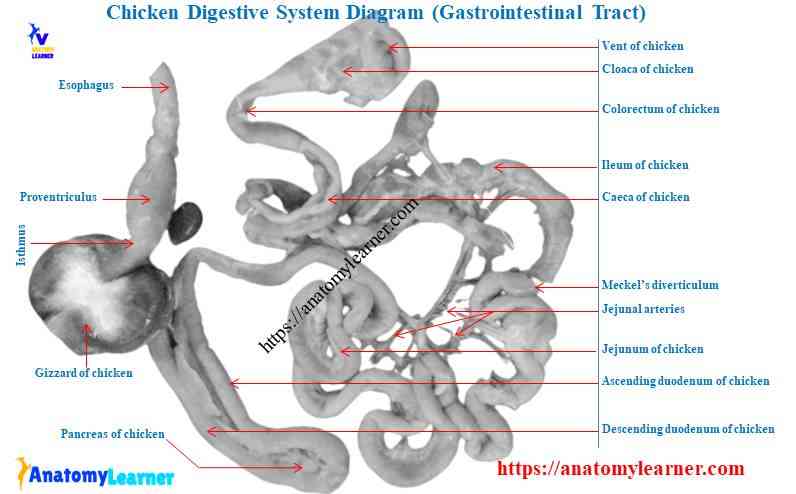
Organs and parts of the chicken digestive system
The chicken digestive system includes the mouth cavity, pharynx, alimentary canal, and accessory glands. You will find beak, tongue, oropharynx, and different salivary glands in the oral cavity. The alimentary consists of the esophagus, stomach, intestine, and cloaca.
- The mouth cavity of a chicken – consists of beak, tongue, salivary glands
Component of the alimentary canal of a chicken (digestive tract of any bird) –
- The esophagus of a chicken – includes the cervical part, crop, and thoracic part,
- A stomach of a chicken – consists of the glandular proventriculus and muscular ventriculus or gizzard,
- The small intestine of chicken – includes the duodenum, jejunum, and ileum,
- A large intestine – consists of caeca, caecal tonsil, and (showed in the diagram),
- The cloaca of a chicken – includes coprodeum, urodeum, and proctodeum.
Now, I will discuss the anatomy of the organs of a chicken with labeled diagrams.
Mouth cavity of the chicken digestive system
In mammals, the oral cavity joins with the pharynx caudally and has a clear distinction. But in chicken, the mouth cavity and pharynx constitute a combined cavity that surrounds dorsally and ventrally by a beak. The oral cavity of the chicken digestive system is almost cone-shaped (but may vary within the species).
There is no cheek, lips, teeth present in the mouth cavity of a chicken. From the mouth cavity, I will discuss the beak, tongue, pharynx, and salivary glands.
The beak of a chicken
You know, there is no teeth and lip in the mouth cavity of a chicken. These structures functionally replace with the hard keratinized beak. The beak is the distinctive feature of all bird species. You will find a considerable variation in the shape of the beak in different species of bird. The beak of the chicken is an adaptation for feeding and also for fighting.
You will find two parts in the beak of a chicken – short upper beak and lower beak. The maxilla and mandible are from the bony foundation for the upper and lower beaks.
The upper beak of a chicken is short, narrow, and pointed. It covers the fused premaxilla and extends caudolaterally on the maxillary bones. You will find a ventrally directed operculum on either side of the upper beak that partially closes the external nostril.
You will find a dorsal median ridge on the upper beak (known as culmen). Again, the ventral midline sharp edge of the upper beak is tomium. In the newly hatched chick, the culmen remain a small pointed process (known as egg tooth). But the egg tooth will lose shortly after hatching. The lower beak of a chicken covers the dentary bones of the mandible.
“The external appearance of the beak may vary with the chicken species. This structure in duck is familiar as a bill. It is flattened and spoon-like shaped in a duck.”
Bill tip organ of a bird
You may hear this term bill tip organ of a bird. In most chickens, the tip of the beak contains multiple aggregations of sensory receptors.
These receptors form a complex sensory structure (bill tip organ) in chickens. They are houses within the touch papillae. There are cylindrical papillae embeds within the keratinized tissue of unguis maxillaries and mandibular.
Do you want to know the functions of these bill tip organs of a chicken? The bill tips organ is using for the selection and assessment of prehended feedstuffs. It (bill) also plays an essential role in the plumage care of the chicken.
The bill tip organ is well-developed in duck and goose. But you will not find any bill tip organ in pigeons and sparrows.
The touch papillae are present only in the lower beak of a chicken.
The floor of the chicken mouth cavity
From the floor of the chicken mouth cavity, you might identify the below-mentioned structures. The mucous membrane of the chicken mouth cavity has a stratified squamous epithelium.
- The apex, body, and root of the chicken tongue
- A glottis and a laryngeal mound of chicken
- The lingual papillae of chicken
- A hyobranchial apparatus of chicken mouth
- The pharyngeal papillae in the floor of a mouth cavity of a chicken
- A torus linguae of chicken
- The large paired rostral submandibular salivary gland on the floor
The tongue of a chicken is a rigid triangular structure comprised of a fixed base and a considerable free apex. It attaches with a well-developed hyoid bone. Rostrally on the dorsal surface of the chicken tongue, there is a median groove.
“The shape of the tongue may vary within the avian species, according to the diet.”
At the dorsal surface of the tongue body and root, you will find the transverse row of caudally directed lingual papillae. A median fold of the mucous membrane of the floor is the lingual frenulum.
The body of the chicken tongue supports by the bones and muscles. You will find more prominent features of muscle at the root of the tongue. Again, the ventral part of the tongue support by a keratinized plate.
The hyobranchial apparatus of a chicken differs markedly from the hyoid apparatus of mammals. You will find two groups of muscle in the hyobranchial apparatus of a chicken.
The roof of the chicken mouth cavity
The roof of the chicken mouth cavity forms with hard plate. Let’s find the below mention structures from the roof of the chicken mouth cavity.
- The palatine ridge (lateral and median)
- A part of nasal conchae of chicken and (narrow slit-like choncal opening)
- Infundibular cleft and pharyngeal papillae
- The opening of maxillary, lateral palatine, median palatine glands
The chicken lacks a soft plate; thus there is no clear distinction between the roof of the mouth cavity and the pharynx. There is a choncal slit that extends longitudinally in the midline in the caudal half of the rigid plate. A short infundibular cleft lies just caudal to the choncha. Anatomically, it locates in the pharynx that connects with the middle ear of a chicken.
The mucous membrane of the hard plate has several macroscopically visible ridges and papillae. These ridges extend longitudinally on the hard plate of the chicken. You will find a short median ridge on the rostral third of the plate. There is also a lateral ridge on each side of the midline of the plate (at the level of the junction of the narrow and expansive part of choncal slit).
You will also find short transverse rows of papillae directed caudally on the roof of a chicken mouth cavity. They remain to the midline and medial to the lateral ridge.
There are also two rows of papillae present immediately rostral to the chonical slit. The most caudal row of papillae of each side of the midline is well developed. They extend between the narrow and broad part of the choncal slit and the lateral ridge.
The pharynx of the chicken digestive system
You know there is no clear demarcation between the mouth cavity and pharynx of the chicken digestive system. Most of the chicken pharynx roof divides by a short, median, longitudinal opening (infundibular slit). The mucous membrane is a stratified squamous epithelium and slightly thicken at the edge of the slit.
You will find caudally directed and irregularly distributed papillae on the roof of the pharynx. There are some large papillae present immediately caudal to the slit at the junction with the esophagus.
The infundibular slit is the common opening of the right and left auditory tube of the chicken. It leads to the funnel-shaped median infundibular cavity dorsally.
The common duct of the auditory tube opens into the dorsal surface of the infundibular cavity.
The rostral floor of the pharynx is forming by the fixed base of the tongue in the chicken. You will find a pronounced laryngeal mound surrounds the slit-like glottis, the entrance to the laryngeal cavity.
The surface of the laryngeal mound covers the well-defined pharyngeal papillae, particularly on its caudal end. You will also find some ducts of salivary glands that open at the base of the laryngeal mound.
Salivary glands of chicken
The well-developed salivary glands of the chicken form an almost continuous layer in the wall of the mouth and pharynx. You will find maxillary, palatine, rostral lingual, and rostral submandibular glands that open into the mouth cavity of a chicken.
Again, the sphenopterygoid, caudal lingual, caudal submandibular, and laryngeal gland opens into the pharyngeal cavity of a chicken.
You will find paired maxillary and palatine salivary glands on the mucosal surface of the hard plate. The maxillary glands are close to the midline in the rostral part of the middle third of the plate.
Again, palatine glands’ medial and lateral group extends longitudinally on each side of the choncal slit.
Esophagus of chicken
The esophagus of the chicken digestive system is a flexible, thin-walled tube that extends from the laryngeal mound to the proventriculus. So, it locates in between the oropharynx and the glandular part of the stomach. You will find two distinct segments in the chicken esophagus – the cervical and thoracic segments.
The cranial cervical esophagus is shorter and lies midline dorsally to the trachea. The mid to lower cervical region (caudal to fifth cervical vertebrae) lies on the right slide of the neck between the jugular vein, vagus, and thymus dorsally. Here, the trachea lies ventral to the esophagus of the chicken.
Immediately cranial to the thoracic inlet, the esophagus of chicken returns to the midline and enlarges ventrally to form a saccular diverticulum (crop). Here, the crop lies ventrolateral to the esophagus. Again, the esophagus surrounds by the components of cervical and clavicular air sacs within the thoracic region of the body cavity.
The thoracic segment of the chicken esophagus is shorter than the cervical segment. It extends caudally dorsal to the trachea and the base of the chicken heart.
The thoracic part of the chicken esophagus covers by the cervical air sac dorsally. It also covers by the clavicular air sacs ventrally. Caudal to the third rib goes between the cranial thoracic air sac to the medial surface of the left lobe of the liver.
Fine, at the level of the fourth rib, the esophagus opens into the proventriculus.
The thickness of the chicken esophagus wall increases caudally. You will find an esophageal tonsil at the junction with the proventriculus.
The esophagus is supplying with the mandibular arteries, descending esophageal arteries, and broncho-esophageal arteries. Again, the nerve supply provides by the glossopharyngeal and hypoglossal nerves.
Crop anatomy of a chicken
The crop of the chicken digestive system is a unique structure. It forms by the dilation of the esophagus immediately before its entry into the thoracic cavity.
There are crop channels present in the wall of its. The anatomy of the crop wall is similar to that of an esophagus. It contains a mucosal crop gland identical to those found in the esophagus. These crop glands locate within the vicinity of the crop channel.
The crop has several functions in chicken. Generally, it permits temporary storage of feed meal as well as softening and predigestion of poorly digestible foodstuffs. The forceful contraction of the muscles of the crop and crop channel propel the feed into the stomach.
“The crop of duck and goose are a simple spindle-shaped dilatation of the esophagus. In pigeons, the crop divides into two large sacs. It produces crop milk that contains mucosal epithelial cells.”
The esophagus of other bird
The cervical part of the turkey esophagus is twice in length as that of the thoracic region. In duck and goose, the position of the esophagus is similar to that of a chicken. It has a small spindle-shaped crop immediately cranial to the thoracic inlet.
You will find more numerous mucosal glands in a duck than in a chicken. The lymphoid tissue thickens the wall of the caudal part of the esophagus to form an esophageal tonsil.
Stomach from the chicken digestive system
The stomach of the chicken digestive system has two distinct parts separates by a constriction. It consists of a small cranial glandular stomach (proventriculus) and a large caudal muscular part (ventriculus, gizzards)
First, I would like to summarize the chicken stomach, and then I will go through the detailed anatomy of two different parts of the stomach.
There are two distinct parts – glandular stomach and muscular stomach,
The glandular stomach is a spindle-shaped elongated tubular structure that connects with the muscular stomach by a constriction (isthmus).
Several papillae project into the lumen of the glandular stomach at the apex of which open the excretory ducts of the small submucosal glands.
The muscular stomach is like a biconvex disc that is popularly known as gizzard,
The wall of the muscular stomach is thick and muscular in chicken,
You will find two orifices on the dorsolateral aspect of the gizzard for communication with the glandular part of the stomach and duodenum,
There are some longitudinal parallel folds and few transverse folds present on the surface of the mucosa membrane of the muscular stomach.
Fine, let’s discuss the detailed anatomy of these two parts from the chicken stomach.
The glandular stomach of chicken (proventriculus)
The glandular stomach of a chicken is an elongated, spindle-shaped organ that directs craniocaudally. It extends from the fourth thoracic vertebrae to the seventh thoracic vertebrae.
This glandular part of the chicken stomach is the continuation of the esophagus without a clear anatomical boundary.
Caudally, it joins with the muscular part of the esophagus (gizzard) by a distinct lighter color constriction (isthmus). The left and ventral surface of the proventriculus is close to the liver. You will find an impression of the left lobe of the liver on the glandular stomach. The right side is caudodorsally close to the spleen.
The cranial part of the dorsal surface separates from the ventral surface of the lung by the cranial thoracic air sac. Again, the left abdominal air sac separates the caudal part of the dorsal surface from the ovary and left oviduct.
How will you differentiate the wall of the chicken glandular stomach from the esophagus? The glandular stomach of chicken is thicker than that of the esophagus. Again, the lumen diameter is a little different (more prominent in the glandular stomach).
Grossly, the internal surface of the glandular stomach is reddish. Microscopically, you will find simple squamous epithelium lining on their mucosa. Again, numerous low, broad visible papillae are projecting into the lumen of the glandular stomach.
At the apex of each papilla, the excretory ducts open the lamina propria. You will find concentrically arranged plicae and sulci surrounding the opening of the glands.
“Feed only remains the glandular stomach of a chicken for a short time. The rhythmic contraction force the ingesta into the gizzard, where mixing and chemical breakdown of these feedstuffs.”
Isthmus (between the glandular and muscular stomach)
The glandular stomach of chicken typically separates from the muscular part by a narrow gastric isthmus. You will not find any type of papillae, plicae, or glands in this area (isthmus). In the chicken isthmus, you will find a high amount of elastic fiber and a relatively thin muscular layer.
So, you will quickly identify the isthmus region of a chicken and differentiate it from the glandular part of the stomach.
Muscular stomach part of a chicken
The muscular stomach is a large biconcave-shaped organ in the chicken digestive system. Sometimes this organ refers to as a masticatory organ in chickens as it replaces the functions of the teeth. It locates between approximately at the level of the third and fourteen lumbosacral vertebrae.
The craniocaudal diameter is grater than its dorsoventral diameter. Its craniocaudal axis directs somewhat ventrally and to the right in the left ventral part of the body cavity.
Well, you will find the following parts in the muscular stomach of a chicken –
- The body of the muscular stomach,
- A craniodorsal blind sac and a caudoventral blind sac of muscular stomach,
These two blind sacs of the muscular stomach protrude from its two extremities. The glandular stomach opens into the craniodorsal blind sac of the muscular stomach. Again, you will find the duodenal opening on the right surface of the craniodorsal blind sac. It locates at the level of the third lumbosacral vertebrae.
You will also find a significant relationship of the muscular stomach with some visceral organs of chicken –
The left surface of the muscular stomach closes to the left lobe of the liver cranially,
Caudally, it separates from the abdominal wall by the ventral hepatic celomic cavity,
The dorsal part of the right surface separates from the large intestine by the left abdominal air sac.
The ventral part of the muscular stomach closely relates with the ascending and descending parts of the duodenum and the pancreas.
Muscles of chicken muscular stomach
The wall of the muscular layer consists of predominately smooth muscle.
They are dark-colored smooth muscle and well developed in chicken. You will find four different semi-autonomous muscles in the wall of a muscular stomach of a chicken.
Two lateral muscles (the dorsal and ventral muscles) of the body of the stomach, and
The two intermediate muscles (the craniodorsal and caudoventral muscles) of the blind sacs of the stomach,
These four muscles attach to the extensive aponeuroses in the right and left walls of the muscular stomach.
The dorsal muscle extends between the aponeuroses and over the dorsal surface of the muscular stomach. Again, the ventral muscle similarly distributes to the ventral surface of the stomach. You will find a thicker dorsal muscle caudally and ventral thinner muscle cranially.
The two intermediate muscle extends between the aponeuroses over the blind sacs. They are thinner than that of the lateral muscle of the muscular stomach of the chicken.
In addition, the craniodorsal intermediate muscle is continuous with the dorsal lateral muscle of the muscular stomach. In contrast, the caudoventral intermediate muscle is continuous with the ventral lateral muscle.
The stomach of a chicken is supplying with the branches of the celiac artery.
Intestine from the chicken digestive system
The intestine of the chicken digestive system is relatively shorter than that of a mammal. You will find the villi in all the segments of the large and small intestine of a chicken. The digestion and absorption of nutrition take place in the small intestine of a chicken. Again, the caecum is responsible for the breakdown of cellulose. In addition, the reabsorption of water takes place in the last part of the large intestine and in the cloaca.
The different parts of the small and large intestine of a chicken are showing below. I will discuss the anatomy of every single part of the small and large intestine with a labeled diagram.
But, before going to intestine anatomy, I would like to introduce one of the exciting terms, gut-associated lymphatic tissue (GALT), with you.
The chicken’s digestive tract comprises diffuse lymphatic tissue that extends throughout the entire mucosa of the stomach and intestine. This refers to the gut-associated lymphatic tissue. In the chicken, the aggregated lymphatic nodules are familiar as the Peyer’s patches.
Fine, let’s discuss the anatomy of different segments from both the small and large intestines of a chicken.
Small intestine from the chicken digestive system he
The chicken digestive system’s small intestine comprises the duodenum, jejunum, and ileum, as in mammals. But you will not find any clear distinction among these segments of the small intestine. It is tough to differentiate the jejunum and ileum with their distinct morphological feature. Sometimes, these segments refer as the jejunoileal in chicken.
Duodenum of chicken
The duodenum of a chicken is light to grayish-red that forms a loop with the proximal descending and distal ascending part. The descending part of the chicken duodenum extends from the cranial part of the right surface of the gizzard caudoventrally. It crosses to the left side of the muscular stomach and then bends dorsally to join with the ascending part.
The ascending part of the duodenum extends cranially and ventrally immediately dorsal to the descending part. It bends dorsally, crosses the cranial mesenteric artery, and joins with the jejunum ventrally.
The descending part of the chicken duodenum is most ventral. Most of the ascending part is close to the jejunum on the right. You will find the pancreas that lies in between the two parts of the chicken duodenum.
The pancreatic and the bile ducts of chicken open into the ascending duodenum opposite the cranial part of the muscular stomach.
“You might also know the opening of the bile duct and pancreatic ducts of different animals.”
Both parts of the chicken’s duodenum are held together by the narrow folds of the mesentery. You will find two types of a ligament in the chicken duodenum –
- The suspensory ligament of duodenum and
- The hepatoduodenal ligament,
In addition, the lumen of the chicken duodenum is more comprehensive than that of the other part of the small intestine. You will find long villi on the mucosa membrane of the chicken duodenum. Histologically, this mucosa of the chicken duodenum lines with the simple columnar epithelium with goblet cells.
Jejunum anatomy of a chicken
The jejunum of the chicken digestive system anatomy arranges in a number of short garland-like colis at the border of the long dorsal mesentery. But you may find a straight portion in the proximal and distal part of the chicken jejunum.
The proximal part of the chicken jejunum joins with the duodenum, close to the cranial mesenteric artery. It extends caudally as several loosely arranged loops lying on each other in the right part of the body cavity.
The distal part of the chicken jejunum joins with the proximal part of the ileum. You will find a short blind remnant of the yolk sac (Meckel’s diverticulum) located on the ansa axialis.
The ansa axilis is an intestinal loop in the middle of the jejunoileal, opposite to the longest second branch of the cranial mesenteric artery.
This rudimentary structure often considers as the boundary between the jejunum and ileum of a chicken. But their is a problem and that is – there are no distinguishable morphological characteristics between these parts of the small intestine. In addition, the presence of Meckel’s diverticulum may be variable in the different aged chicken.
The mucosa membrane of the chicken jejunum is almost similar to that of a duodenum. But you will find shorter villi in the mucosa of chicken jejunum. Again, the wall of the chicken jejunum is thicker than that of a duodenum.
Anatomy of chicken ileum
The ileum of chicken is yellowish to reddish-gray, which continues with the jejunum. It extends cranially dorsal to the ascending duodenum of the chicken. Again, it bends dorsally and caudally just opposite to the spleen. At the seventh lumbosacral vertebrae level, the ileum of the chicken continues with the last part of the large intestine (shown in the diagram).
There is a small and short constriction between the ileum and the last part of the large intestine of a chicken. The ileum of a chicken also closely connects with the muscular stomach and the left abdominal air sac on the left.
The mucous membrane of the chicken’s ileum is similar to the jejunum. But the wall is much thicker than that of the jejunum of a chicken.
The small intestine in other birds
You may find a slight variation on the small intestine of the bird digestive system compared to the chicken. In duck and goose, the duodenum is essential possesses the same structure as that of a chicken. The ascending and descending segments of the duodenum are much broader in duck than that of a chicken.
The jejunum of the duck and goose arranges in five to eight long, narrow close loops at the edge of the dorsal mesentery. You will find the longest loop of duck’s jejunum just opposite the distal part of the cranial mesenteric artery. The Meckl’s diverticulum of a duck opens into the longest of these jejunal loops.
The final loop of both the jejunum and ileum of a duck refers as the supraduodenal loop. In addition, the ileum of the duck and goose forms the ascending part of the supraduodenal loop.
Large intestine from the chicken digestive system
You will find a lot of variation in the large intestine of the chicken digestive system compared to the mammals. The large intestine consists of paired ceca and a short straight tube continuous with the ileum and cloaca. So, you will not find any colon in the chicken digestive tract like a mammal.
Caecum of chicken
In contrast to the mammals, the chicken has two long caeca (right and left). They begin in the transition between the ileum and the last part of the large intestine. These caeca also connect with the terminal part of the ileum by a well-defined ileocecal ligament.
In the ceca of a chicken, you will find three different parts – proximal, middle, and distal parts. The proximal portion is short, light red that posses a narrow lumen and a relatively thicker wall. The long bluish-green to gray-green middle part is more comprehensive, and the wall is thinner.
Again, you will find a short, light red distal part that posses an expanded structure with a pointed extremity. The lumen of these three different parts is mostly wider than in other parts of a chicken’s intestinal tract.
The mucous membrane of this part is similar to that of the small intestine of a chicken. You will find well-developed villi in the proximal part, whereas the shorter and broader villi are present in the middle position.
The wall of each cecum is thinner than in other parts of the intestinal tract of a chicken. It also contains lymphoid tissue that mainly develops in the proximal portion, a cecal tonsil.
Last part of the chicken large intestine
The last part of the chicken’s large intestine is a short, light gray to green color that continues with the ileum cranially. It extends caudally as an almost straight tube to the cloaca. In females, it locates near the middle of the abdominal cavity close to the dorsally and the muscular stomach ventrally.
The mucosa membrane of this last part of the large intestine is similar to that of the small intestine. You will also find the villi in the last part of the large intestine of a chicken. The wall of this part is thicker than in the small intestine.
In goose, it is difficult to distinguish the three parts of ceca as in the chicken. However, the proximal part is thicker-walled, narrow, and lighter in color. In duck, you will find a sphincter at the wall of the proximal portion. Again, the proximal part is thinner than in the chicken.
Anatomy of the chicken cloaca
The cloaca is the common passes for the digestive system of chicken and the uro-zenital system. Two mucosal folds divide this structure into three compartments – coprodeum, urodeum, and proctodeum. However, these compartments are difficult to distinguish in any bird like a chicken.
The coprodeum is the first compartment and may be separated from the last part of the large intestine by a mucosal fold. The mucosa of this compartment contains villi; they are usually boarding and becoming shorter caudally. The last part of the large intestine merges with the coprodeum without any distinct boundary.
The urodeum is the smallest of these three compartments. It marks from other compartments by the internal folds (both cranial and caudal).
The uro-zenital ducts open on the dorsolateral wall of the urodeum compartment. In addition, the ureter opens dorsally, and the other duct opens laterally.
The final compartment (proctodeum) is also short in chicken. You will find cloacal bursa (bursa of Fabricius) that lies at the dorsal aspect of the cloaca of a chicken. It opens at the proctodeum at its dorsal surface.
The bursa of Fabricius is a spherical structure in chicken and appears as a diverticulum from the proctodeum. It presents a narrow and irregular lumen and a thick wall. The wall accommodates lymphoid tissue in the form of small lobules in ten to twelve folds. Therefore the external surface is found to be ridged.
However, the bursa of Fabricius regresses with the advancement of the age of the chicken. The dorsal wall of the proctodeum also accommodates the dorsal proctodeal gland.
Glands associated with the chicken digestive system
As in mammals, the intestine of a chicken is also has a connection with the liver and pancreas. I will provide a little information on the anatomy of a chicken’s liver, pancreas, and gallbladder.
Liver of a chicken
The liver of a chicken is conspicuously large that surrounds the hepatic peritoneal sac. It covers a large portion of the median trabecular of the sternum, and its slides are in contact with the sternal ribs.
The color of the chicken liver is red-brown to light brown, and consistency varies from soft to firm. You will find two separated lobes (left and right) that join cranially in the midline.
The left lobe of the chicken liver is prism-shaped, and the right one is heart-shaped. In addition, the left lobe of the chicken liver is usually smaller than the right lobe and extends between the level of the third thoracic and fourth lumbosacral vertebrae. Its caudal part divides by a cranially directed fissure on the caudal border of the lobe into caudoventral and caudodorsal parts.
The right lobe of the chicken liver extends from the third thoracic to the fifth lumbosacral vertebrae. Its cranial part extends further dorsally than that of the left lobe.
The parietal surface of the chicken liver is smooth, convex, and close to the ventral and lateral body walls. Again, the visceral surface of the chicken liver is irregularly concave and posses many impressions of the visceral organs.
The bile duct and blood vessels passes the chicken liver at a transverse fissure on the visceral surface. You will find a fusiform gallbladder that lies on the visceral surface of the right lobe of chicken liver.
“You will not find any gallbladder in pigeon and parrot.”
The hepatocystic duct extends from the right lobe of the liver to the gallbladder of a chicken. Again, the cysticocentric ducts join with the gallbladder to the distal part of the ascending limb of the duodenum.
Gallbladder of chicken
As you know, the gallbladder of a chicken lies on the visceral surface of the right lobe of the liver. Bile from each liver lobes of the chicken drains by the hepatic ducts.
The two ducts pass towards the hepatic portal and unite to form the common hepatocentric duct. This continues to the duodenum and is the functional equivalent of the common bile duct of mammals.
The structure of the wall of the gallbladder of a chicken is also similar to that of mammals. That bird lacks the gallbladder (like pigeon, parrot); the branch of the right hepatic duct opens directly into the duodenum as the right heaptocentric duct.
The pancreas of a chicken
The pancreas of a chicken lies within the mesoduodenum between the two limbs of the duodenum. It is usually a pale yellowish to reddish color in chicken.
The pancreas of a chicken has dorsal, ventral, and splenic main lobes. These lobes are thin, long, and extend longitudinally in the dorsal mesentery joining the ascending and descending parts of the duodenum.
The splenic lobe is small, and its cranial part lies close to the spleen. You will find three main excretory ducts in the pancreas of a chicken. The two pancreatic ducts open with the bile duct into the ascending part of the duodenum.
Frequently asked questions on chicken digestive organs
In this part of the article, I will enlist some commonly asked questions on the organs of the chicken digestive system. These questions might be helpful for you to know more about the chicken digestive tract anatomy.
How does a chicken’s digestive system work?
If you read this article, you might find your answer to how the digestive system works in a chicken. The crop of a chicken permits temporary storage of ingesta as well as softening the poorly digested feeds. Again, the proventriculus of the chicken stomach stores the feed for a short time. The feed will reach into the muscular stomach of the chicken with the help of rhythmic contraction of muscle layers of the proventriculus.
Again, the feedstuffs are grinding by the masticatory organs (muscular stomach) of the chicken. In addition, the intestine of chicken has an essential role in the digestion of feed particles (described above).
What digestive system does a chicken have?
The chicken has a typical avian digestive system that makes them different from other monogastric animals. They possess a mouth cavity and a short gastrointestinal tract in their digestive system. You will also find some glands like the liver, pancreas associated with their digestive system.
What type of digestive system do chickens have?
As I told you previously, the chicken posses a typical avian digestive system with a more or slight variation on the organs compared to the mammals. A significant variation is present in the stomach and large intestine of a chicken.
How fast is a chicken’s digestive system?
This depends on the different factors of a chicken like – age, weight, egg production. It also depends on other factors like the temperature of the environment, feed texture, energy content of the feed, water quality of the feed, level of a key nutrient, and more. Typically, it takes four hours to twelve hours to digest the feed particle in a chicken digestive tract. If you provide more hard grain, then it will take more time to digest it.
Why does the chicken have a monogastric digestive system?
What is good for the chicken digestive system?
Conclusion
I hope you got a basic idea about the organs of a chicken digestive system. There are many variations present in the mouth cavity and the digestive tract of a chicken compared to that of mammals. The beak is the unique structure present in the mouth cavity of a chicken compared to that of mammals. A crop, another unique structure of chicken, is the saccular diverticulum of the esophagus. There is a glandular proventriculus and muscular gizzards in the stomach of a chicken.
Again, there is a lot of difference in the structure of the large intestine of a chicken compared to mammals. If you want to learn more details about the anatomy of the chicken digestive system organs, I recommend you read the book. Again, all the chicken digestive system organ anatomy labeled diagrams might be helpful for you to identify all the organs practically from the actual sample.