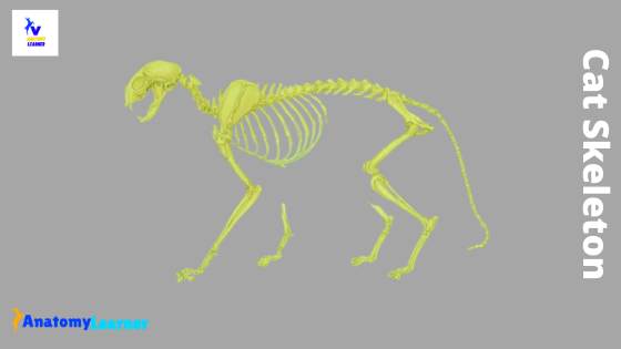
The skeleton of all mammals, including the cat, supports the body and protects the deeper soft tissue structure. There are not so many differences found in the cat skeleton compare to the dog. In this article, I will discuss the osteological features of bones from cat skeleton anatomy.
If you a beginner in learning veterinary anatomy, you may get confused to identify the cat bones from other animal’s bones. But here, I will provide you the special identifying features of every single bone of the cat skeleton. I will try to show you these identifying features with a cat skeleton labeled diagram.
So, after finishing this article, you will identify the cat’s bones with their special identifying features. You will also make a difference between a cat skeleton and other mammal’s skeleton.
Cat skeleton anatomy
Before going to the detailed anatomy of bones, I would like to provide you the most peculiar features of a cat skeleton. If you like these unique features and love to learn more, then you may continue this article. I found the following exceptional features in the skeletal system of a cat.
- The temporomandibular joint of the cat is a synovial joint and contains an articular disc.
- Cat skull has a short fascial and palatal region compares to other mammals.
- The skull is oval elongated in shape, and has strong, highly curved zygomatic bones.
- There is an incomplete orbital rim in cat skull anatomy.
- Premaxilla is a separate bone in cat skull anatomy.
- Masseteric fossa present in the mandible of a cat (also found in a dog).
- There may lack nuchal ligament in between the atlas and cranial thoracic vertebrae.
- The spine of the cat scapula divides the lateral surface into two halves – supraspinous and infraspinous fossa.
- The acromion process of scapula bone is more complex and has a pointed coracoid process than other mammals.
- The humerus of the cat possesses supracondylar foramen on the distomedial aspect.
- The radius and ulna of a cat are entirely separated bones in the skeleton.
- There are seven carpal, five metacarpal, and five digits in the forelimb of cat anatomy.
- The ischium bone of the cat is twisted, and the boney floor of the pelvis is V-shaped.
- There is no supracondyloid fossa in the cat’s femur bone.
- The patella is comparatively longer and has only one patellar ligament.
- The tibia and fibula of a cat have also separated bones in the skeleton.
There are other many differences in the different bones of cat anatomy. I suggest you read the full article to know the osteological features of different bones from a cat.
Cat skeleton bones
There are 230 bones in cats, and most bones are identical to those in a dog. You know, in every skeleton, there are axial and appendicular parts. The axial skeleton of the cat corresponds to the part associated with the midline and includes the skull, vertebrae, ribs, and sternum. Again, the appendicular skeleton of the cat comprises these bones located in the limbs. I will try to cover the following bone anatomy from the cat skeletal system.
Osteological features of skull, vertebrae, ribs, and sternum of cat
Exceptional osteological features of hindlimb bones of cat (scapula, humerus, radius and ulna, carpal, metacarpals, and phalanges bones)
Identifying osteological characteristics of hindlimb bones of cat (ox coxae, femur, tibia, and fibula, tarsal, metatarsal bone, and phalanges)
“Here, you will find the specific osteological features in every bone from a cat. If you know the general osteological features of an animal’s bones, then it will very easy for you to compare their features with other mammals. I suggest you read the general osteology if you have no idea on osteology.”
Cat skeleton skull
The skull of a cat attaches to the spinal column at the atlas and consists of more than 29 bones (may vary). Most of these cat’s bones found in bilateral pairs. But the unpaired bones of the cat locates on the medial plane.
Most of the skull bones of the cat join by the suture type articulation. You will find the more exceptional feature in the temporomandibular joint of the cat. This temporomandibular joint is a synovial type of joint and consists of an articular disc and mandibular symphysis.
The cat skull has a short fascial and palatal region as compare to the other common domestic mammals. Inside the cat skull, you will find some air-filled pockets known as sinuses. The cat has sizeable frontal sinus, sphenoid sinus, and maxillary sinus. If you perform a median section of the cat skull, you will easily find the frontal sinus and sphenoid sinus.
The orbital cavity of the cat is incomplete, and you will find a short orbital ligament at the lateral portion of the orbit. This short ligament joins with the zygomatic process of the frontal bone and the frontal process of the zygomatic bone. For your kind information, a cat has a solid and curved zygomatic process like a dog.
The cranium of the cat skull is proportionally equal in size to other mammals. You will find many foramina in the cat skull that allow the existence and entry of different blood vessels and nerves. The foramina of a cat is clustered on the skull’s ventrolateral and caudal surface, as found in the dog.
There are nuchal crest, external occipital protuberance, and round tympanic bullae find in the cat skull. The tympanic bullae contain an incomplete bony septum that divides the middle ear cavity into tympanic and endotympanic parts.
Bone identification from cat skull
I am not going to the detailed description of every single bone from the cat skeleton skull. Instead, I prefer to show you the most important bones of the cat skull.
Bone identification from the face region of cat –
- The incisive bone of the cat
- A nasal bone of the cat
- Lacrimal bone of cat skull
- Zygomatic bones of cat and its process
- Infraorbital foramen of cat skull
- The palatine bone of the cat
Bones from the cranium of a cat skull –
- A frontal bone of the cat
- The parietal bone of the cat
- Bregma of cat skull
- The occipital condyle and nuchal crest of cat skull
- Temporal bone and pterygoid bone of the cat
Mandible of cat skull anatomy
There is no remarkable difference in the mandible of cat anatomy except in temporomandibular articulation. You will find the same osteological features in the mandible of a cat as found in a carnivore.
You will find the masseteric fossa in the mandible of the cat. Let’s try to identify the following other osteological features from the cat mandible.
- Body and ramus of cat mandible
- Masseteric fossa of cat
- Angular, condylar, and coronoid processes of cat mandible
- Mental and mandibular foramen in cat mandible and
- Mandibular symphysis of cat mandible
Vertebrae from cat skeleton anatomy
You will find seven cervicals, thirteen thoracic, seven lumbar, three sacral, and eighteen to twenty caudal vertebrae in the vertebral column of a cat. The vertebrae of a different region of a cat skeleton display the typical characteristics found in the same region of other domestic mammals.
The atlas and axis of the cat show more specialization and less resemble the other vertebrae. You will find an extensive palpable transverse process or wing in the atlas of the cat. In the axis, there are long dorsal spinous processes and prominent dens in the cranial end.
You will find transverse foramina in the transverse process of all cervical vertebrae of the cat (except the seventh cervical vertebrae). The cat lacks a nuchal ligament extending between the cranial thoracic vertebrae and the atlas.
The thoracic vertebrae of the cat possess the extensive dorsal spinous process. The height of these dorsal spinous processes gradually decreases caudally. You will also find the distinguished mamillary process in the thoracic vertebrae of cat anatomy.
There present relatively long lumbar vertebrae in cats, and they possess the cranially directed transverse process. You will find the mammillary process and accessory process in the lumbar vertebrae of the cat.
There are three sacral vertebrae fuse to form the single sacrum bone. The original sacral dorsal spinous process remains individually distinct after the fusion of the bodies and transverse process.
The number of caudal vertebrae of a cat may vary in different species. Ideally, you may find eighteen to twenty caudal vertebrae in cat vertebral column anatomy. The caudal vertebrae of a cat possess only the body and gradually lose their processes. You may miss caudal vertebrae in some species of cat.
Ribs and sternum anatomy of a cat
There usually are thirteen pairs of ribs in a cat skeleton. These ribs tend to be short but rather broad at the cranial thorax. The head and tubercles of the cat ribs articulate with thoracic vertebrae. But, the head and tubercle of the first ribs of the cat articulate only with the first thoracic vertebrae.
The head of the second rib articulates with the caudal body of the first thoracic vertebrae and the cranial body of the second thoracic vertebrae. Again the tubercle of the second rib articulates with the transverse process of the second thoracic vertebrae of the cat.
First to ninth ribs of the cat articulate directly with the sternum (sternal ribs). The cartilages of ribs, ten to twelfth, unite to form the costal arch of the caudal thorax. The last rib is a floating rib in which cartilage remains separate from the others.
There are eight bony sternebrae in the cat sternum. The first part of the sternum is the manubrium sterni, and the last part is the xiphoid process. The first costal cartilage of the cat’s rib directly articulates with the manubrium sterni.
The appendicular skeleton of cat anatomy
The appendicular skeleton consists of the bones from the forelimb and hindlimbs. You will find different long straight long bones in the limbs of the cat skeleton. The osteological features of these bones may vary due to the small size of the domestic cat.
I will show you the osteological features from the thoracic limbs and pelvic limbs of a cat –
- Thoracic limb (scapula, humerus, radius – ulna, carpal, metacarpal, and phalanges bone of cat) and
- Pelvic limb (consists of hip bones, femur, tibia- fibula, tarsal, metatarsal, and phalanges bones)
Scapula and humerus of cat
The scapula of the cat is triangular, and the spine divides the lateral part into two halves. The dorsal border of the cat scapula positions dorsal to the thoracic spinous process.
The acromion process is complex in a cat rather than the other domestic mammals. You will find the hamate and suprahamate processes in the acromion process of cat scapula anatomy.
A pointed coracoid process may find on the supraglenoid tubercle of the cat scapula. There also presents an infraglenoid tubercle at the distal end of the caudal border of the cat scapula.
“If you want to learn the details anatomy of animal scapula, then go to the general anatomy section. That guide will help you to understand the scapular anatomy easily and help you to differentiate the cat scapula from others.”
The humerus bone of a cat is comparatively long and less twisted. It is the most robust bone in the cat skeleton and posses a round head. You will find a supracondylar foramen on the distomedial aspect of the cat humerus bone. But in some cat species, this supracondylar foramen may absent. The median nerve and brachial artery pass through this supracondylar foramen.
On the craniodental aspect of the cat humerus, there present the radial fossa. Medial to this radial fossa, you will also find the coronoid fossa. The supratrochlear foramen is not present in the cat humerus bone.
In the proximal part of the lateral aspect of the cat humerus, there is a tricipital line. Along this tricipital line, you will find prominent tuberosity (known as deltoid tuberosity). Let’s find the following osteological features from cat humerus –
- Head, lesser and greater tubercle
- Crest of the greater tubercle
- Tricipital line and deltoid tuberosity
- The brachial groove of the lateral aspect
- Coronoid, radial fossa, trochlea, and capitulum
Anatomical features of radius and ulna bones from cat skeleton
The radius and ulna bones of the cat skeleton remain separate in their entirety. Proximally, the radius bone of the cat positions cranial and slightly lateral to the ulna bone. Distally, the radius bone is positioned medially to the ulna bone.
You will find narrow interosseous spaces that extend throughout the length of the bones. Other osteological features of the radius and ulna bones of a cat are similar to other animals. From the cat radius bone, you might identify the following osteological features.
- Head and shaft of the cat radius bone
- Radial tuberosity of cat radius and
- Styloid process of cat radius (medial aspect)
Again, cranial part of the olecranon process of ulna bone, there are two small tubercles. Let’s find out the following osteological features from the cat ulna bone.
- Anconeal process and olecranon tuberosity of cat ulna bone
- Lateral and medial coronoid process
- A trochlear notch of the cat ulna bone
- Articular circumference of cat ulna and
- Lateral styloid process of the ulna bone.
Carpus, metacarpal, and phalanges of cat’s forelimb
There is seven carpus bone in cat forelimb that arrange in two rows (proximal and distal row). You will also find the sesamoid bone on the medial aspect of the carpus bone in a cat.
There are also five metacarpal bones and five digits in the forelimb of the cat. The first digit contains two phalanges, and the remaining digits contain three phalanges. You will also find sesamoid bones at the palmar aspect of the metacarpal bones. The osteological features of metacarpal bones are similar to the dog bones.
The middle phalange of the cat is oblique and beveled. Again the distal phalanges is slightly modified.
Now, I will discuss the bones from the pelvic limb of the cat. But I would like to remind you again to read the basic anatomy of different bones from the general anatomy section.
Hip bones of a cat skeleton
A hip bone of the cat skeleton consists of ilium, ischium, and pubis bones. The left and right ossae coxarum forms the pelvis (fusion of ilium, ischium, and pubis bones).
The lateral part of the ossae coxarum is essentially parallel to the longitudinal axis of the cat. If you view from a craniocaudal direction, you will find the V-shaped bony floor of the pelvis.
The gluteal surface of the ilium is concave like the dog. The ischium is somewhat twisted, and the ischial tuberosity is almost flat.
From the cat hip bones of ossa coxae, you might identify the following osteological features. I will show you all these structures with the cat hip bone labeled diagram.
Acetabular fossa and acetabular notch (the notch is wide in cat)
Iliac creat of cat ilium bone
The concave gluteal surface of cat ilium bone
Cranial and caudal dorsal iliac spines
Tuber sacralae and tuber coxae
Cranial ventral iliac spine of cat
Alar spine of cat ilum bone
Ischiatic spine and greater ischiatic notch
Ischiatic tuberosity and ischiatic arch of cat ischium bones
Caudal ramus of cat pubis bone
If you want to know more about the animal hip bone, then read the details guide from the general animal anatomy section.
Femur of cat
There is no marked difference in the cat femur anatomy in comparison to other carnivores. The supracondyloid fossa is usually absent in cat femur bone.
In the proximal part of cat femur you will find – greater trochanter, lesser trochanter, trochanteric fossa, intertrochanteric crest, and third trochanter. Again, there are medial trochlear ridge, trochlear groove, lateral trochlear ridge, and condyles in the distal part of the cat femur bone.
A ligament of the head of the cat femur is present as an intracapsular ligament at the hip joint. The Patella of the cat is more pointed at the distal end. There is only one patellar ligament in a cat. But in the stifle joint of the cat, there are collateral ligament, cranial and caudal cruciate ligaments, medial and lateral menisci, transverse ligament, and meniscofemoral ligament.
Tibia – fibula, carpus, and metacarpal of a cat skeleton
The other bones from the hindlimb of the cat skeleton are similar to other carnivores (except tibia –fibula bones). The tibia and fibula of the cat remain separate throughout life. The tibial crest of the cat tibia bone is very prominent.
The upper part of the tibia bone is prismatic, and the lower part is cylindrical. Again the fibula of a cat is a long and thin bone that extends the whole length of the tibia bone.
There are seven bones in the carpus of the cat that arrange in two rows. You will find one rudimentary metacarpal bone and four complete metacarpal bones in the hindlimb of the cat.
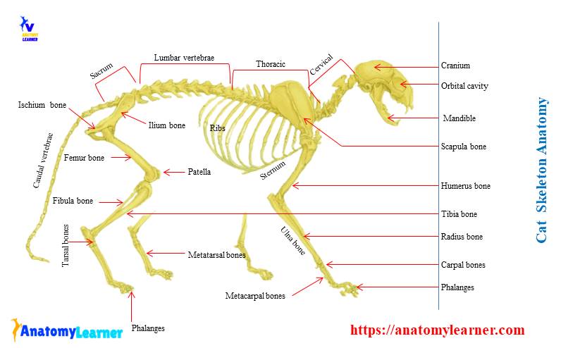
Cat skeleton anatomy labeled diagram
Now, I will show you all the bones from the cat skeleton with a diagram. If you find any mistakes in this cat anatomy labeled diagram, please let me know.
I hope this cat skeletal system anatomy labeled diagram might help you understand and identify all the cat’s bones. If you need more labeled diagrams of cat anatomy, you may follow anatomy learners on social media.
“I will update the information and images of cat skeletal anatomy on demand or if necessary. You will get the updates of cat skeletal system anatomy here or on social media.”
Frequently asked questions on cat skeletal anatomy
In this part of the article, you will find the answers to frequently asked cat skeletal systems. If you have your questions about a cat, please let me know.
Why are cats so bendy?
The vertebrae of the cat have some unique features from the vertebrae of other animals. These vertebrae of the cat have a unique, flexible, and elastic disc that gives the cat more flexibility.
Are cat’s bones strong?
Yes, the cat’s bones are so strong than other animals. Even the kitten’s bones are also more robust than other animals.
What types of skeletons do cats have?
You will find all types of bones in the cat skeletal system like – long, short, flat, irregular, and sesamoid bones.
What is the strongest bone in a cat’s body?
All the bones of a cat are comparatively strong than other carnivores. But the femur of a cat is the strongest bone in a cat’s body.
Conclusion
This simple guide might help you to get the basic anatomy of a cat skeleton. I always suggest you read the osteological features of any bones from a cat with a helpful diagram or model. The cat skeletal anatomy labeled diagrams that provide in this article might help you a lot. But if you need more cat anatomy labeled diagrams, please let me known.