
The cow ear anatomy consists of external, middle, and internal parts. It is the organs related to hearing and equilibrium, and are considered as vestibulocochlear organs of the animal’s body. Here, I will describe the detail anatomical features of the cow’s external, middle, and internal ear with diagrams.
Quick answer: the cow ear anatomy consists of 3 natural divisions – external, middle, and internal. The external division contains the auricle and the external acoustic meatus, where the middle division contains the tympanic cavity and the auditory ossicles. Finally, the internal division consists of the osseous and membranous labyrinth.
Now, I will describe all the above-mentioned contents from the external, middle, and internal divisions of the cow’s ear. I will also compare the anatomical features of the ear among different animal species at the end of this article.
Cow ear anatomy
It is better to know the special features of the cow ear anatomy before going for a details description. The following are the special anatomical features that you should know and identify from the cow ear –
- A cow’s ear naturally divides into 3 divisions: external, middle, and internal.
- The auricle or pinna of the cow’s external ear comprises 3 cartilages (conchal, scutiform, and annular)
- An external acoustic meatus extends from the conchal cavity to the tympanic membrane.
- The tympanic cavity is located in the middle ear and comprises auditory osicles (malleus, incus, and stapes)
- The lateral membranous wall of the tympanic cavity consists of the vestibular (oval window) and cochlear (round window) windows.
- Again, the internal ear of a cow consists of a complex membranous sac (the membranous labyrinth) and a series of body cavities (osseous labyrinth).
- The osseous labyrinth consists of the vestibule, the cochlea, and the semicircular canals, and
- The membranous labyrinth consists of the utricle, the saccule, the semicircular ducts, and the cochlear duct.
Parts of the cow’s ear
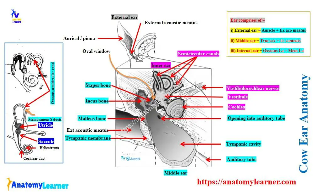
For description purposes, the cow ear divides into 3 parts or divisions –
- External ear of cow: consists of the auricle / pinna and the external acoustic meatus.
- Middle ear of cow: consists of the tympanic cavity and its contents (auditory ossicles and tympanic membranes), and
- Inner ear of cow: consists of osseous labyrinth (semicircular canals, vestibule, and cochlea) and membranous labyrinth (semicircular ducts, utricles, saccule, and cochlear ducts)
Here, the labeled diagram (Figure 1) shows the different parts and features of the cow ear anatomy. You should identify and memorize these features from the cow ear diagrams.
Cow ear features identification
Now, let’s enlist the anatomical features that you have identified previously from the diagram –
From the external ear of the cow:
- Auricle or pinna,
- External acoustic meatus
- 3 cartilages of the auricle (conchal, scutiform, and annular), and
- Conchal cavity
From the middle ear of the cow:
- Tympanic cavity or eardrum,
- Auditory ossicles (malleus, incus, and stapes),
- Oval window and round window (vestibular and cochlear windows),
- Auditory or Eustachian tube,
Finally, from the inner ear of the cow:
- Semicircular canals, vestibule, and cochlea,
- Urticle, saccule, semicircular ducts, and cochlear duct,
- Scala vestibuli, scala tympani, and helicotrema (will be identified under the membranous labyrinth of the inner ear)
Cow external ear anatomy
Here, I will describe two parts of the cow’s external ear –
- Auricula: this is a funnel-like organs that collect sound waves together with its muscles, and
- External acoustic meatus: It conveys the waves to the tympanic membrane. Another name for the tympanic membrane is the eardrum, which separates the canal from the cavity of the middle ear.
Auricle or pinna of the external ear
The auricle is attached by its base around the osseous external acoustic meatus. The opening of the auricle is directed laterally, and the long axis is partially vertical. It possesses 2 surfaces (convex and concave), 2 borders (rostral and caudal), a base, and an apex (Figure 2).
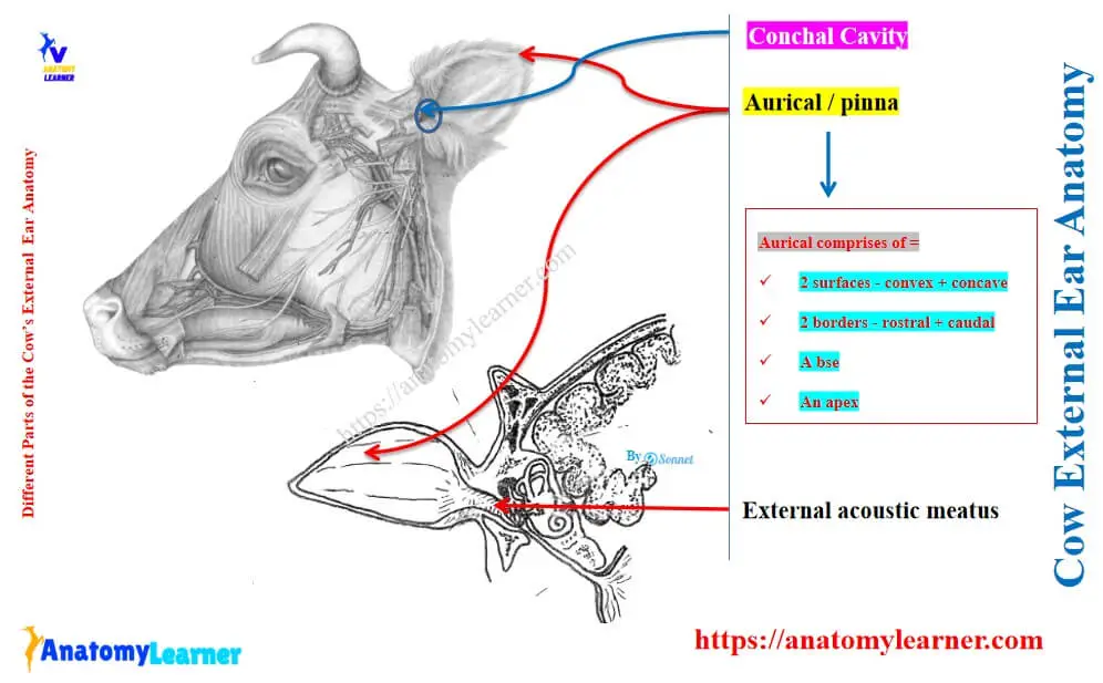
Surfaces: the convex surface of the auricle is also known as the dorsum that faces medially. It is widest in its middle, and the lower part is almost circular.
Again, the concave surface is the reverse of the dorsum and presents several ridges. These ridges of the concave surfaces subside towards the apex of the cow’s ear.
Borders: the rostral border of the auricle is largely convex. But it becomes concave near the apex and divides into 2 diverging parts. The caudal border of the auricle is convex.
Base and apex: the base attaches to the osseous external acoustic meatus of the petrous part of the temporal bone of the cow’s skull. You will find the parotid gland that overlaps the base ventrally and laterally. The apex of the auricle is flattened and pointed.
Cartilages of the auricle of the cow ear
The cartilages of the cow’s ear auricle are chiefly elastic and form the shape of the ear. They are arranged in the complicated arrangement of the cow’s ear muscles. You will find 3 cartilages in the cow’s ear structure –
- Auricular or conchal cartilage (larger),
- Scutiform cartilage (irregular quadrilateral plate), and
- Annular cartilage (incomplete small ring),
Auricular cartilage of the cow’s ear
This auricular cartilage determines the actual shape of the cow’s ear. Thus, you can easily determine its form in a general way without dissection.
The base part of the auricular cartilage (conchal) coils and forms a tube that encloses the cavity of the concha. This cavity is known as the conchal cavity of the external ear of the cow.
The medial surface of the auricular cartilage is strongly convex and shows some prominences. These prominences are the conchal eminence of the external ear.
You will find a narrow, pointed prolongation at the lower part of the medial margin of this cartilage. This narrow prolongation is the styloid process of the auricular cartilage.
Scutiform cartilage of the cow ear
The scutiform cartilage of the cow ear is a small, irregular, quadrilateral plate. It lies on the temporalis muscle just cranial to the base of the auricular cartilage.
The rostral end of this cartilage is thin and rounded. Again, the caudal end of the base is wider and thicker in the cow ear.
Annular cartilage of the cow’s ear pinna
The annular cartilage of the cow’s ear is also quadrilateral, but it curves to form an incomplete (three-fourths) of a ring. This cartilage encloses the osseous external acoustic meatus. It helps to form the cartilaginous part of the external acoustic meatus of the cow’s external ear.
External acoustic meatus of the cow’s external ear
The external acoustic meatus extends from the conchal cavity to the tympanic membrane of the middle ear. It directs medially, ventrally, and slightly rostrally toward the middle ear.
This external acoustic meatus consists of a cartilaginous part and an osseous part.
- Cartilaginous part: this part is the lateral one-third and is formed by the lower part of the auricular and annular cartilages.
- Osseous part: It is formed within the temporal bone.
These two parts of the external acoustic meatus are united by an elastic membrane to form the complete tube.
You will find 3 – 4 cutaneous ridges that run parallel with the border of the conchal cartilage. Here, the skin between ridges and below is thin and supplied with numerous sebaceous glands.
Again, the inner part of the acoustic meatus, the skin becomes thinner and tightly attaches to the underlying bone and cartilage. Thus, if there is any problem in the acoustic meatus, it is very painful.
The cartilaginous part is supplied with huge, colied ceruminous glands. These glands of the external ear secret cerumen or earwax.
Cow middle ear anatomy
You know the cow’s middle ear anatomy comprises –
- Tympanic cavity, and
- Its contents (auditory ossicles and auditory tube / eustachian tube),
Exceptional: only in the horse, you will find 2 remarkable diverticula of the latter, which are known as the Guttural pouches.
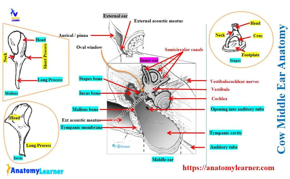
Here, diagram (Figure 3) shows the different structures of the middle ear of the cow.
You will find variation in the structure of the dog and cat ear compared to the ruminant ear. The following article might help you learn the details of the dog and cat ear anatomy in detail.
- Dog ear anatomy – external, middle, and internal ear features, and
- Cat ear anatomy – external, middle, and inner ear with diagrams,
Now, let’s discuss the tympanic cavity, auditory ossicles, and auditory tube from the middle ear of the cow.
Tympanic cavity of the cow’s middle ear
The tympanic cavity is an irregular biconcave space in the tympanic and petrous part of the temporal bone of the cow skull. It is located between the tympanic membrane and the internal ear.
This cavity is lined with the mucous membrane and communicates with the cow’s pharynx through the auditory tube. It contains the chain of auditory ossicles, muscles, air, and parts of the chorda tympani. Ossicles receive the vibration of the tympanic membrane and transmit it to the internal ear.
Parts of the tympanic cavity
The tympanic cavity consists of 3 parts or recesses –
- A main part or atrium,
- Epitympanic recess, and
- Relatively large ventral recess in the tympanic bulla,
All these parts or recesses are identified in the labeled diagram of the cow’s middle ear.
The main part or atrium lies immediately to the medial aspect of the tympanic membrane. Here, the epitympanic recess is located above the level of the tympanic membrane and contains the upper part of the malleus and the greater part of the incus bone.
The boundary of the animal’s tympanic cavity
The tympanic cavity of the cow’s middle ear is bounded by the following –
- Laterally by the lateral membranous wall,
- Caudally by the mastoid wall,
- Rostrally by the carotid or tubal wall,
- Dorsally by the tegmental wall (roof), and
- Ventrally by the jugular wall (floor),
The lateral membranous wall is formed by a thin tympanic membrane. This membrane consists of two windows –
- Vestibular window (oval window), and
- Cochlear window (round window),
The vestibular window is an opening which closed by the footplate of the stapes bone and its annular ligament. Again, the cochlear window is located below and caudal to the preceding window (oval window). This window is an irregular oval opening and is closed by a thin membrane that separates the cow’s tympanic cavity from the scala tympani of the cochlear.
The rostral carotid or tubal wall is narrow and pierced by a slit like tympanic opening of the auditory tube. Again, the tegmental wall of the roof is crossed in its medial part by the facial nerve.
Auditory ossicles of the cow’s middle ear
The auditory ossicles are the chain of bones that extends from the lateral to the medial wall of the tympanic cavity. You will find the following 3 ossicles in the middle ear of a cow –
- Malleus or hammer bone,
- Incus or anvil , and
- Stapes or stirrup bone,
Here, the malleus is the first one and attaches to the inner surface of the cow’s tympanic membrane. Again, the last bone (stapes) is fixed in the vestibular window of the middle ear.
Malleus or hammer bone
Malleus is the largest bone in the cow’s middle ear and consists of the head, neck, handle, and 2 processes.
Head: the head of the malleus is located in the epitympanic recess. It is smooth, convex above and rostrally. You will find a concave facet on its caudo-medial aspect of the malleus. This facet is for articulation with the body of the second bone (incus).
Neck: This is the constricted part below the head, and its medial surface is crossed by the chorda tympani nerve.
Handle: the handle of the malleus directed ventrally, inward, and rostral from the neck. It attaches along its entire length to the tympanic membrane.
Processes: there are 2 processes – long or rostral, and lateral or short. Here, the long process is pointed and projects rostral from the neck and attaches to the petrotympanic fissure. Again, the short process attaches to the upper part of the tympanic membrane.
Incus or anvil bone
The incus is the second bone and is located chiefly in the epitympanic recess. This bone resembles a tooth with two divergent roots. It consists of a body and two processes (long and short).
The body of the incus projects downward from the body and curves inward. You will find a small nodule on the extremity of the long process, and known as the os lenticulare. The head of the stapes bone articulates with the os lenticulare. The short process projects caudally and attaches to the wall of the epitympanic recess.
Stapes or stirrup bone
This is the third or last bone of the auditory ossicles and consists of the head, neck, two crura, and a base or footplate. Here, the head directs outward and articulates with the os lenticulare.
Two crura are rostral and caudal and directed inward from the head. They join at the end of the base by a thin membrane. Finally, the footplate occupies the vestibular window of the lateral membranous wall.
Auditory tube or eustachian tube of the cow’s middle ear
It is the tube that extends from the tympanic cavity to the pharynx, which equalizes pressure on the two surfaces of the tympanic membrane. It is directed rostrally, ventrally, and slightly inward.
The caudal part of the auditory tube lies at the medial side of the root of the muscular process of the petrous part of the cow’s temporal bone. It communicates with the cranial part of the tympanic cavity through a small slit like opening. Again, the pharyngeal opening lies on the caudodorsal part of the lateral wall of the cow’s pharynx.
Exception: in horses, there are guttural pouches, which are situated between the base of the cranium and the atlas dorsally and the pharynx ventrally. These are the larger mucosal sacs, each of which is a ventral diverticulum of the auditory tube.
Cow inner ear anatomy
The cow inner ear anatomy comprises –
- Membranous labyrinth: It is a complex membrane sac that contains fluid. It supports the auditory cells and the peripheral ramifications of the auditory nerve.
- Osseous labyrinth: This is a series of cavities in the petrous part of the temporal bone and encloses the membranous part.
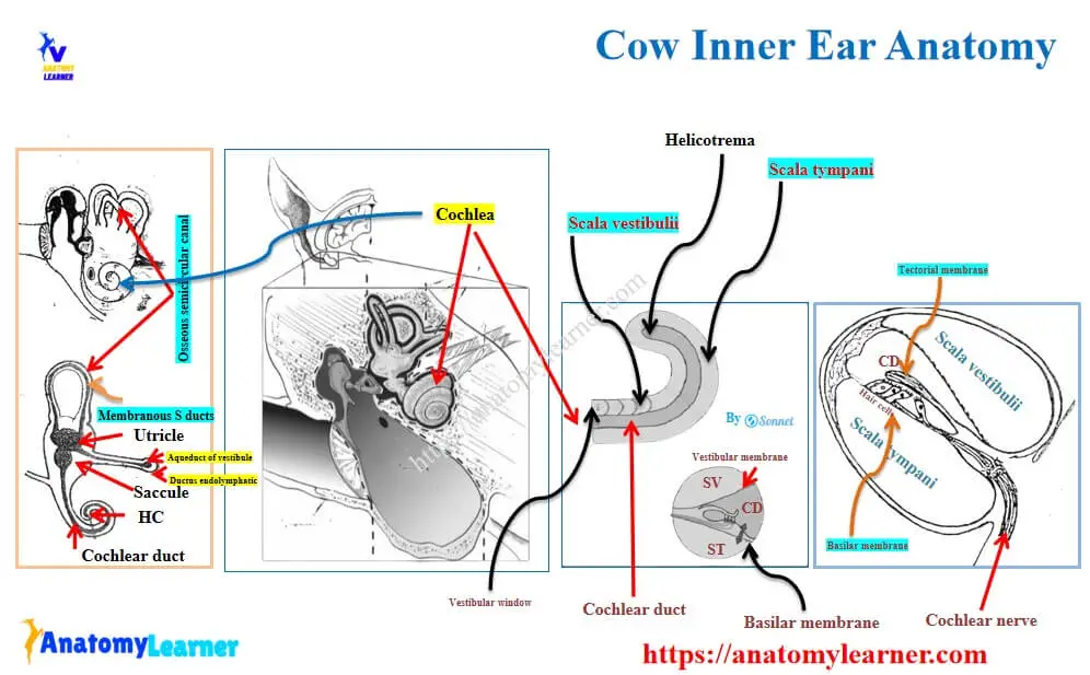
Let’s discuss both the osseous and membranous labyrinth from the middle ear of the cow. Here, the diagram (Figure 4) shows the different parts and features of the osseous and membranous labyrinth of the cow’s inner ear structure.
Osseous labyrinth of the cow’s inner ear
The osseous labyrinth is located in the petrous part of the temporal bone and just medial to the tympanic cavity. It consists of 3 divisions –
- Vestibule: a middle part,
- Cochlea: an anterior part, and
- Semicircular canals: a posterior part,
Vestibule of the osseous labyrinth
The vestibule is the center segment of the osseous labyrinth in the cow’s inner ear. It communicates anteriorly with the cochlea and posteriorly with the semicircular canals.
The vestibule of a cow’s inner ear is a small, irregular ovoid cavity. Its lateral wall separates it from the tympanic cavity and contains the vestibular window.
The medial wall corresponds to the fundus of the internal acoustic meatus. The medial wall is crossed by oblique ridges, which are known as the vestibular crest.
The vestibular crest separates the vestibule into 2 recesses: anterior (small) and posterior (larger) depressions. Here, the anterior small recess lodges the saccule of the membranous labyrinth. Again, the posterior larger depression lodges the utricle of the membranous labyrinth.
The anterior wall of the vestibule is pierced by an opening that leads into the scala vestibuli of the cochlea. Again, the posterior wall possesses 4 openings for the semicircular canals.
The lower part of the vestibular crest presents a small slit like posterior opening. This is known as the vestibular aqueduct (identified in the diagram).
Osseous semicircular canals of the inner ear
Semicircular canals are three in number in the cow’s inner ear structure –
- Anterior: nearly vertical,
- Posterior: also vertical, and
- Lateral: nearly horizontal,
Each semicircular canal forms about 2-3 circles. These semicircular canals are situated posterior and dorsal to the vestibule. They communicate with the vestibule through 4 openings.
Here, the inner end of the anterior and the dorsal end of the posterior canal unite to form the common canal. Again, the ampullate end of the anterior and lateral semicircular canals has the common opening.
The Cochlea structure of the cow’s inner ear
The cochlea is the cranial part of the osseous labyrinth. It has a short, blunt cone and apex. Here, the short blunt cone corresponds to the anterior part of the fundus of the internal acoustic meatus. Again, the apex of the cochlea is directed outward, forward, and downward.
The cochlea consists of a spiral canal termed as osseous modiolus (appearance of a shell of snail). In ruminants, the spiral canal forms two and a half turns around the central column.
There is a thin plate of bone that projects from the osseous modiolus that divides the cochlear cavity into –
- Scala vestibulii (upper one), and
- Scala tympani (lower one)
Here, the diagram (Figure 5) shows the scala vestibuli and scala tympani from the cow ear’s cochlea. These two scalae communicate through the opening at the apex and are termed the helicotrema.
Close to the beginning of the scala tympani, there is a small canal that opens behind the internal acoustic meatus. This opening communicates between the scala tympani and the subarachnoid space.
Membranous labyrinth of the cow’s inner ear
The membranous labyrinth lies within the osseous labyrinth and attaches to the latter by delicate trabeculae. It conforms more or less closely to the osseous labyrinth of the inner ear.
You will find 4 divisions in the structure of the membranous labyrinth of the cow’s inner ear structure –
- Utricle (larger sac),
- Saccule (small, spherical),
- Semicircular ducts, and
- Cochlear duct,
Here, the diagram (Figure 6) shows the different parts and features of the membranous labyrinth from the cow’s inner ear structure.
Utricle of the membranous labyrinth
The utricle is the larger sac that lies in the posterosuperior part of the vestibule. It receives the opening of semicircular ducts and small utriculosaccular ducts at its lower part.
Saccule of the membranous labyrinth
The saccule is located in the spherical recess of the vestibule. There is a ductus reuniens that proceeds to open into the cochlear ducts.
You will find another duct (endolymphatic duct) that passes from the posterior part of the saccule and joins with the utriculosaccular ducts. Together, they traverse the vestibular aqueduct and terminate under the duramater on the caudal part of the medial surface of the petrous part of the cow’s temporal bone.
Semicircular ducts of the membranous labyrinth
The semicircular ducts lie in the osseous semicircular canal of the osseous labyrinth. They occupy only one-fourth of the osseous cavities.
Cochlear ducts of the membranous labyrinth
The cochlear duct is a spiral tube located within the cochlea. It begins at a blind end of the vestibule and ends at a second blind end. They together attached to the apex of the cochlea.
The vestibular part of the cochlear ducts connects the saccule by the ductus reunies.
What are the differences in the middle ear structure between a cow and a horse?
Answer: The structure of the middle ear between cows and horses is almost similar, except for having guttural pouches in horses. The guttural pouches are the only features in the horse and are located between the cranium bone and pharynx.
The auditory or eustachian tube of the horse shows the ventral diverticulum near the pharynx. These two (lateral and medial) diverticula are the mucosal sacs and are termed the guttural pouches.
What is the chorda tympani?
Answer: The chorda tympani is the branch of the facial nerve of the animal. This structure enters the middle ear of the animal through the posterior canaliculus.
It crosses the tympanic cavity between the malleus and incus auditory ossicles. Finally, it reaches the anterior wall and makes it exist by the anterior canaliculus.
Why is the auditory tube of an animal’s ear important?
Answer: The auditory tube maintains the atmospheric pressure in the middle ear of an animal by its communication with the pharynx. Thus, a throat problem occasionally spreads to the middle ear.
Conclusion
So, the cow ear anatomy describes all the features of the external, middle, and inner divisions. Here, the 3 cartilages from the cow’s external ear should be identified practically.
The anatomy of the tympanic cavity from the middle ear of a cow is important as it contains auditory ossicles. Finally, the anatomical features of the osseous and membranous labyrinth might be known properly to understand the hearing function and equilibrium.