The cow skull anatomy comprises cranial and facial bones. Most of these bones are irregular in shape and united immovably with the help of sutures (except the mandible). You will find some paired bones and single bones in the cow skull anatomy.
The cranial bones of the cow or ox skull from the cranial cavity accommodate the brain, different parts of orbit, middle, and internal ears. Again, the facial bones of the cow or ox skull possess some essential structures that are practically important.
Here, I will show you all the bones of the ox or cow skull anatomy (large ruminant) with the labeled pictures. Then I will also show you the essential parts or structures of these bones to identify them quickly at your anatomy laboratory.
So, if you are interested to learn the details of the cow skull bones and identifying all the structures from these bones, you may continue this article.
Cow skull anatomy
The skull of a cow or ox is more clearly pyramidal than that of the horse. It is relatively shorter but broader than these of a horse. You will also find some differences in different structures and bones of a cow skull anatomy compared to a horse or a dog.
The cranium of the cow skull is quadrangular and larger externally than in the horse. However, I will provide some information at the end of the article to help you identify the skull of different animals.
Before starting learning the cow skull anatomy, you might have good pieces of knowledge on the cranial and facial bones. Let’s enlist the bones from the cow skull –
- The cranial bones (both single and paired), and
- The fascial bones from the skull (both single and paired)
Name and number of the bones from the cranial part of the cow skull –
- Occipital bone (single) – possess three parts
- Frontal bone (paired)
- Temporal bone (paired) – possess two defined parts
- Parietal bone (paired)
- Interparietal bone (paired)
- Sphenoid bone (single) – includes two parts
- Ethmoid bone (single)
These are the cranial bones from the ruminant skull anatomy. All these bones are shown in the ruminant skull labeled picture.
Now, let’s enlist the fascial bones from the ox or cow skull –
- Lacrimal bone (paired)
- Malar or zygomatic bone (paired)
- Maxilla bone (paired)
- Pre-maxilla bone (paired)
- Palatine bone (paired)
- Nasal bone (paired)
- Pterygoid bone (paired)
- Turbinate bone (paired) and
- Vomar bone (single)
I hope you can identify all these bones with the help of labeled ox skull anatomy and ruminant skull anatomy video. Again, if you want to know more about these bones, let’s continue this article till the end.
Bones and parts of cow skull anatomy
In this part of the article, I will go with a short description of every bone from the cow skull anatomy. Here, I will try to show you the essential structures from every single bone of the cow skull (large ruminant).
I am sure you could identify all the bones from the cranial and fascial parts of the cow skull. But, as a veterinary student, you might have a good piece of knowledge on the different essential structures of these bones.
“I am not going to repat the list of bones from the cranial and fascial parts of the cow skull.”
First, I will show you all the structures from all cranial bones of the ruminant skull anatomy. Then, I will go with the detailed structures or parts of the fascial part of the cow (ruminant) skull.
An occipital bone of cow skull
The occipital bone of the ox skull anatomy forms the ventral and caudal parts. You may divide the ox occipital bone into three parts for better understanding.
The three parts of the occipital bone are – lateral mass, basioccipital, and the supraoccipital part.
From the lateral mass of occipital bone, you might identify the following essential structures –
- The condyle of the occipital bone
- A foramen magnum
- The paramastoid process of occipital bone
- A condyloid fossa of occipital
- The hypoglossal and condyloid foramen of the occipital bone
Again, from the basilar part of the occipital bone, you might identify the following structures –
- The basilar tubercle of the basilar part, and
- A jugular foramen
In addition, there is a squamous or supraoccipital part in the occipital bone of the ox. You will find the following essential structures in the squamous part of the cow occipital bone –
- The external occipital protuberance, and
- A mastoid foramen
I hope you can identify all the structures as mentioned above from the different parts of the cow occipital bone with the help of the labeled diagram. But, if you want to know more about the anatomy of these structures from the occipital bone, continue the below-mentioned part.
Lateral mass of the cow occipital bone
Do you know how the condyle looks? Well, you will find two condyles in the caudal surface of the ruminant skull anatomy. These condyles are oval-elongated structures, broad above and narrow below. The condyle articulates with the concave cranial surface of the atlas vertebra.
You will find a larger foramen between these two condyles of the cow skull (ventral surface). This is the foramen magnum of the cow skull that contains the spinal cord, the spinal root of the cranial nerve, spinal arteries, and vein.
Again, you will find a large projection (process) on either side of the corresponding condyles at its external aspect. This large projection is known as the paramastoid process of the cow skull. In addition, the condylar fossa locates in between the condyle and paramastoid processes.
“Look at the ventral surface of the skull, you will find a depression in between the condyles and paramastoid process on the both side (condyloid fossa).”
In the condyloid fossa, a small foramen contains the hypoglossal nerve. This foramen is known as the hypoglossal foramen of the cow skull anatomy. Again, behind the hypoglossal foramen, you will find another passage (foramen): the condyloid foramen.
Basilar part of the cow occipital bone
The basilar part of the cow occipital bone is short, wide, and very thick. It helps to form the posterior part of the floor of the cranial cavity. The cerebral surface of the basilar part is deeply concave.
Again, the basilar part of the occipital bone joins with the post-sphenoid bone. Here, you will find a prominent crest in the sphenoidoccipital bone. Two sizeable muscular tubercles present and ventrally mark the junction with the basisphenoid bone.
In addition, the lateral border of the basilar part forms the medial boundary of the jugular foramen. The petrioocipital fissure is very short and narrow.
The supraoccipital or squamous part of the ox occipital bone
The supraoccipital, interparietal, and parietal bones fuse before birth and separate from the lateral part of the occipital bone by a transverse suture. You will find a rough tuberosity at the center of the external surface, known as the external occipital protuberance.
This occipital protuberance provides attachment to the ligamentum nuchae. Again, there is some rough depression present on the external surface for muscular attachment. You will also find a median external occipital crest that extends ventrally from the protuberance.
In addition, there is a mastoid foramen that locates on either side of the squamous part of the occipital bone. The cranial surface of the squamous part of the occipital bone is concave and rough.
The frontal bone of ruminant skull anatomy
The frontal bone of the most ruminant skull anatomy is very extensive and forms about one-half of the entire length of the skull. First, you might identify the most important structures from the frontal bone.
Here, in the labeled skull diagram, you will find the following essential structures from the frontal bone –
- A large central intercornual protuberance
- The corneal processes of the frontal bone
- A supraorbital groove and process (with foramen)
You know, there are two frontal bones present in the cow skull. The frontal bone possesses a parietal and lateral border. You will find a large central intracorneal protuberance in between the parietal border of both frontal bones.
This intercornual protuberance is the highest point of the cow skull anatomy. Again, you will find a cornual process at the junction of parietal and lateral borders of the frontal bone. The cornual process supports the horn and elongated conical form that greatly vary in size, length, curvature, and direction.
There is a zygomatic process between the nasal and parietal margin which is very short and joins with the frontal process of the zygomatic bone. You will find a supraorbital groove on the frontal bone of the cow skull. Within this supraorbital groove, there is a presence of supraorbital foramen.
In addition, the orbital part of the frontal bone is extensive and perforated caudally by orbital opening and ventrally by the ethmoidal foramen. The frontal bone also contains the frontal sinus, which is very extensive in a cow.
The temporal bone of cow skull anatomy
Temporalis are the paired bones in the cow skull anatomy that form the cranial cavity’s lateral wall. Each temporal bone of the cow skull has two parts – squamous and petrous part.
You might identify the following vital structures from the squamous and petrous parts of the ruminant temporal bone. Fine, let’s identify the structures from the squamous part of the temporal bone –
- A body and the zygomatic process of the temporal bone
- The temporal fossa (partially formed by the body of temporal bone)
- A retroarticular process, and
- The post glenoid foramen
Again, from the petrous part, let’s try to identify the following structures –
- The external acoustic meatus
- A stylomastoid foramen
- The tympanic bulla and hyoid process
The squamous section of the temporal bone is relatively small and divides into two parts by temporal crest. You will find a body and a zygomatic process in the squamous part of the cow temporal bone. The body helps to part from the temporal fossa.
The zygomatic process of the temporal bone is short and weak and articulate with the zygomatic bone. Again, the zygomatic process joins with the process of malar bone and presents post-glenoid foramen behind the glenoid cavity.
The petrous section of the temporal bone is the short but most complex bone in the cow skull. You will find the external acoustic meatus, tympanic bulla, stylomastoid foramen, and internal acoustic meatus within the petrous part of the temporal bone.
The tympanic bulla is large and laterally compressed in the cow skull. It separates from the occipital bone by a narrow opening (petro-occipital fissure). Again, you will also find the temporal meatus within the temporal bone of the cow skull.
Parietal and interparietal bones of the ruminant skull
The parietal bone of the ruminant skull, located at the dorsal part of the caudal wall, bend sharply rostrad along the lateral wall. This bone fuses with the occipital bone below and frontal bone above.
You will find an upper horizontal part and a lower vertical part in the parietal bone of the ruminant skull. Again, the outer surface of the parietal bone is concave and helps to form the temporal fossa.
A prominent external sagittal crest in the parietal bone continues with the temporal crest ventrally. So, you might learn and identify the following structures from the parietal bone of a cow skull –
- The temporal fossa (partially formed by the parietal bone), and
- A parietal crest and temporal crest
In addition, the interparietal bones are paired but fused before or shortly after birth with the parietal and supraoccipital bones.
The sphenoid bone of ox skull anatomy
The sphenoid bone of the ox skull anatomy forms the floor of the cranial cavity. You will find two defined parts of the sphenoid bone – basisphenoid and presphenoid parts.
From the basisphenoid part of the sphenoid bone, you might identify the following structure –
- The pituitary fossa
- Pterygoid crest and process
- The formane ovale and foramen orbitorontundum
Again, from the pressphenoid part of the temporal bone, you might identify the optic foramen and other structures.
At the medial of the basisphenoid bone of cow skull anatomy, you will find deep sella turcica (pituitary fossa). The wing of the sella turcica is small but forms the prominent thick pterygoid crest. Two foramina occur on either side of the pterygoid crest.
You will find a sizeable rounded foramen orbitorontundum rostrally. Again, caudal and ventral to its center, you will find the foramen ovale. The formen ovale takes place in the middle and lateral notches of the caudal border of the pterygoid wing.
The anterior part of the sphenoid body possesses a central ridge that joins with the crista Galli. Again, the wing of the presphenoid is thick and overlapped by the frontal bone. You will find the optic foramen at the anterior part of the pre-sphenoid bone.
An ethmoid bone of cow skull
This is the single cow skull that forms the anterior part of the floor of the cranial cavity. It also includes the partition between the cranial and nasal cavity of the ruminant skull.
You will find the horizontal and perpendicular parts in the ethmoid bone of the cow skull. The horizontal part of the ethmoid bone comprises the median crest (crista Galli), right and left olfactory fossa, and cribriform plates. Again, the cribriform plate is a bony network between the cranial and nasal cavity of the cow.
In addition, the perpendicular part of the ethmoid bone forms the dorsal portion of the osseous nasal septum. You will find the ethmoidal cell in the lateral masses of the perpendicular ethmoid bone.
Cow skull parts (fascial bones)
I hope you got a good idea of the bones from the cranial part of the cow skull. In this part of the article, you will know about the details of the facial bones of the cow skull anatomy. In the fascial part of the cow skull, you will find more than nine important bones (eight single and one paired).
“As I already enlisted the fascial bones from the cow skull before, so I am going to skip the list.”
What should you do now? First, you should identify the bones from the fascial part of the cow skull. Then, you might know and identify the structures of the specific bone belongs.
Lacrimal bones of the ox skull
The lacrimal bone of the ox skull is extensive and located at the anterolateral portion of the skull. Superficially, it articulates with the frontal bone, ventrally maxilla bone, anteriorly nasal bone, and posteriorly malar or zygomatic bone.
You will find an extensive fascial part that is concave and bears no lacrimal process. The orbital part of the lacrimal bone bears a very large and thin protuberance (lacrimal bulla). You will find a small lacrimal fossa for the lacrimal sac just caudal to the orbital margin.
Malar or zygomatic bone of ruminant skull anatomy
In most ruminant skull anatomy, you will find a long malar or zygomatic bone. This malar bone helps in the formation of the orbit of the ruminant.
From the malar or zygomatic bone, you might identify the following two essential structures –
- The frontal process (shown in diagram) of the zygomatic bone, and
- A temporal process of the zygomatic bone
The frontal process turns dorso-caudally and joins with the zygomatic process of the frontal bone. Again, the temporal process continued caudally and overlapped by the zygomatic process of the temporal bone. Thus, it helps the information of the complete zygomatic arch.
Maxilla bone of the cow skull
The maxilla is the most prominent bone in the fascial part of the cow skull. This bone helps form the upper jaw, the roof of the oral cavity, and the floor of the nasal cavity. You will find a relationship with the nasal, premaxilla, lacrimal, malar, and palatine bones with the maxilla bone.
The maxilla bone is relatively shorter but broader and higher than the horse. You might identify the following essential structures from the maxilla bone of the cow skull.
- A fascial tuberosity (at the lateral surface)
- The infraorbital foramen (single or double)
- A maxillary tuber (small, laterally compressed)
- The interalveolar border and sockets
- A palatine process of the maxilla bone
The fascial tuberosity of the maxilla bone locates just above the infraorbital foremen or the lateral aspect of the maxilla bone. Again, the infraorbital foramen locates at the lateral part of the maxilla bone at the level of the first cheek tooth. You know, this foramen is the external opening of the infraorbital canal.
You will find a small, laterally compressed maxillary tuber in the maxilla bone. Here in the maxilla bone, the zygomatic process is concise.
There are concave interalveolar borders present in the cow maxilla bone. The palatine process of the cow is broader but somewhat shorter than that of the horse. You will find a maxillary foramen at the medial side of the lacrimal bulla.
The maxillary sinus of the cow skull is very short and undivided.
Pre-maxilla or incisive bone of the ox skull anatomy
The body of the incisive (pre-maxilla bone) is thin and flat in the ox skull anatomy. Pre-maxilla forms the anterior part of the upper jaw of an ox or cow. Each premaxilla bone consists of a body, a nasal process, a palatine process, and a palatine fissure.
So, you might identify the following essential structures from the premaxilla bone of a cow or ox skull –
- The nasal and palatine processes, and
- Palatine and interinsisive fissures of the premaxilla bone
The nasal process is short, convex laterally, and does not reach the nasal bone of the cow skull. Again, the palatine process is narrow and grooved on its nasal surface. In addition, the palatine fissure is vast in the ox.
Palatine bones of cow skull anatomy
The palatine bone is the more extensive bone in the cow skull anatomy. It possesses two defined parts – horizontal and perpendicular palatine bones. The horizontal part of the cow palatine bone forms one-fourth or more of the hard palate. Again, the perpendicular part forms the caudal part of the lateral wall of the nasal cavity.
So, there are many important structures that you may identify from the palatine bone of the cow skull. Fine, I will enlist some of these important structures that you should identify from the palatine bone of the cow skull.
- The horizontal and perpendicular part of the palatine bone of a cow
- Major and minor palatine formina
- A palatine canal and palatine sinus
- The sphenopalatine foramen
You will find the major palatine foramen near the junction of the palatine bone with the maxilla bone. Again, you will find some minor palatine foramina on the palatine bone of the cow skull. The palatine groove is not very distinct in the horizontal part of the palatine bone.
You will find the palatine canal within the horizontal part of the palatine bone. In addition, the perpendicular component of the palatine bone is quadrilateral and has a thin plate. This bone joins with the pterygoid bone along its dorsal border.
You will also find sphenopalatine foramen at the dorsal edge of the palatine bone of the cow. The ethmoid and sphenoid bones help in the formation of this sphenopalatine foramen.
A nasal bone of a cow
The nasal bones of the cow are straight in its length but strongly curved from side to side. These nasal bones of the cow skull do not fuse laterally with the adjacent bones. You will find a pointed caudal extremity that fits into the notch between frontal bones.
The cranial end of the nasal bone is broader and divided into two parts by a deep notch. You will find a small extension of the frontal sinus within this nasal bone of the cow.
A pterygoid bone of ox skull
The pterygoid is also a paired bone present in the ox skull. Each bone is in the form of a thin plate. The medial surface of the pterygoid bone forms the lateral wall of the nasal cavity. Again, the lateral surface articulates with the palatine and pterygoid process of the sphenoid bone.
You will find a ventrally directed pterygoid hamulus in the pterygoid bone of the ox skull. This pterygoid hamulus is in the form of a hook.
Turbinate and vomer bones of a cow skull
You will find two turbinate bones (nasal conchae – dorsal and ventral) in each of the nasal cavities. These turbinate bones are formed by the thin plate of the bony net.
The dorsal turbinate bone of the cow skull anatomy is less cribriform and fragile. This dorsal turbinate is most expansive in the middle and small at either end. It joins with the ethmoidal crest of the nasal bone and is curved ventrally.
Again, the ventral turbinate bone of the cow skull is shorter and much broader. This bone joins with the maxilla by a basal lamella and slopes ventrolaterally.
You will also find the ethmoid turbinates bone between the dorsal and ventral turbinate bones at their caudal ends.
On the other hand, vomar is a single, thin plate located at the median plane. This vomar bone extends from presphenoid bone to premaxilla bone.
Again, this vomar bone takes part in the formation of the nasal septum. The proximal end of the vomar bone joins with the presphenoid bone. In addition, the distal end of the vomar is grooved and joins with the palatine process of the premaxilla bone.
Mandible of the cow
The mandible of the cow consists of two halves and forms the lower jaw. These two halves of the mandible fuse incompletely by the mandibular symphysis. You will find a body and two rami in the anatomy of the cow mandible.
Here, I will not provide a detailed description of the anatomy of the cow mandible. Instead, I prefer to show you the essential structures from the cow mandible.
Body of the cow mandible
The body of the cow mandible is the horizontal part of the bone and consists of two portions – anterior and posterior. Again, the anterior portion of the body possesses two surfaces – lingual and labial surfaces.
The lingual surface of the anterior body is concave, and the labial surface is convex. In addition, the posterior portion extends from the corresponding side of the body and possesses two borders, two surfaces, and two extremities.
The most important structure of the body of the mandible is the mental foramen. This mental foramen locates at the anterior aspect of the lateral surface. This mental foramen is the external opening of the mandibular canal.
Ramus of the cow mandible
Ramus is the verticle part of the cow mandible and possesses two surfaces, two borders, and two extremities. There are different structures that you might identify grossly from the ramus of the cow mandible.
Now, I will enlist some of the structures that you should identify from the ramus of the mandible –
- The condylar process of the mandible
- A coronoid process of the mandible
- The neck of the mandible
- A mandibular formaen with mandibular canal
- The mandibular notch, and
- A groove for the lingual nerve
You know, the condylar process locates behind and joins with the squamous part of the temporal bone. Again, the coronoid process projects upward and inward into the temporal bone.
The mandibular notch locates in the depression between the coronoid process and condyle. In addition, the constricted part below the condylar process is known as the neck of the mandible.
On the medial surface of the mandible, you will find a groove for the lingual nerve and mandibular foramen. The mandibular foramen is the external opening of the mandibular canal on the medial aspect.
Skull of horse anatomy
Here, I will not provide the detailed anatomy of the horse skull. If you want to learn the detailed anatomy of the horse skull, you may read the article from here. Okay, let’s learn some essential features from the horse skull.
The skull of a horse is long and four-sided. You will not find any cornual process in the frontal bone of the horse. The foramen lacerum is more extensive in the horse skull. The zygomatic process joins with the malar bone as well as the supraorbital process.
You will find a single interparietal bone in the horse skull located centrally. Again, the parietal bone of the horse skull help information of the roof of the cranial cavity. The fusion of the two halves (two lateral) of the horse’s mandible is complete.
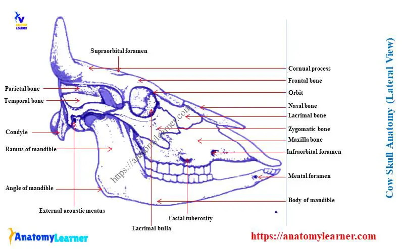
Labeled cow skull anatomy
Again, in this part, I will show you all the bones with a labeled cow skull anatomy diagram. You may also get help from the cow skull bones identification video.
If you need more labeled diagrams or labeled cow skull pictures, you may join with anatomy learners on social media.
Frequently asked questions on cow skull.
In this part of the article, I will try to solve the common inquiries on cow skull bones. If you have any other questions on the cow skull, please let me know.
What are the singles and paired bones of the cow skull?
There are four single and twelve paired bones present in the cow skull. The single bones of the cow skull are – occipital, sphenoid, ethmoid, and vomer. So, the rest bones of the cow skull are paired.
Conclusion
I think you will identify all the single and paired bones from the cow skull anatomy. You know, the cow skull comprises two essential parts – the cranial cavity and the fascial part. Most of the skull bones of the cow are irregular in shape and united with the help of sutures.
The cow’s cranial bones or ruminant skull form the cranial cavity, parts of the orbital and nasal cavity. Again, the facial bones also help form some of the essential structures of the body’s different systems. All of the cranial and facial bones structures are shown with the labeled cow skull anatomy diagram.