The dog ear anatomy consists of the three distinguished portions – the external, middle, and internal ears. These ears are the vestibulocochlear organs associated with hearing and equilibrium in a dog. Here, I will show you all the dog’s external, middle, and internal ear anatomy features with the labeled diagrams.
You will also find the detailed anatomical description of the dog ear canal, cartilage, and others in this article. Again, I will try to solve the common inquiries on the dog ear anatomy at the end of this article.
Dog ear anatomy
As a veterinary student or pet dog practitioner, you might have a good piece of knowledge on dog ear anatomy. You may divide the dog ear into three portions for the description purpose – the external, middle, and internal ear.
The external ear of a dog consists of the auricle or pinna and the external auditory meatus. Again, in the middle ear, you will find the tympanic membrane, tympanic cavity, and three tympanic ossicles. There you will also find the ligaments and muscles associated with the auditory ossicles.
In addition, the internal ear of a dog consists of a bony labyrinth that accommodates an almost similar-shaped membranous labyrinth structure. In the bony or osseous labyrinth, you will find three important structures (vestibule, cochlea, and semicircular canals).
The external ear of a dog is responsible for collecting the sound from the environment. Again, the middle part of a dog ear helps conduct the collected sound into the inner ear. Finally, the inner or internal ear has the important functions of hearing and equilibrium.
What should you do now to learn the details anatomical facts of the dog ear? Simply, you should try to identify the basic structures from the three parts of the dog ear with the help of below-mention labeled diagrams. Then, you should learn the detailed anatomy of these structures from the three different parts of the dog ear.
Dog ear anatomy external
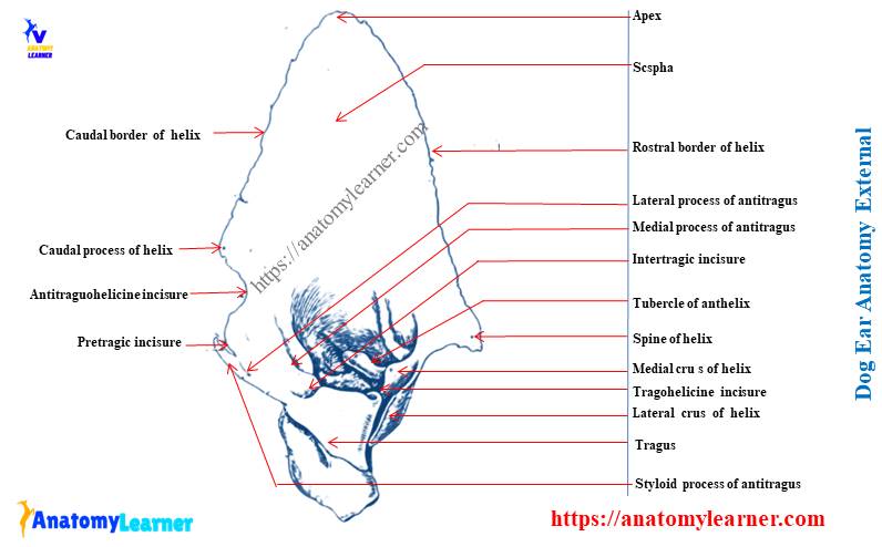
The anatomy of the dog external ear consists of skin-covered cartilages, auricle, and external acoustic meatus. It is a somewhat funnel-shaped structure but may vary between species and within the domestic breed.
The outer part of the dog ear considers as the sound gathering structure. Again, the sound conducts through the external acoustic meatus to the tympanic membrane of the dog’s inner ear.
Now, I will show you the different structures from the auricle or pinna, external acoustic meatus with the diagrams of dog ears. Let’s try to identify the following structures from the outer part of a dog ear.
- Three types of cartilages of the dog ear (auricular, annular, and scutiform cartilage)
- The cranial and caudal border of the helix
- A caudal process of the helix
- The spine or distal crus of a helix
- The lateral and medial crus of the helix
- A tubercle of the anthelix
- The scapha of the auricular cartilage
- Targus of the auricular cartilage
- The different process of the antitragus (styloid, lateral, and medial)
- The intertragic incisures
- A tragohelicine incisures
- The antitragohelicine incisures structure and
- A pretragic incisures
All the structures as mentioned above from the dog’s external ear are shown in the labeled diagram. If you want to know the detailed anatomy of these structures from the dog ear, you may continue this article till the end.
All these structures are from the auricular cartilage of the dog ear. So, you may jump to the dog ear cartilage anatomy to know details about them.
Auricle or pinna of the dog ear
The auricle or pinna is the externally visible part of the external ear of a dog. It is roughly an elongated funnel-shaped structure with two surfaces, two borders, a base, and an apex. But the shape of the auricle or pinna of the dog varies with the breed.
The cranial surface of the pinna of a dog is concave, ridged, and possesses the opening of the external ear. On the other hand, the posterior surface of the dog pinna is convex that covers hairy skin. The upper and lower borders of the dog pinna are thick.
You may find the blunt or pointed apex (vary with the dog breed) in the dog auricle or pinna. The base of the dog pinna is broad and attaches to the external auditory process of the petrous temporal bone.
Dog ear cartilage anatomy
In dog ear cartilage anatomy, you will find three types of cartilages – auricular, annular, and scutiform. The auricular cartilage is large and forms the shape of the dog ear. Again, the scutiform cartilage is a small quadrilateral shape that attaches to the cranial aspect of the base of the auricular cartilage.
In addition, the annular cartilage is a small incomplete ring that attaches to the bony external acoustic meatus. With the help of other cartilages, it forms the cartilaginous portion of the meatus.
Now, let’s know the detailed anatomical features of the three different types of cartilage from the dog ear.
Auricular or conchal cartilage of dog ear
Auricular is a single sheet of cartilage that covers the visible part of the dog ear anatomy. It is thin and pliable at its distal end, thicker and less pliable on its proximal end. The distal three fourth portion of this cartilage is roughly elliptical in the outline, whereas the proximal portion forms the tube.
The free margin of the auricular cartilage is slightly folded to form the helix. On the concave surface of the auricle, you will find a transverse fold with the prominent tubercle (known as anthelix). Again, the triangular area between the helix and anthelix is the scapha.
You will find a dense, irregularly quadrangular plate of cartilage (tragus) opposite to the anthelix. It forms the lateral boundary of the initial portion of the dog ear canal.
The antitragus is a thin, elongated leaf-like cartilage that locates caudal to the tragus. It separates from the tragus by an important notch, intertragic incisures. The antitragus is from the caudal boundary of the dog ear canal.
You will find two prominent processes in the antitragus (medial and lateral). The end part of the lateral antitragus process forms a sharp styloid process.
You will find some prominent processes, borders, and crus in the helix of a dog’s auricular cartilage. The cranial and caudal borders of the helix are the prominent features of the dog ear. You will find a deep antitragohelicine incisure just near the caudal process of the helix.
The cranial border of the helix continues to form the spine of the helix. You will find the lateral and medial crus of the helix near the spine. These structures from the dog auricular cartilage are separated from the tragus by the tragohelicine incisures.
Scutiform cartilage of the dog ear
The scutiform cartilage of the dog ear is a small, boot-like cartilaginous plate. It locates in the rostroauricular muscles medial to the dog ear. You will find several muscles attached to the scutiform cartilage that helps to move the dog ear.
The scutiform cartilage of the dog ear considers as sesamoid cartilage that is intercalated in the auricular muscles. You will find some fatty cushion deep to the scutiform cartilage of a dog.
Annular cartilage of dog ear
The annular cartilage of the dog ear anatomy is interposed between the auricular cartilage and the bony external acoustic meatus. It is a thin sheet of cartilage that helps to form the incomplete tube.
The free end of the annular cartilage meets on the caudal side of the external acoustic meatus. Again, the proximal end of the annular cartilage overlaps the bony external acoustic process.
The annular cartilage of the dog ear helps to move the auricle freely. It contributes to the elliptical outline of the external acoustic meatus of the dog ear.
The external acoustic meatus of the external dog ear
The external acoustic meatus of the dog ear is the canal that forms the base of the auricle to the tympanic membrane. This canal is bounded by the annular cartilage and the tubular part of the auricular cartilage.
The external acoustic meatus canal is slightly curved ventrally and cranially in a dog ear. The lateral one-third of this canal is cartilaginous, and the medial two-third is osseous.
You may find very few fine hairs in the skin at the entrance of the external acoustic meatus. There are also large sebaceous and sudoriferous glands present in the external acoustic meatus of a dog. These glands help to secrete the cerumen or ear wax.
The dog’s external ear infection is generally very painful because the skin is firmly adherent to the underlying bone and cartilage.
Dog middle ear anatomy
In the dog middle ear anatomy, you will find three important features – the tympanic membrane, tympanic cavity, and auditory ossicles. Here, I will show you the important features from the dog’s middle ear with the labeled diagrams.
The tympanic membrane surrounds the tympanic cavity of the dog middle ear externally. Again, the tympanic cavity connects with the nasopharynx through the auditory or Eustachian tube.
The tympanic cavity of a dog ear possesses a small, dorsal epitympanic recess and a large, ventral tympanic bulla. You will find three important auditory ossicles in the middle portion of the tympanic cavity of a dog’s ear.
The tympanic membrane of the middle ear of a dog
The tympanic membrane of the eardrum of a dog covers the entrance to the tympanic cavity. It also separates the middle ear cavity from the external acoustic meatus of the dog ear.
This eardrum is a thin, semitransparent, four-layered membrane that is somewhat oval. If you view this membrane externally, it looks somewhat concave. This is because of traction on the medial surface by the manubrium of the malleus.
You will find two different parts (pars flaccida and pars tensa) in the tympanic membrane of a dog ear. The pars flaccida is a small dorsal triangular portion that lies between the short lateral process of the malleus and the margin of the tympanic incisure.
Again, the pars tensa is the rest of the tympanic membrane that attaches peripherally to the fibrocartilaginous annulus. If any injury occurs on the pars flaccida, it may heal quickly. But, if any injuries occur in the pars tensa, it is very hard to heal.
You will find a most depressing point (umbo membrane tympani) just opposite the distal end of the manubrium of the malleus. If you view it from the lateral aspect, you will find a light-colored streak (stria mallearis). This stria mallearis runs dorsocaudally from the umbo membrane tympani to the pars flaccida.
The tympanic membrane of a dog ear consists of four layers. You will find a thin, stratified squamous epithelium lining on the membrane’s outer surface that continues with the lining of the external acoustic meatus.
Again, the mucosa is lining inside the membrane range from simple squamous to cuboidal or columnar epithelium. Between these epithelium layers, you will find tunica propria that comprises collagen and elastic fibers.
The tympanic cavity of the dog ear anatomy
In the middle ear anatomy, you will find the oblique space (tympanic cavity) between the petrosal and tympanic parts of the temporal bone (tympanic bulla). It divides into three major parts – dorsal epitympanic recess, middle tympanic cavity proper, and ventral fundic part within the tympanic bulla.
The epitympanic recess is dorsal to a frontal plane through the external acoustic meatus. This is a very small part that is occupied almost entirely by the head of malleus and incus.
Again, the tympanic cavity proper is that part just opposite to the tympanic membrane. It is irregular quadrangular shape but maybe flatten laterally by the tympanic membrane. Here, you will find the cochlear window at the caudal portion of the tympanic cavity.
The ventral portion of the cavity within the tympanic bulla may be compared in shape to the interior of an eggshell. It has an elliptical opening on the side that faces dorsally.
This structure of the dog middle ear communicates with the tympanic cavity through the elliptical opening. You will find a bony eminence (promontory) on the medial wall of the tympanic cavity. It lies opposite to the tympanic membrane medial to the epitympanic recess.
You will find a vestibular window (oval window) located on the dorsolateral surface of the promontory just medial to the pars flaccida. The oval window fits with the footplate of the stapes bone.
In the rostral extremity of the tympanic cavity proper, you will also find the ostium of the auditory tube. The ossicles (three in number) form a short chain across the dorsal part of the tympanic cavity.
The auditory or Eustachian tube of the dog ear
The auditory tube is a short tube that extends from the nasopharynx to the rostral part of the tympanic cavity. It is a bony tube that is rostrally formed by the squamous part of the temporal bone. Again, the floor of the auditory tube forms by the tympanic part of the temporal bone.
The medial wall of the auditory tube forms by the plate of hyaline cartilage. In addition, the rostral end of the cartilage curves medially to form the short hook.
The auditory tube of the dog lines with the pseudostratified ciliated epithelium with goblet cells.
Auditory ossicles of the dog middle ear
Another most important structures of the dog middle ear anatomy are three auditory ossicles. These auditory ossicles are three small bones that transmit air vibration from the tympanic membrane across the middle ear cavity to the inner ear.
The malleus is the most lateral and largest auditory ossicle in the dog’s middle ear. Again, the most medial one is stapes. Between the malleus and stapes, you will find the incus that articulate with both bones.
The manubrium of the malleus of the dog’s middle ear attaches to the tympanic membrane. In addition, the base of the stapes attaches to the margin of the vestibular window by the ring of ligamentous fibers.
The malleus of the dog middle ear
The dog’s middle ear’s malleus consists of a head, wide and thin neck, and a manubrium or handle. You will find the three-sided manubrium in the malleus of the dog middle ear. The side embedded in the substances of the tympanic membrane is wider and smooth.
At the base of the manubrium of the malleus, you will find a muscular process of the malleus bone that extends medially and slightly rostrally. Again, there is a long rostral process present in the tympanic membrane of the dog ear.
It extends directly rostrad from the neck of the malleus. You will also find a short lateral process just opposite to the rostral process of the malleus bone.
This is the most dorsal attachment of the manubrium to the tympanic membrane of the dog middle ear anatomy. Again, the malleus’s head articulates with the incus bone’s body in the epitympanic recess.
The incus of the dog auditory ossicle
The incus bone is smaller than the malleus, and the shape is like a human bicuspid tooth with divergent roots. It lies caudal to the malleus in the epitympanic recess.
You will find a head, two crura, and a process in the incus of the dog’s middle ear. The head of the malleus of the dog’s middle ear articulates with the body of the incus.
Two transverse folds form the caudal limit of the tympanic recess and where the crura locate. The short crura point caudally into the fossa includes dorsal to the ridge. Again, the long crura also direct caudally but present a small bone.
You will find a lenticular process in the short crura that extends rostrally and somewhat medially.
The stapes of the dog ear (auditory ossicle)
The stapes is the innermost bone in the dog middle ear anatomy. It is (stapes) also the smallest bone in the body of a dog. The stapes of the dog’s middle ear consist of a head, neck, two legs, a footplate, and a muscular process.
This stapes lies in a horizontal plane with the base medially. The base is articulate with the cartilage, which covers the edge of the vestibular window.
The head of the dog ear stapes articulates with incus through the lenticular process. Again, the base of the dog stapes articulate with the fibrocartilaginous ring that covers the edge of the vestibular window.
You will find two crura (rostral and caudal) in the dog’s stapes bone structure. There is a thin stapedial membrane that connects one crus to the other.
You will also find a small muscular process on the caudal crura near the head of the stapes bone. These small muscular processes provide the attachment for the stapedius muscle of the dig ear.
Ligaments of the auditory ossicles
Several ligaments attach the ossicle to the wall of the tympanic membrane of the dog ear. Here, I will enlist the ligaments that are found in the dog middle ear anatomy.
- A short but fairly well-defined lateral ligament of the malleus bone
- The dorsal ligament of the malleus bone
- A rostral ligament of the malleus bone
- The dorsal ligament of the incus bone
- The caudal ligament of the incus bone
- An annular ligament of the auditory ossicles
These are the most important ligament found in the dog’s middle ear. The malleus bone’s lateral ligament connects the malleus’s lateral process to the margin of the tympanic notch. You will find a short rostral ligament of the malleus that connects the rostral process of the malleus bone to the bony tympanic ring.
The body of the dog’s incus is attached to the roof of the epitympanic recess by the dorsal ligament of the incus bone. Again, the caudal ligament of the incus bone connects the short crura of the incus to the fossa includes. In addition, you will find an annular ligament that attaché the base of the stapes bone to the cartilage of the oval window.
Dog ear anatomy internal
The internal ear of a dog locates within the petrous part of the temporal bone. It consists of a bony labyrinth that accommodates an almost similar-shaped membranous structure (membranous labyrinth). The space between the two labyrinths fills with a fluid (perilymph) that resembles cerebrospinal fluid. Now, I will show you all the dog’s internal ear anatomy features with the labeled diagram.
So, you will find two types of labyrinths in the anatomy of the internal ear of a dog. The bony labyrinth consists of a vestibule, cochlea, and semicircular canals. Again, the membranous labyrinth of the internal dog ear consists of the cochlear duct, saccule, utricle, and three ducts of the semicircular canals.
Vestibule of the internal dog ear
The vestibule of the dog’s internal ear is irregular, and an oval bony cavity is placed medial to the tympanic cavity. It is continuous with the cochlea rostrally and semicircular canal caudally. There is an epithelium covering present in the periosteum of the bony labyrinth.
You will find the oval window at the lateral wall of the vestibule of the dog internal ear. The medial wall of the vestibule is perforated for the passage of the filaments of the vestibulocochlear nerve and aqueduct of the vestibule. This aqueduct extends through the petrous part of the dog temporal bone.
The three semicircular canals attaché at the caudal aspect of the vestibule using the five orifices. Again, the cavity of the vestibule lodges with the utricle and saccule of the membranous labyrinth.
The utricle locates on the dorsocaudal depression in the elliptical recess of the membranous labyrinth. Just ventraorostral to the utricle, you will find a spherical recess that contains saccule. There is a vestibular crest that separates the elliptical and s[herical recess.
You will find several groups of small opening (known as maculae cribrosae) that accommodates the nerve of the recess region.
The cochlea of the dog inner ear
The cochlea of the dog inner ear anatomy is a bony spiral canal. It surrounds the cochlear duct in a spiral of three and one-quarter around the central hollow core of the bone (osseous modiolus). A bony spiral lamina projects from the osseous modiolus that makes an incomplete portion within the canal.
The basilar membrane completes the incomplete partition. Again, the basilar membrane extends from the edge of the lamina to the outer wall of the canal. Here, you will find two scalae – scala tympani and scala vestibule.
The osseous or bony spiral lamina begins within the vestibule and ends at the hamulus (hook-like process). In addition, the scalae communicate at the apex of the modiolus by a small opening (helicotrema). The helicotrema forms at the free border of the hamulus.
The longitudinal modiolar canal and the spiral modiolar canal serve to distribute both blood vessels and nerves to the cochlea.
Semicircular canals in the dog inner ear
You will find three (anterior, posterior, and lateral) semicircular canals in the dog inner ear anatomy. These three semicircular canals of the dog inner ear lie caudal and slightly dorsal to the vestibule. The segment of the semicircular canal proximal to the vestibule is known as a crus.
So, you will find two crura in each semicircular canal that communicate with the vestibule. But, the posterior and anterior semicircular canals have a common crus (five total in numbers). There is a dilation (ampulla) present in one crus of each semicircular canal just near the junction of the vestibule.
The anterior (cranial) canal of one ear is roughly parallel to the posterior canal of the opposite ear of the dog. Again, the lateral canal of each side occupies a nearly horizontal plane.
The longest semicircular canal of the dog inner ear is an anterior canal. In addition, the posterior semicircular canal is the smallest in the dog inner ear.
The ampullated end of the posterior (caudal) semicircular canal and the non-ampullated end of the lateral canal are united for a short distance just caudal to the vestibule.
Membranous labyrinth of the dog inner ear anatomy
The membranous labyrinth of the dog inner ear anatomy is smaller than that of the bony labyrinth. It comprises cochlear ducts, saccule, utriculus, and three ducts of the semicircular canals. The membranous labyrinth is also a fluid-filled compartment that is continued within the compartment of the bony labyrinth.
The membranous cochlear ducts of the labyrinth are compared to a hollow, endolymph-filled ribbon. It begins blindly as the vestibular cecum. The utricle is an elongated, slightly flattened sac and somewhat larger than the saccule. They locate at the elliptical recess in the caudodorsal extremity of the vestibule.
Again, the saccule is the more spherical sac in the minute recess rostroventral to the utricle. The endolymphatic duct connects both the saccule and utricle with the endolymphatic sac.
In addition, the membranous semicircular ducts are very similar in shape to the osseous canal. They attach principally along the greater curvature of the osseous canal by connective tissue trabeculae.
Dog ear anatomy diagram
Again, I will show the summary of the dog ear anatomy with the labeled diagram. In this diagram, I tried to show you the most important structure from a dog ear’s outer, middle, and inner parts.
If you need a more labeled diagram on the dog ear, you may get it on social media of the anatomy learner.
Frequently asked questions on dog ears.
Now, you will find some answers to the common inquiries on dog ears. If you have any inquiries on the dog ear, please let me know.
What are the parts of a dog’s ear?
In a dog ear anatomy, you will find three parts: the external ear, middle ear, and inner ear. I have already discussed the anatomical features of the three different parts of the dog ear in this article. If you want to learn these anatomical features in detail, read the full article till the end.
You know the external ear of a dog comprises pinna or auricle and external acoustic meatus. Again, the middle part of the dog ear consists of the tympanic membrane, tympanic cavity, and auditory ossicles. In addition, you will find cochlear ducts, saccule, utricle, and ducts of the semicircular canals in the inner part of the dog ear.
What is the flap on the dog’s ear?
What is the Targus on a dog?
It is an irregularly quadrangular plate of cartilage opposite to the anthelix.
What dogs have Henry’s pocket?
Where is the ear bulb located on a dog’s ear?
What is a pinna on a dog?
The externally visible part of the dog’s ear is the pinna. Another name of the dog pinna is auricle.
Conclusion
I think this short guide might help you to learn the basics of dog ear anatomy. You might learn anatomical facts from the dog’s inner, middle, and external ear with the labeled diagrams. From the dog external ear anatomy, you might have a good piece of knowledge on the three different types of the cartilage of the pinna.
Again, you should know the tympanic membrane, tympanic cavity, and auditory ossicles from the dog middle ear anatomy. It is also essential to have a good piece of knowledge on the cochlear ducts, semicircular canals, and ducts from the dog inner ear.