The cow esophagus anatomy consists of musculomembranous tube-like features. You will find two distinct parts in the cow esophagus that are short and wider compared to the horse.
Here, I will show the cow esophagus course along with its structure. You will also know the cow esophagus’s relationship with various neck organs on its courses.
Quick overview: cow esophagus is a musculomembranous tube that courses from the pharynx to the atrium ventriculi of the stomach. It has two parts – cervical and thoracic, but no abdominal part. Here, the cervical part cow esophagus courses left lateral to the neck at the third to sixth cervical vertebrae level.
After completing this guide, you will differentiate the cow’s esophagus from the horse’s and dog’s esophagus. Let’s continue this guide to learn the course of the cow esophagus with its important anatomical facts.
What is the esophagus of a cow?
Esophagus is the musculomembranous tube-like structure of a cow. It passes along the structure of the cow’s neck and thoracic region above the heart.
Finally, it terminates on the atrium ventriculi of the compound stomach of the cows. The esophagus is considered the most important organ of the digestive system of a cow.
You may also know the other organs of the cow’s digestive system from the below-mentioned article of an anatomy learner –
- Dog digestive system diagram – list of primary and accessory organs
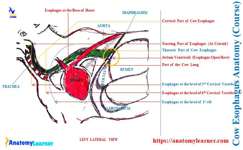
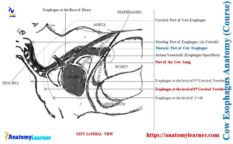
Here, the diagram shows the cervical and thoracic parts of the cow esophagus.
Cow esophagus anatomy
First, let’s get a basic idea of the whole structure of the cow esophagus. You may identify the below-mentioned features or parts from the cow esophagus –
- Starting part to the cervical part of the cow esophagus,
- The left lateral cervical part of the cow esophagus (at the level of the 3rd to 6th cervical vertebrae),
- Convex ventral and convex dorsal curvature of the esophagus,
- Esophagus passes the esophageal hiatus of the diaphragm, and
- The terminal part of the cow esophagus on the atrium ventriculi,
The diagram identifies all these features or structures from the cow esophagus.
In the cow esophagus anatomy, you might describe the followings –
- A course of the cow esophagus,
- Relationship of the various organs from the neck and thorax with the cow esophagus,
- Structure of the cow esophagus, and
- Blood and nerves are supplied to the cow esophagus,
Let’s see the summary of the anatomical facts of the cow esophagus from Table 1 –
| Cow esophagus anatomy | Characteristics |
| Course | Start from the pharynx and end on the stomach |
| Left lateral location | At the level of the third to sixth cervical vertebrae |
| Relationship (at the neck) | Various surfaces of the trachea Carotid covering Recurrent laryngeal nerve Thymus Tracheal lymphatic trunk Deep cervical lymph node |
| Relationship (at thorax) | Diaphragm Base of heart Tracheal bifurcation Aortic arch and aorta The junction between the rumen and reticulum |
| Structure | Have four coats – Fibrous tunica adventitia Muscular tunica muscularis Submucosal layer, and Tunica muscosa membrane |
| Blood supply | Branch of thyroid artery Carotid and broncho-esophageal arteries |
| Innervation | Pharyngeal branch of the vagus Recurrent laryngeal nerve The internal branch of cranial laryngeal |
Now, you will know the details of the cow esophagus anatomical facts.
What are the three parts of the esophagus?
Typically, the esophagus of an animal shows 3 (three) parts – cervical, thoracic, and abdominal part. You will find the cervical, thoracic, and short abdominal parts in the dog esophagus.
But, the cow esophagus does not show any abdominal part in their courses. This is because the rumen and reticulum of the compound stomach are in close contact with the diaphragm.
You know, get the basic idea on the compound stomach of the cows from a below-mentioned article of anatomy learner –
- Cow stomach anatomy – rumen, reticulum, omasum, and abomasum with diagram,
Again, if you see the course of the horse’s esophagus, you will find a small abdominal part. The diameter of the cow or other animal’s esophagus increases from the pharynx to the atrium ventriculi.
Thus, the cervical part of the cow esophagus shows less diameter compared to the thoracic part.
What is the length of the esophagus in a cow?
Quick answer: the average length of the esophagus is about 90 centimeters in a medium-sized cow. Here, the cervical part of the cow esophagus is about 40 centimeters, whereas the thoracic part is 50 centimeters.
And the abdominal part of the cow esophagus is practically absent. You will see 4 centimeters in diameter in the cervical part of the cow esophagus. But, this diameter may increase up to 6 centimeters in the thoracic part of the esophagus.
Small ruminants like sheep or goats have short esophagus. The length of the sheep or goat esophagus is about 40 centimeters with 2 centimeters diameters.
Here, Table 2 shows the length of the animal’s esophagus with its diameters –
| Animals Esophagus | Length (Centimeter) | Diameter (Centimeter) |
| Ox / Cow | 90 | 4 – 6 |
| Sheep/ Goat | 40 | 2 – 3 |
| Horse | 140 | 4 – 7 |
Cow esophagus course
The course of the cow esophagus shows several changes of direction. You will find the changes of direction on the cricoid cartilage (CC), at the level of the third to sixth cervical vertebrae, at the level of the first rib, at the level of the heart’s base, and aortic arch.
A short course of cow esophagus: I will show you the short course of the cow esophagus in Table 3 –
| Landmarks | Cow esophagus course |
| At the level of the cricoid cartilage | Start the course of the esophagus |
| The first part of the trachea | Lies dorsally |
| Third to sixth cervical vertebrae | Incline to the left surface of the trachea |
| At thoracic inlet | Goes dorsolaterally |
| At the level of the first ribs | Make the first curvature |
| Cranial mediastinum | Passes dorsal over the base of the heart |
| At bifurcation of trachea | Gives second curvature |
| Aortic arch | Esophagus goes to the right of the median plane |
| At the caudal mediastinum | It goes ventral to the aorta |
| At the level of 8th or 9th intercostal space | Pass through the esophageal hiatus |
| Shallow vault between rumen and reticulum | Terminate esophagus |
The provided information and the labeled diagram might help you to understand the course of the bovine or cattle esophagus. Now, let’s describe the course of the esophagus in detail.
What is the course of the esophagus in animals (cow/goat/bovine)?
The cow esophagus starts at the level of the lamina of the cricoid cartilage (pharynx). This esophagus lies dorsal to the trachea up to the one-third cranial part of the trachea.
At the level of the third (3rd) cervical vertebra, the cow esophagus inclines to the left surface of the trachea. And you will find this relation until the esophagus reaches the sixth cervical vertebra.
At the level of the thoracic inlet (TI), the cow esophagus slopes dorsad slightly to the dorsolateral surface of the trachea. Then it undergoes a slight curvature that forms the convex ventrally.
The cow esophagus now enters the mediastinum (space between the lungs). Within this mediastinum, the esophagus passes dorsally over the heart and bifurcation of the trachea.
You may understand how the mediastinum is formed in the cow’s thoracic cavity from the below-mentioned article –
- Cow lung anatomy – bovine right and left lungs structure with diagram,
At the level of tracheal bifurcation, the esophagus makes its second curvature (convex dorsal). It crosses the right face of the (AA) aortic arch of the cow’s heart.
You may learn the features of the aortic arch of the cow’s heart from the below-mentioned article –
After the aortic arch of the heart, the esophagus becomes straight and passes back to the caudal part of the mediastinum. You will find the esophagus just ventral to the aorta.
Now, the esophagus passes through the esophageal hiatus (EH) of the diaphragm (at the level of the 8th or 9th intercostal space). Finally, the esophagus opens into the shallow vault between the rumen and reticulum of the cow’s compound stomach.
Relationship of cow esophagus with other organs
In the course of cow esophagus anatomy, you will find various relationships with the other organs. The cervical part of the cow esophagus is related to the following organs –
- Dorsal and left lateral aspects of the trachea,
- A carotid artery with its covering,
- Recurrent laryngeal nerves,
- Thymus and tracheal lymphatic trunks, and
- Deep cervical lymph nodes,
At the level of the first rib of the cow thorax, the esophagus has contact with the followings –
- Cervicothoraic ganglion,
- Costocervical trunk, and
- Thoracic ducts,
You will get a little idea of the thoracic ducts of a cow from the below-mentioned article –
Within the mediastinum, you will also find a relationship of the esophagus with the dorsal and ventral trunk of the vagus nerve. Again, the larger caudal mediastinal lymph nodes have contact with the cow’s esophagus.
Structure of the bovine esophagus
You will find four distinct coats or layers in the structure of the bovine esophagus. A fibrous layer named tunica adventitia, the muscular coat, a submucosal layer, and the mucous membrane layer is present in the structure of the bovine esophagus.
Here, the mucous membrane of the cow esophagus is pale and covered with the lining epithelium. But, you will not understand the lining epithelium of the esophagus grossly. It will be better to understand the total structure of the different layers of the cow esophagus from the below-mentioned article –
- Esophagus histology – four different layers description from the actual microscope slide and diagram
The mucous membrane of the cow esophagus is loosely attached to the muscular coat. Again, the muscular coat of the cow esophagus shows the various arrangement pattern.
Innervation and blood supply to cow esophagus
The arteries that supply blood to the cow esophagus are as follows –
- Branches from the thyoird artery,
- The common carotid artery and reticular arteries, and
- Bronchoesophageal artery of the cows,
You will also find the below-mentioned veins that drain blood from the cow’s esophagus –
- Branches of the cranial and medial thyroid veins,
- The caudal part of the external jugular veins, and
- Caudal esophageal vein of the cows,
The pharyngeal branch of the vagus, recurrent laryngeal, and internal branch of cranial laryngeal nerves innervate the structure of the cow esophagus. Sometimes the branch from the glossopharyngeal nerve also innervates the cow’s esophagus.
Ruminant or goat esophagus anatomy and course diagram
Now, let’s see the changes in the direction of the esophagus course from the ruminant or goat. Here, I tried to show you the course of the ruminant esophagus (goat) with the diagram.
I showed you the dorsal and lateral relationship of the goat esophagus with the trachea. Again, the first and second curvatures of the esophagus after the lateral relationship are also shown in the diagram.
The relationship of the goat or ruminant esophagus with the base of the heart and aortic arch is also shown. Finally, the diagram shows the opening of the goat esophagus on the atrium ventriculum (area between the rumen and reticulum) of the stomach.
Conclusion
The course of cow esophagus anatomy is essential to understand its changed direction. But, the structure and relationship with other neck and thorax organs might help you know the cow esophagus features.
You will not find the abdominal part in the cow esophagus anatomy compared to horses and dogs. Again, a little difference may be found in the structure between the cow’s and horse’s esophagus.