The cow hoof anatomy is a cornified modification of the epidermis that lies under the vascular layer and covers the end of the digits. You will find two primary and two accessory hooves on each limb of a cow. The leading hoof of a cow comprises three parts – periople, wall, and sole. Here, I will show you the anatomical features of a cow hoof with a labeled diagram.
The cornified tissue of a cow hoof consists of some vital structures like corium, coronary band, and wall. You will see the hoof wall easily when the cow is standing. Again, a concave keratinized sole attaches to the palmar or plantar surface of the third phalanges.
In addition, you will find the third phalange, ligaments, tendons, vessels, and synovial structure in the cow hoof anatomy. So, if you are interested to know these details anatomical features of the cow hoof, let’s continue this article.
Cow hoof anatomy
The cow hoof is the hard covering of the ends of the digits. It is the modification of the epidermis of that region. First, I would like to summarize the anatomy of a cow hoof in short. Here, I will describe the peripole, wall, and sole from a cow hoof.
The peripole is a soft, flat, and light-colored band that surrounds the coronary border of the hoof. Do you know what a coronary band or border is? Excellent, the coronary band is an area where the hairy skin changes to the hoof.
The coronary band is thin, and at the heels, it widens to cover the significant convexity of the bulb. So, here you got the two new term heels and the bulb of the hoof. You will learn details about these terms from the cow hoof later.
The wall of a cow hoof represents two surfaces – axial and abaxial. You will find a convex abaxial surface that presents some transverse ridges. Again, the axial surface of the cow hoof is slightly concave. These two surfaces join at the toe and become thin towards the bulb of the wall.
“You will find the toe and bulb from the cow hoof labeled diagram. Again, you will know details about these structures from the cow hoof description section.”
The sole is a slightly concave structure that places at the cow hoof’s ventro-palmar (ground surface). Caudally the sole is continuous with the peripole of the bulb.
What is the corium of cow hoof?
The corium of a cow hoof is a highly vascular and modified structure that adopts the shape of the corresponding outer covering. You will find different parts of corium in a cow hoof-like perioplic corium, laminar corium, coronary corium, and solar corium.
Again, the laminar corium divides into sensitive and insensitive laminae in a cow hoof. All these different coriums and laminae are identified from the cow hoof structure.
The laminar corium of a cow hoof receives the coronary lamellae of the wall. Again, the corium supplies nutrition to the cow hoof.
Bones and digital cushions of cow hoof
In the anatomy of a cow hoof, you will find the third phalanx (distal phalanx) and navicular bone in each limb. The navicular bone is the distal sesamoid bone of a cow’s hind or forelimbs. The distal or third phalanx is a triangular-shaped structure in each digit of a cow.
You will also find a bit distal extremity of the second phalanx in the structure of a cow hoof. Again, a digital cushion is a modified subcutaneous tissue that locates in between the corium of the bulb and the flexor tendon.
In a cow hoof, you will find this digital cushion as a fatty elastic tissue whose primary function is to minimize the concussion.
Identification of different structures from a cow hoof
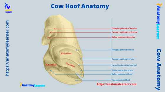
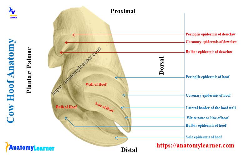
Let’s identify some essential structures from a cow hoof, both a sagittal section and an external view. These might provide a good conception of the cow hoof structure. Fine, let’s try to identify the below-mentioned structures from the cow hoof –
- The perioplic epidermis of the hoof
- Coronary epidermis or wall of the hoof
- The solar epidermis of the hoof
- A bulbar epidermis of a hoof
- The white line of the cow hoof
- Dorsal border, lateral border of the hoof wall
- A perioplic dermis or corium
- Coronary corium or epidermis
- Laminar dermis or corium
- Solar corium or solar dermis
- The bulbar dermis or bulbar corium
- A digital cushion
- Third phalanx and navicular bone of the hoof structure
- Structures of the accessory hoof of the cow (identified)
You will also find some of the other structures from a cow hoof-like –
- Lateral digital extensor tendon of the hoof
- Common digital extensor tendon of a hoof
- Tendon of superifical digital flexor
- Deep digital flexor tendon of the cow hoof
- A navicular bursa of the cow hoof
I will use different diagrams and show you all the structures mentioned earlier from the cow hoof. The first cow hoof labeled diagram shows the external features you might know first. Here, you might identify the periople, different types of periople, external wall (epidermis), sole, parts of sole, and bulb of the sole.
Again, I used a sagittal sectioned cow hoof labeled diagram, where I tried to identify most of the structures. Please try to identify all of these structures so carefully so that you may identify them from the actual cow hoof sample quickly.
In addition, all the structures from the accessory hoof are identified in the labeled diagram.
Accessory digits of cow hoof
You will find two extra digits above and behind the corresponding leading hoof. Each comprises a hard conical covering (epidermis) and a corium (dermis).
Again, the corium covers the nodular vestigial phalanx that connects to the third phalanx and distal sesamoid bone (navicular bone) and the elastic connective tissue pad (digital cushion) of the heel by some fibers.
You will also find the following features in the accessory hoof structure of a cow –
The epidermis of the accessory hoof of a cow consists of –
- A perioplic epidermis of an accessory hoof,
- The coronary epidermis of an accessory hoof, and
- A bulbar epidermis of the accessory hoof.
Again, the dermis (corium) of the accessory hoof of a cow consists of –
- Perioplic demis of the accessory hoof,
- The coronary dermis of the accessory hoof,
- Wall or parietal dermis of the accessory hoof,
- The solar dermis of the accessory hoof, and
- The bulbar dermis of the accessory hoof.
All these features from an accessory hoof of a cow are identified in the labeled diagram.
Cow hoof anatomy corium
The corium of a cow hoof is a vascular layer that lies under the epidermis. You will find a coronary band where the hairy skin changes to the hoof. The cow hoof anatomy corium consists of perioplic corium, coronary corium, laminar corium, solar, and bulbar corium.
So, you will find the following segments of the corium in the hoof structure of a cow –
- A perioplic corium of a hoof,
- The coronary corium of a hoof,
- A laminar corium of a hoof,
- The solar corium of a hoof, and
- A bulbar corium of a hoof.
Now, I will provide a little information on these coriums of the cow hoof. But, you may know more about this corium in the cow hoof description section of this article.
The perioplic and coronary corium
The perioplic corium is a thin band at the coronary band and is associated with the epidermis layer. It produces a thin, waxy periople on the surface of the hoof wall (proximal end). Again, you will find a wide band that underlies the portion of the epidermis and form the bulk of the hoof wall. This wide band of the cow hoof is the coronary corium.
You will find the prominent papillae in the coronary corium that interdigitate with the coronary epidermis. These coronary papillae form the tubular configuration of the cow hoof wall. Another name for the cow hoof wall is stratum medium.
But, these tubular papillae are very hard to identify grossly. Under the light microscope, you may identify the tubular papillae from the surrounding intertubular papillae.
So, the tubular and intertubular papillae form the wall of the cow hoof (stratum medium).
The laminar corium of the hoof
The longitudinal leaves of the corium cover the convex surface of the digital phalanx (third or distal phalanx). These longitudinal leaves of the outer surface of the distal phalanx are laminar corium. You will find two segments in the laminar corium of a cow hoof – sensitive laminae and insensitive laminae.
The segment of the laminar corium, which is well innervated, is the sensitive laminae of the hoof. Again, the sensitive laminae interdigitate with epidermal laminae, not innervated. This non innervated segment of the laminar corium is the insensitive laminae of the cow hoof.
You will find thousand of interdigitating laminae on the large surface of the hoof that also create a strong connection between the distal phalanx and the hoof wall. The inflammation of the sensitive laminae of the cow hoof is widespread, termed laminitis.
The solar and bulbar coriums of a cow hoof
The sole of a cow hoof is a concave keratinized plate that attaches to the palmar or plantar surface of the distal phalanx. It includes the entire ground surface of the cow’s hoof. The concavity of the sole of a cow hoof allows the wall to bear the most weight of the body.
So, the segment of the vascular corium that covers the sole is the solar corium. Again, the segment of this vascular corium that covers the bulb of the heels is the bulbar corium.
The cow hoof labeled diagram identifies all these structures (solar and bulbar corium).
Segments of cow or cattle hoof anatomy
The cow or cattle hoof anatomy is somewhat different from those of a horse hoof. Now, I will discuss the different segments of a cow hoof structure like the perioplic segment, coronary segment, sole segment, wall segment, and bulbar segment. Again, if you want to know the different segments from the horse hoof, please read the following article from the anatomy learner.
- Anatomical features of different segments of a horse hoof with a labeled diagram
You will find a fully developed hoof on each leading digit of a cow’s legs (digits III and IV). It comprises the modified thick skin and strongly cornified epidermis. Again, the hoof surrounds the skeletal and soft structures of the distal phalanx of each digit of a cow leg.
So, in this part of this article, I will try to cover the followings –
- Three layers of the modified skin of the cow hoof,
- Features of the perioplic segment of the cow hoof,
- Anatomy of the coronary segment of the hoof,
- Features of the wall segment of a hoof,
- A sole segment of a cow hoof, and
- The bulbar segment of a cow hoof.
Let’s discuss these segments and layers from a cow hoof structure.
Three layers of the modified skin of a cow hoof
The hairless skin covering the cow hoof is distinctly modified and possesses three different layers: the subcutaneous, epidermis, and dermis. These three hoof layers possess almost similar features to the typical skin layers. But, in the hoof, these layers become modified into different segments.
You will not find the subcutis in the sole and wall of a cow hoof. But, in the other part of the cow hoof, you will find the subcutis in the form of an immovable cushion. It consists of a three-dimensional network of longitudinal, transverse, and oblique connective tissue fibers with the adipose tissue.
The subcutaneous tissue in the bulbar region of a cow hoof is modified and forms the special thick cushion. This unique thick bulbar cushion of the cow hoof absorbs the shock.
In the epidermis of a cow hoof, you will find the dermal papillae and lamellae. They form a rigid, tubular structure in the different segments of the cow hoof except for the wall. But, in the wall, the dermal papillae and lamellae form the complicated lamellar structure in the hoof wall.
Again, the dermis of the cow hoof consists of a deep reticular layer and a more superficial papillary layer. The superficial papillary layer of the hoof dermis bears dermal papillae. But, you will not find dermal papillae in the wall of the cow hoof.
The papillae arise either from a smooth surface or parallel dermal ridges. In the wall segment of a cow hoof, you will find the parallel dermal lamellae. These parallel dermal lamellae direct from the proximal end to the distal end.
A perioplic segment of a cow hoof
You will quickly identify the different segments of a cow hoof when you will remove the capsule. If you don’t know what the capsule of a cow hoof is, you may know it from the next section of this article.
You will find the perioplic segment of a cow hoof next to the hairy skin. There is a coronary segment at the distal part of the perioplic segment. Again, the ground surface represents the sole and bulbar segment in a cow hoof anatomy. In addition, the wall segment of a hoof is easily identifiable at the lateral aspect of the hoof while in the standing condition of an animal.
The perioplic segment of the cow hoof is broad and surrounds the dorsal and palmar or plantar aspect. You will find a slightly convex perioplic cushion at the dorsal and abaxial aspects of the hoof. But, you will not find the perioplic cushion at the axial aspect of the hoof.
Do you know who forms the perioplic cushion in the cow hoof? Fine, the subcutis tissue forms the perioplic cushion in a cow hoof. On the palmar or plantar aspect, this cushion continues with the digital cushion of the bulb of the heels.
Here, you will find two structures in the perioplic segment – perioplic dermis or corium and perioplic epidermis. The perioplic dermis or corium covers the subcutis and bears the finely distally directed perioplic papillae. Again, the perioplic epidermis covers the dermis and forms hard tubules on the dermal papillae.
You will also find the soft perioplic part that grows distally as the external layer of the cow hoof wall.
A coronary segment of a cow hoof
So, the coronary segment of a cow hoof is distal to the perioplic segment. It extends to the level about halfway down the hoof. You will find a wide and slightly convex coronary cushion in the coronary segment of the cow hoof. Again, this coronary cushion of the cow hoof is made of subcutis with fatty tissue.
The vastness and thickness of the coronary cushion decrease at both sides of the palmar and plantar aspect of the cow hoof. Here in the coronary segment, you will see two essential parts – the coronary corium or dermis and the coronary epidermis.
The coronary corium contains the fine coronary conical papillae round off the end. They are thicker at their base and directed horizontally. Again, at the apical part, these conical papillae incline distally in the direction of growth.
In addition, the coronary epidermis of the cow hoof from the hard tubules corresponded to the dermal papillae. This coronary epidermis forms the middle layer of the hoof wall.
You will find the thickest and unpigmented hard tubules in the middle layer of the coronary segment. The thinner tubules are present in the outer layer of the coronary segment.
Wall segment of a cow hoof anatomy
The wall segment of a cow hoof anatomy is easily visible, and the broader part of a hoof. It is distal to the coronary segment of the hoof and of about equal width. You will not find any subcutaneous tissue or fatty tissue in the structure of the wall of a cow hoof.
Another name for the wall segment of a cow hoof structure is the lamellar segment. Here, you will find the two essential features – the lamellar dermis and the lamellar epidermis. The lamellar dermis or corium contains the proximodistally oriented dermal papillae.
These dermal papillae of the lamellar corium of the cow hoof structure are smooth, but you will not find the secondary lamella. You will also find the lamellar epidermis in a cow hoof wall structure. This lamellar epidermis contains the epidermal lamellae between the dermal lamellae.
The epidermal lamellae form the middle layer of the hard wall of the cow hoof. You will find two essential segments in the laminar corium and insensitive laminar corium in the laminar corium. When the laminar corium is innervated, that is known as the sensitive corium of the cow hoof wall.
Again, the insensitive laminar corium is not innervated by the nerve or blood supply.
The sole segment of the hoof
You know the sole segment of a cow hoof is a keratinized concave structure that attaches to the palmar or plantar surface of the distal phalanx of each digit. The sole of a cow hoof divides into a body and axial and abaxial crura. You will not find any subcutaneous tissue or fatty tissue in a sole structure.
The sole segment of a cow hoof diagram shows two essential features – solar dermis or corium and solar epidermis. The lower transverse ridge is found in the sole soler dermis or corium region. Again, the solar epidermis of the sole segment contains the hard tubules.
The sole segment of the cow hoof becomes crescent and narrow at the white zone or line. Let’s know the features of the white line of a cow hoof in short.
White zone or white line of a cow hoof
A narrow band is typically slightly lighter in color than the rest of the cow hoof wall. This slightly lighter color is the cow hoof’s white zone or white line. The white line is useful as a landmark for driving nails in shoeing. An adequately directed nail started at or outside the white line will not touch any sensitive structures of the hoof.
So, you will find the white line surrounds the wall segment and presents the external, middle, and internal parts. The outer part of the white line is easily visible as a bright white wide stripe. It consists of the basal segment of lamellae and flanking proximal cap.
Again, the middle part of the white zone consists of an intermediate section of hard lamellae with the distal cap that lies between them. In addition, the internal part consists of crests of the lamellae and terminal tubular structure.
The white zone of a cow hoof has axial and abaxial crura that lie between the unpigmented coronary zone and sole. Again, the axial crus ends halfway between the apex of the hoof and the palmar or plantar surface. The abaxial crus extends further to the basal part of the bulb. Here, the white zone becomes distinctly wider and turns inwards.
A bulbar segment of the hoof
The bulbar segment of a cow hoof lies palmar or plantar surface to the sole. It extends back to the haired skin of the cow limbs. You will find a digital cushion made of subcutaneous and adipose tissue. This digital cushion helps distinguish the bulb from the sole of the cow hoof.
The apical part of the bulb is less thick than the basal part. Again, you will find two essential features in the structure of a bulb from a cow hoof labeled diagram – bulbar dermis or corium and a bulbar epidermis.
The bulbar dermis of the bulb covers the digital cushion and bears dermal papillae. Again, the bulbar epidermis covers the dermis and consists of hard tubules. The apical part of the bulb is more prominent in the cattle, sheep, and goat hooves.
Cow hoof capsule
In the cow hoof anatomy, you will also find a capsule surrounding the middle phalanx’s distal end, the distal interphalangeal joint, and the distal phalanx. You will also find a connection between the common digital extensor tendon and the deep digital flexor tendon with the hoof capsule.
A navicular bone (distal sesamoid bone) also serves as the trochlea for the deep digital flexor tendon. The navicular bursa reduces the friction between them.
In the cow hoof capsule, you will also find the cornified lamina. This lamina of the capsule consists of an abaxial part, a dorsal border, and an axial part that faces the interdigital space. The dorsal part of the capsule is thicker than that of the apical part.
Other structures of the hoof or foot
Here, in the other structures from the hoof or foot of a cow, I would like to describe the bones, cartilage, tendon, ligaments, and synovial structures in a concise form. If you are interested to learn these structures from the hoof or foot of a cow, you may continue this part of the article.
- Bones and cartilage of the foot or hoof,
- Tendon and ligaments of the hoof, and
- Synovial structure of the cow hoof.
Let’s discuss these structures from the foot or hoof of a cow.
Bones and cartilage of the foot or hoof
In the foot or hoof structure, the three phalanges include the proximal, middle, and distal phalanx. The other name of the proximal, middle and distal phalanges are long pastern bone, short pastern bone, and coffin bone, respectively.
The proximal phalanx articulates with the cannon bone (the metacarpal bone) and forms the fetlock joint. Again, the proximal phalanx joins with the middle phalanx and forms the paster joint. Finally, the middle phalanx joins the distal phalanx and forms the coffin joint.
You will find four (two for each digit) proximal sesamoid bones at the palmar or plantar surface of the fetlock joint. Again, you will find two (one for each digit) distal sesamoid bones at the palmar or plantar aspect of the coffin joint.
You will find the collateral cartilages that connect with the distal phalanges of each digit of a cow.
Tendons and ligaments of the hoof
You will find different tendons and ligaments in the anatomy of the cow hoof. A common digital extensor tendon passes down the dorsal aspect of the metacarpus and inserts into the extensor process of the distal phalanx.
Again, a long digital extensor tendon inserts into the distal phalanx of the cow hoof. The lateral digital extensor tendon inserts on the proximal end of the proximal phalanx of the cow foot.
You will find a deep digital flexor tendon that passes down the palmar or plantar surface of the metacarpal bone and inserts on the distal phalanx. There are also superficial and deep digital flexor tendons in the structure of a cow hoof.
Again, you will find different ligaments in a cow hoof or foot structure. Here, I will enlist some of the crucial ligaments present in the cow hoof structure.
- Medial and lateral collateral ligament of the fetlock, pastern, and coffin joints,
- A suspensory ligament in the cow hoof,
- The sesamoids ligaments for the proximal sesamoid bones,
- Medial and lateral collateral sesamoids ligaments,
- Two short sesamoids ligaments of the hoof,
- Straight, oblique, and cruciate sesamoid ligaments of the cow hoof,
- Medial and lateral collateral ligaments,
- Palmar and plantar annular ligaments of the hoof, and
- Proximal and distal digital annular ligaments of the cow hoof.
You will find the details description of these hoof ligaments in the syndesmology section of anatomy learner.
Cow hoof infection
You will find different infections or abnormalities in the cow hoof. Here, I will discuss the common infection or abnormalities with the cow hoof. Most commonly, you will find the following problems in the cow hoof –
- Laminitis of the cow hoof,
- Sidebone condition in a cow hoof,
- Tendonitis in the cow hoof, and
- A navicular problem in a cow hoof.
Let’s discuss these common problems in a bit of a cow hoof.
Sidebone of a cow hoof: Direct trauma or chronic injuries may ossify the cow hoof’s collateral cartilage, which produces a condition called the sidebone. If there is any injury to the coronary band of the cow hoof, it may lead to an infection of the collateral cartilage.
Laminitis in the lamina of the hoof: Inflammation of the sensitive laminae leads to very painful laminitis. The sensitive laminitis is associated with the hoof wall and distal phalanx. In this case, the distal phalanx may lose or rotate downward. Thus the hoof wall grows abnormally and produces an irregular flared and up-curled toe.
Tendonitis and navicular problem: The inflammation of the flexor tendon commonly occurs in cow hoof and lead to tendonitis. Again, you will find a variety of navicular problems in a cow hoof, including the erosion of articular cartilage of navicular bone, bursitis of the navicular bursa, adhesion between the deep digital flexor tendon and navicular bone, and erosions of the navicular bone.
Cow claws
The claws of a cow (declaws or accessory hoof) are reduced digits II and V. They attach without synovial joint by a fascial ligament at the level of the fetlock joint.
These declaw do not reach the ground and remain a caudomedial or caudolateral aspect of the fetlock joint. They are short conical structures and are composed of the same modified skin layer as the leading hoof.
You may find two phalanges in the claws of a cow. But, sometime, you may find only one phalanx in the dewclaws of the cow.
Dairy cow hoof anatomy diagram
Now, I will show you again the different features from the dairy cow hoof anatomy labeled diagram. The main three parts (perioplic, wall, and sole) of a cow hoof are shown on the labeled diagram. Again, all other segments like different types of corium, perioplic epidermis, laminar epidermis, solar epidermis, digital cushion, and bulb of the heels are shown on this diagram.
In addition, the sagittal section of the cow hoof shows different essential features. You may get more cow hoof labeled diagrams on social media for anatomy learners.
Frequently asked questions on a cow hoof.
I hope this frequently asked questions section on cow hoof might help you get your desired inquiries. Here, I tried to provide you with a short answer to all the questions that cow hoof structure learners commonly ask.
What kind of hoof does a cow have?
You will find a cloven hoof in a cow that consists of two claws. This type of hoof also may find in the sheep, goats, and dear. You will find the fully developed hoof in a cow on both leading digits III and IV on each limb.
What is a cow’s hoof made of?
You know cow hoof is the hairless covering of the end part of each digit. It consists of modified skin that possesses subcutis, epidermis, and dermis like the standard skin layer.
You will also find the different segments in the structure of a hoof-like periople, corium, wall, sole, and bulb of heel. All the structures of the cow hoof are already described in this article. You may read the full article to acquire basic knowledge on the cow hoof.
Do cows feel pain in their hooves?
Yeah, cows feel pain in their hooves if there something is wrong. If sensitive laminitis, tendonitis, sidebone condition, or navicular problem occurs, cows feel pain in their hooves.
Why do cow hooves need to be trimmed?
If you don’t trim the cow hooves, overgrowth of the hoof may occur. This overgrowth of hoofs leads to lameness and impairment in their motion permanently. Again, the overgrowth of the cow hoof may cause bacterial lameness.
Conclusion
So, this is concise and essential information on the cow hoof anatomy with a labeled diagram. I hope you got the description of the basic structure like periople, different parts of the wall, and sole from a cow hoof. The leading hoof of a cow consists of modified hairless skin that possesses subcutis, epidermis, and dermis.
Again, in the accessory hoof anatomy of a cow, you will also find almost similar features to the main hooves. There are also bones, cartilages, ligaments, tendons, and synovial features in the structure of a cow hoof.