There are two or more pairs of parathyroid glands located on the posterior surface of the thyroid gland. You will find two main types of cells (chief and oxyphils) in the parathyroid gland histology slide. Here in this article, I will show you all the structures of a parathyroid gland with a microscope slide image and a labeled diagram.
I will also provide the essential identification points of the parathyroid gland histology slide under the light microscope. Again, you will find the detailed histology of the oxyphil and chief cells of the parathyroid gland with their functions in this article.
So, if you are interested to learn the parathyroid gland histology with the microscope slide image and labeled diagram, this might be an excellent guide for you.
Parathyroid gland histology
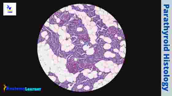
Each parathyroid has a thin connective tissue capsule that sends septae into the interior gland substance. The septae merge with the reticular fibers that support the parenchyma of a parathyroid gland. Again, the parenchyma of the parathyroid gland histology consists of secretory endocrine cells that arrange in a cluster or anastomosing cords between the sinusoidal capillaries.
The endocrine cells of the parathyroid gland slide are of two types – chief or principal cells and the oxyphil cells. I will show you the difference between these two parathyroid gland cells so that you may identify these two types of endocrine cells so quickly under a light microscope.
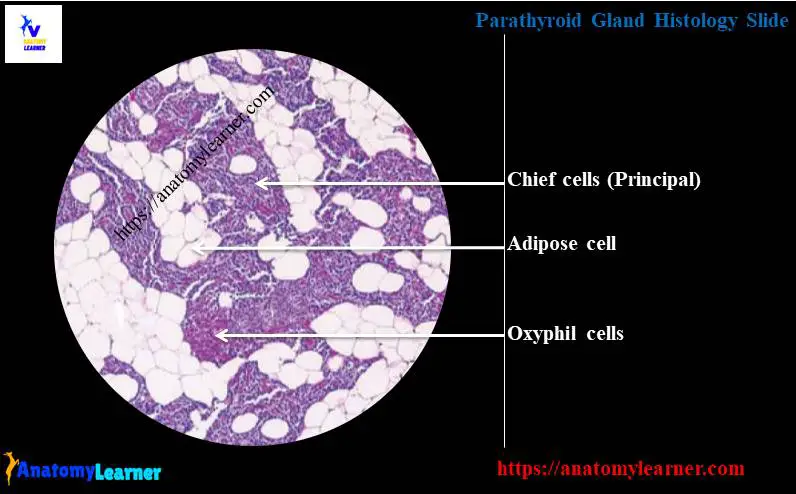
Typically, the chief or principal cells of the parathyroid are smaller but numerous than the oxyphils cells. Again, the oxyphil cells are more prominent and increase the number with the age of an animal.
You will find the adipose tissue infiltration in the parenchyma of the parathyroid gland in older animals. Sometimes, the fatty tissue constitutes more than fifty percent of the parenchyma of the aged animal.
However, let’s know the structures you might identify from the parathyroid gland histology slide under the light microscope.
- A connective tissue (thin) capsule of the parathyroid gland
- The thin trabeculae that merge with the reticular fibers of the parenchyma
- The secretory endocrine cells of the parathyroid glands (chief and oxyphil cells)
- Numerous blood vessels (vein and capillaries)
- Thyroid follicles with follicular and parafollicular cells (optional)
“Sometimes, you may find the thyroid gland and parathyroid gland in the same histology slide.”
Fine, now, you may try to identify all the structures of the parathyroid gland with the help of labeled microscope images.
Parathyroid gland histology slide identification points
You may be asked to identify the parathyroid gland microscope slide under the light microscope at your laboratory. I will share some essential identification points for the parathyroid gland histology slide. I hope these identification points might help you identify the parathyroid gland so quickly under the microscope.
- The provided tissue section shows the secretory endocrine cells that arrange into clumps and anastomosing cords.
- There are numerous small basophilic chief cells present in the tissue sample.
- Presence of a small number of larger eosinophilic oxyphil cells in the provided tissue sample.
- Again, the tissue sample shows numerous adipose tissue (fat cells) in its parenchyma.
- The secretory endocrine cells surround by numerous blood vessels.
So, this is a parathyroid gland histology slide. But sometimes, you may find the thyroid and parathyroid glands in the same microscope slide.
I think you got the basic idea on the parathyroid microscope slide. If you want to learn more about microscopic features of the parathyroid gland, you may continue this article.
Parathyroid gland histology guide
In parathyroid gland histology, you will find two basic structures – connective tissue stroma and the parenchyma. The stroma of the parathyroid gland includes the connective tissue capsule, trabeculae, and the reticular framework that support the parenchyma.
Again, the parenchyma of the parathyroid gland includes the secretory endocrine cells. These secretory endocrine cells are of two types – chief or principal cells (basophilic) and oxyiphil (eosinophilic).
Hey, if you don’t know the term stroma and parenchyma of a gland, this little information is for you. But if you have a good piece of knowledge on stroma and parenchyma, you may skip this part of the article.
The stroma includes the connective tissue framework of any gland made with the connective tissue fiber, cells, matrix, and blood vessels. Again, the parenchyma is the secretory unit or lobule or lobe of that gland that locates on the stroma.
So, to describe the parathyroid gland, you might mention the histology of the following structures –
- A capsule and trabeculae of the parathyroid gland
- The stroma of the parathyroid gland and
- The parenchyma of the parathyroid gland
Now, I will describe all the structures from the parathyroid gland histology slide one by one.
The stroma of the parathyroid gland
You know there are two parathyroid glands, one superior and one inferior, on either side of the thyroid gland. So you will find four parathyroid glands in an animal. Sometimes, you may find eight parathyroid glands in an animal.
The parathyroid glands are small oval structures, yellowish-brown in color, and separated from the thyroid gland by a thin connective tissue capsule. But, sometimes, the parathyroid gland may be embedded in the thyroid gland.
The connective tissue framework of the parathyroid gland forms the stroma that includes the capsules and supporting connective tissue fibers. Again, the connective tissue capsule sends the thin septae into the interior of the parathyroid that merges with the reticular fiber of the stroma.
In addition, the adipose tissue and the reticular fibers provide a bed for the parenchyma and blood vessels.
The parenchyma of the parathyroid gland
The parenchyma of the parathyroid gland consists of cells arranged in cord and clump.
Numerous sinusoids lie in a close relationship to these secretory endocrine cells. There are two types of cells – smaller chief or principal cells and larger oxyphil cells.
The chief cells of the parathyroid gland slide are involved in the synthesis and secretion of parathyroid hormone (parathormone). This secretion increases the calcium and decreases the phosphate levels in the blood. But the functions of the oxyphils cells are unknown to the researchers.
However, let’s discuss the histology of the chief and oxyphils cells of the parathyroid gland slide. So that you may get help to differentiate the chief cells from the oxyphils cells under the light microscope.
Chief cells and oxyphil cells histology
In the parathyroid gland histology slide, you will find much numerous chief (principal) cells than the oxyphil cells. You will see the chief cell as a small rounded (polygonal) with the vesicular nucleus under the light microscope. The cytoplasm of these principal cells is clear and maybe basophilic or eosinophilic.
You may find some glycogen and adipose tissue deposition in the cytoplasm of the principal cells. Thus these cells sometimes look clear. So, there are numerous possibilities to see the light, dark, and clear chief cells in the parathyroid gland slide under the light microscope.
Again, if you see these chief cells under the electron microscope, you will find the abundant endoplasmic reticulum and well-developed Golgi complex. You will also find the small secretory granules in the cytoplasm of the chief cells of the parathyroid gland.
These granules of the chief cells are located near the adjacent sinusoids of the cytoplasm. But this feature may not be prominent in the parathyroid gland’s inactive principal or chief cells.
“In the routine stain, the granules of the principal cells will take the pale color.”
In addition, you will find fatty tissue and glycogen in both the active and inactive cells of the parathyroid gland. But the amount of the glycogen and adipose tissue is more significant in the inactive cells than that of the active chief cells of the parathyroid.
Functions of the chief cells of the parathyroid
The primary function of the chief cells of the parathyroid gland is synthesizing and secreting parathyroid hormone (parathormone). You know the parathormone increase calcium and decrease the phosphate in the blood.
This parathormone increases bone resorption by stimulating osteoclastic activities. It also increases the calcium resorption from the renal tubules of the kidney. Again, it helps in enhancing calcium absorption from the gut.
Oxyphil cells of the parathyroid gland histology slide
The oxyphil cells of the parathyroid gland histology slide are larger than that of the chief cells. You will see the less numerous oxyphil cells in the parathyroid gland slide than the chief cells. The nucleus of the parathyroid gland’s oxyphil cells is smaller and stains more intensely than those of the chief cells.
You will also find numerous granules in the cytoplasm of the oxyphil cells, taking a more strong stain with the acidic dye. If you take the parathyroid tissue sample from the young animal, you may not find the oxyphil cells in the microscope slide image.
Parathyroid gland anatomy
The parathyroid glands are the oval structure, yellowish-brown in color, located on the posterior surface of the thyroid gland. The external or superior parathyroid glands of cattle are usually small and located cranial to the thyroid lobes.
Again, the superior parathyroid of cattle is a disc or elongated in outline. In addition, the inferior parathyroid of cattle is smaller than that of the superior. The position of these inferior parathyroids of cattle varies from a connective tissue site on the caudal edge of the thyroid to an area near the midline beneath the thyroid isthmus.
These inferior parathyroid are embedded in the thyroid tissue on its medial dorsal surface. Again, the parathyroid gland of a sheep drives from the third branchial pouch found at the level of the tracheal bifurcation.
In a horse, you will find the superior parathyroid glands on the dorsal medial edge of the thyroid. Again, the inferior parathyroid glands of the horse lying in the thyroid lobes on their medial surface, usually near the dorsal edge.
The shape of the horse parathyroid may vary from a spherical, flat, disk-shaped structure. In addition, the color of the horse parathyroid varies from straw yellow to a reddish yellow or even brown-red.
Parathyroid gland histology slide labeled and diagram
I will show you the two microscope slide images – only the parathyroid gland section and parathyroid with the thyroid gland section. In the parathyroid gland histology labeled diagram, I will show you all the structures.
In the diagram of the parathyroid gland, cells are arranged in the anastomosing cord and clump instead of the follicle. The section shows cells’ clump and anastomosing cord that fill with colloid substances.
Again, the section shows the larger and less numerous oxyphil cells (they contain the acidophilic cytoplasm and dark nucleus). These oxyphil cells are found as single or a small clump in the parathyroid gland parenchyma.
In addition, the section also shows the small and more numerous chief cells. These chief cells exhibit a pale and slightly acidophilic cytoplasm.
I tried to show you the parathyroid gland with the thyroid on the other diagram. So, here the tissue sections show a thin connective tissue septum that separates the two glands. Different types of follicles with colloid and follicular cells are present in the diagram.
Again, the same tissue section shows the chief and oxyphil cells of the parathyroid gland. There are also numerous blood vessels in the tissue section that surround the parathyroid gland’s secretory cells.
The sample tissue section and diagram also show the numerous fat cells (adipose tissue). You may join anatomy learner on social media for a more updated labeled diagram on the parathyroid gland.
Parathyroid gland microscope slide image drawing
This is a straightforward task to draw the microscope slide image of the parathyroid gland. I know you have a good piece of knowledge on all the structures of parathyroid histology. Now, you may follow the simple drawing guide for the parathyroid.
First, you might draw the different follicles of the thyroid gland (if you want to draw the parathyroid gland with the thyroid in the same focus). Let’s draw all the follicular cells and the parafollicular cells of the thyroid gland.
It would be best to draw a thin connective tissue septum between the thyroid follicles and the parathyroid gland. Let’s draw some thin septae that will enter into the parathyroid substances.
Now, try to draw the parathyroid gland histology slide’s main two types of cells. For that, you might draw numerous small cells that contain the pale nuclei (chief cells).
Now, try to draw some oxyphil cells in the sample drawing that appear in a clump. You may provide less number of oxyphil cells in the picture.
Finally, it would help if you drew some of the fat cells in the parenchyma of the parathyroid gland. Let’s provide some blood vessels around the parathyroid gland’s two types of primary cells.
Frequently asked questions on parathyroid gland slide
Fine, let’s solve the common inquiries on the parathyroid gland microscope slide. But, if you read the full article (parathyroid gland), you may skip this part (question and answers). Again, please let me know if you have any other questions about the parathyroid gland.
What is the histology of parathyroid?
I have already described the detailed histology of the parathyroid gland with the labeled diagram and microscope slide images. So, you may read this article from the beginning to the end to get the basic idea of the parathyroid gland.
But, I would like to share the summary of the parathyroid histology with a few lines. In the parathyroid microscope slide, you will find a thin connective tissue capsule that will send the thin trabeculae into the interior part of the gland.
In the stroma of the parathyroid, you will find the reticular fiber framework and blood vessels. Again, the parathyroid’s parenchyma consists of the endocrine cells instead of follicles. You will find the colloid substances in the endocrine cells of the parathyroid gland.
There are mainly two types of cells present in the parathyroid microscope slide – the chief and oxyphil cells. Generally, the parathyroid microscope slide images will show more numerous small chief cells and fewer larger oxyphil cells.
Again, the parathyroid parenchyma will show the adipose tissue and numerous blood vessels surrounding the endocrine cells.
I hope you can understand the histological features of the parathyroid gland. But, again, I will request you to read the full article to get a piece of basic knowledge on the parathyroid gland.
What is the structure of the parathyroid?
The structures of the parathyroid gland include the connective tissue stroma and the parenchyma. You know most of the glands of the animal body contain the same stroma and parenchyma. You will find the basic idea of the stroma and parenchyma at the beginning of this article.
However, the stroma of the parathyroid gland includes:
- The thin connective tissue capsule.
- Numerous thin septa into the interior substance of the gland.
- Numerous blood vessels.
Again, the numerous thin connective tissue separate merges with the stroma’s reticular fiber that supports the parathyroid gland’s parenchyma.
In addition, the parenchyma of the parathyroid gland consists of numerous endocrine glands (including the chief or principal and oxyphil cells). There is numerous small chief cell (with pale acidophilic cytoplasm) and less large oxyphil cells (acidophilic cytoplasm with a dark nucleus) present in the parenchyma of the parathyroid gland histology slide.
In the parathyroid gland microscope slide structure, you will also find numerous adipose tissue infiltration on the parenchyma. In the young parathyroid gland, the percent of adipose tissue infiltration is less. Again, in the older animal, you will find more adipose tissue infiltration on the parenchyma of the parathyroid gland microscope slide.
What tissues make up the parathyroid?
I think you already got your answer – the tissue that makes up the parathyroid gland. Okay, again, I will share the little information on the tissue that made up the parathyroid gland. If you notice the parathyroid gland microscope slide, you will find the connective and adipose tissue.
Again, the main component of the parathyroid is the secretory endocrine cells. You already have a good piece of knowledge on the endocrine cells of the parathyroid – the chief and the oxyphil cells.
If you want to learn more about the chief or principal cells and the oxyphil cells, you may read them from the above section of this article. I hope you will get the basic idea to help you identify the chief and oxyphil cells under the light microscope.
What type of tissue is the parathyroid gland?
What are the parathyroid gland and its functions?
The parathyroid is an endocrine gland of animals that is derived from the endoderm of the pharyngeal pouches. The parathyroid gland’s endocrine cells (chief and oxyphil) are involved in the synthesis and secretion of parathormone.
The parathormone increases the calcium level in blood in three different ways. It stimulates the osteoclast to increase the rate of bone reabsorption, promoting the liberation of calcium from the bone matrix.
It also influences the distal convoluted tubules of the kidney to increase the tubular resorption of calcium ions from the glomerular filter. Again, it stimulates to secrete the calcitriol, which increases calcium absorption from the small intestine.
What is the most common cells in the parathyroid gland?
The most common cells in the parathyroid gland histology slide are the chief (principal) and oxyphil cells. You will get the detailed histological features of the parathyroid gland’s chief cell and oxyphil cells at the beginning of the article.
If you are interested to know their histological features (principal cell and oxyphil cell), you may read the histology of parathyroid cells section. Again, I would like to share concise histological features of the two main cells of the parathyroid glands with you.
The chief cells of the parathyroid microscope slide are smaller and considered the main cells. They are more numerous in the parathyroid slide than in the oxyphil cells.
The chief cells are three different types – lighter, darker, and clear. In routine stain, these cells of the parathyroid gland take basophilic cytoplasm. But in an electron microscope, you will find the granules and Golgi body in the cytoplasm of the chief cells of the parathyroid.
The oxyphil cells are less in number in the section of the parathyroid slide. They are larger cells, and they contain the eosinophilic cytoplasm. Again, there are different granules present in the cytoplasm of the oxyphil cells of the parathyroid gland slide.
In addition, except for these two cells, you will also find the adipose tissue (fat cells) in the parenchyma of the parathyroid microscope slide.
You may read the other glands histological features from anatomy learner –
- Histological features of the pituitary gland with microscope slide image and labeled diagram
- Adrenal gland histology with microscope slide image and labeled diagrams
- Thyroid gland histology slide with labeled diagram
You may also find more articles on the histology learning section of anatomy learners.
Conclusion
I think you got the basic idea on the parathyroid gland histology slide. Let’s try to summarize the histological features of the parathyroid glands. There are two pairs of parathyroid glands termed superior and inferior parathyroid glands located on the thyroid gland’s posterior surface. You will find a thin connective tissue capsule surrounding the gland and sending septa into the glands’ substances.
Again, the parenchyma of the parathyroid consists of secretory endocrine cells (chief and oxyphils cells) that arrange into the cord or clump. The chief cells are more numerous than the oxyphil cells in the parathyroid gland microscope image.
Sometimes, you will find adipose tissue that infiltrated in the parenchyma of the parathyroid gland. I hope you will identify all the structures of the parathyroid gland histology slide with the help of slide images and a labeled diagram.