The dog abdomen anatomy consists of boundaries of the abdominal cavity with its organs and associated structures. This article might help you with the details of anatomical facts of the abdomen, both male and female.
First, I will try to show you the exact boundary of the dog’s abdomen so that you may also identify it from the live dog. In the abdominal wall of a dog, you will see four significant muscles which are practically necessary.
I will try to show the anatomy of these abdominal muscles in male and female dogs. Again, you will get a details guide on the different essential terms like – linea alba, prepubic tendon, and inguinal canal of the dog in this article.
Finally, you will get a clear idea of the different organs and structures in the dog’s abdomen, their location, and their relationship with each other. I will also try to provide short information on the different essential nerves and vessels that supplies to the abdominal wall or organs.
So, if you are interested to learn (and identifying) the details features of a male and female dog abdomen anatomy, let’s continue this article till the end. I will use different labeled diagrams so that you may easily understand every single part of the canine abdomen anatomy.
Dog abdomen anatomy
The abdomen, the abdominal region of a dog, is that part of the trunk that extends from the diaphragm to the pelvis. In the dog abdomen anatomy, you will see the largest cavity in the body. And that is the abdominal cavity which is made of muscles and bones.
You will see the transverse facia that lines the internal part of the abdomen wall. Again, this transverse facia is covered with the parietal part of the peritoneum.
The abdominal cavity (0f dog) is continuous, with the pelvic cavity caudally by the brim of the pelvis. Here, the dog abdominal cavity of a dog is less voluminous than in a large domestic animal. But, advanced pregnancy enlarged the abdomen in both depth and breadth.
Let’s see the exact boundary of the dog’s abdominal cavity. The cranial boundary of the abdominal cavity of a dog is easily determined by palpation of the last rib and costal arch. Practically, the cranial boundary of the abdomen is formed by the concavity of the diaphragm.
You know, the dog’s abdominal cavity continues with the pelvic cavity caudally. It isn’t straightforward to discover the caudal boundary by surface approach. Because you may only palpate the ventral part of the (pecten osis tuber) of the hip bone.
In the roof of the abdominal cavity, you will see the lumbar vertebrae, lumbar muscles, and part of the diaphragm. The floor of the dog’s abdominal cavity is formed by the two rectus muscles, aponeurosis of the muscles, and xiphoid cartilage of the sternum.
Finally, the lateral wall of the dog’s abdominal cavity is formed by the external and internal obliquus and transverse abdominal muscles. You may find the parietal peritoneum that attaches to the abdominal wall with the help of subserous tissue.
Regions or segments of the dog abdomen
For description purposes, the dog abdomen may divide into nine segments or regions by three transverse and two sagittal planes. The three transverse planes extend from the caudal border of the costal arch to the cranial part of the wing of the ilium bone.
These three transverse planes divide the dog abdominal cavity into three distinct parts – cranial, middle, and caudal abdominal regions. The cranial part is the largest among these three segments of the dog abdomen.
This cranial part of the abdomen extends far cranial between the ribs of each side. Again, the segment is cranially limited by the diaphragm.
The middle part of the dog abdomen extends from the first lumbar vertebra to the fourth lumbar vertebra. But, the exact boundary is complicated to identify in a dog.
Finally, the dog’s abdominal cavity caudal segment is the smallest. It extends from the end of the middle part to the pelvic inlet.
The two sagittal planes divide each cranial, middle, and caudal segment into three divisions. You will see the following three segments of the cranial abdominal regions in a dog –
- The right hypochondriac region,
- A median xiphoid region, and
- In the left hypochondriac region,
So, the median xiphoid region is the unpaired segment in the cranial abdominal part of the dog. The middle abdominal region of the dog divides the following three segments –
- The right lateral region of the abdomen,
- An unpaired median ventral umbilical region, and
- In the left lateral region,
Again, the caudal abdominal segment of the dog’s abdomen divides into the following three regions –
- The right inguinal region,
- A median unpaired pubic region, and
- The left inguinal region,
You should know the different organs that remain in these nine regions of the dog abdominal cavity.
Other unique features of abdominal segments
The middle segment of the dog’s abdominal cavity consists of two lateral regions. Each of these lateral regions includes a fold of the flank. Again, each lateral region of the middle abdomen consists of an expansive paralumbar fossa.
Thes paralumbar foss of the two lateral regions of the abdomen forms a ventral arc and a straight dorsal base. This straight base of the paralumbar fossa locates ventrolateral to the transverse process of the lumbar vertebrae and ilicostal muscles.
In addition to these nine regions, you may find the perpetual and mammary gland regions in the male and female dogs, respectively.
List of dog abdomen organs
Now, I will enlist the organs from the different segments of the dog abdomen. Most of the digestive tract lies within the abdominal cavity, and they can be divided into three major groups – stomach, small and large intestine.
Again, you will find other organs (like kidneys, ureter, bladder, uterus, and others) in the dog’s abdominal cavity. Here, I will provide the list of organs with different segments of the dog’s abdomen.
Right hypochondriac region of dog abdomen: consists of right kidney, caudate and main lobe of the dog liver, gallbladder, and starting part of the duodenum.
The left hypochondriac region of the dog abdomen: consists of the fundus part of the stomach, spleen, and part of the left kidney.
Median xiphoid region of the dog’s abdomen: consists of the liver, stomach, pancreas, part of the aorta, vena cava, and part of the colon.
The left lateral part of the dog’s abdomen:
- Consists of the caudal part of the left kidney
- Part of the dog’s colon
- Different segments of the dog’s small intestine
A right lateral segment of the dog’s abdomen consists of the caudal part of the dog’s kidney, the duodenum, and the cecum.
Median paired umbilical region of the dog abdomen: consists of different segments of the small intestine, mesentery, and different parts of the duodenum.
Left inguinal region of the dog’s abdomen: consists of the left ureter, a few portions of the colon, and a small part of the female organs.
Right inguinal region of the dog’s abdomen: consists of the right ureter, cecum, caudal flexure of the duodenum, and a short part of the large intestine.
The median prepubic region of the dog’s abdomen:
- Consists of the urinary bladder
- Colis of the dog’s small intestine
- Parts of the dog’s uterus (female)
Canine dog abdominal muscle anatomy
The lateral wall of the dog abdomen anatomy consists of four essential muscles. Now, I will describe these canine or dog abdominal muscle anatomy with the labeled diagram. You will see two obliquus muscles (externus and internus), one transverse, and one straight muscle in the abdomen of a dog.
From the external (outer) to the internal part of the dog’s abdomen, you will find the following muscles serially –
- Obliquus externus abdominis muscle of the dog,
- Obliqqus internus abdominis muscle of the dog abdomen,
- Transverse abdominis muscles of the dog’s abdomen, and
- Straight abdominis muscle of the dog abdomen.
The two obliquus abdominal muscles and transverse abdominal muscle from the dorsolateral wall of the dog’s abdomen. In general, these three muscles of the dog abdomen arise from the lateral and medial surfaces of the ribs, lumbar region, and tuber coxae.
Again, you will find the origin of the transverse muscle from the ventral bodies of the lumbar last thoracic vertebra and the lumbar transverse processes. Together, these three muscles of the dog’s abdomen pass from the lateral wall to the ventral wall of the pelvis.
Again, the straight abdominal muscle of the dog extends longitudinally in the ventral abdomen wall on each side of the linea alba. It extends from the external surface of the thorax (lateral surface of the sternum) to the pecten ossis tuber or iliopubic imminence.
Now, I will provide details information on the dog abdominal muscle anatomy with their origin, insertion, fiber direction, and others. Let’s see and identify the dog abdominal muscles from the labeled diagram.
Obliquus externus abdominal muscle of a dog
The obliquus muscle crosses each other at the right angle and forms the oblique girdle in the dog’s abdomen. An expansive sheet of obliquus abdominal muscle in a dog covers the ventral half of the lateral thoracic wall and the ventrolateral part of the abdominal wall.
Fiber direction: the fibers of the obliquus abdominal muscle of the dog run caudoventrally. The caudal part of this muscle is more horizontal than the cranial. You will see a wide aponeurotic part of the fiber at the mid-ventral abdominal wall of a dog.
Origin: The obliquus externus abdominal muscle of the dog has two parts – costal and lumbar. So, the costal part of this muscle originates from the medial part of the fifth to twelfth ribs and the adjacent deep trunk facia. On its origin, you will see the ventral edge of the lattisimus dorsi muscle covering the externus obliquus abdominal muscle.
Again, the lumbar part of the obliquus externus abdominal muscle of the dog arises from the lateral part of the last rib and thoracolumbar fascia.
This obliquus muscle is closely related to the serratus ventralis thoracic and scalenus muscle. Again, you will see a relationship between the iliocostal muscle with the obliquus externus muscle at its caudal part.
Insertion: the obliquus externus muscle of a dog insert into the linea alba, prepubic tendon, and iliopubic imminence. The aponeurosis of the obliquus externus muscle fuses deeply with the aponeurosis part of the straight abdominal muscle at its lateral aspect.
Again, the superficial inguinal ring develops as a slit from the obliquus externus muscle’s aponeurosis. This slit-like inguinal ring occurs at the cranial aspect of the iliopubic eminence.
Action and innervation of obliquus externus muscle
Do you know the term abdominal press in an animal? It is the condition when the abdominal muscle provides compression on the abdominal viscera. Thus, the conditioning aid vital functions such as expiration, urination, and parturition.
The dog’s obliquus abdominal externus muscle also compresses the abdominal visceral along with the other muscles. Contraction of these abdominal muscles also causes the flexion of the vertebral column. Thus, the lateral bending of the vertebral column also occurred.
The lateral branch of the last eight or nine intercostal nerves supplies the obliquus externus abdominal muscle of the dog. Again, you will see the lateral branch of the costoabdominal, iliohypogastricus, and ilioinguinalis nerves that also supply the externus abdominal muscle of the dog.
Obliquus internus abdominal muscle of the dog
The obliquus interns abdominal muscle of the dog is another flat triangular muscle lying medial to the externus abdomen muscle. This muscle covers the lateral and ventral surface of the dog abdomen.
You will see the transverse abdominis muscle at the medial aspect of the dog’s obliquus internus abdominis muscle. Almost the whole obliquus internus muscle is covered by the obliquus externus muscle in a dog abdomen.
Fiber direction: the fibers of the obliquus internus abdominal muscle run cranioventrally. It crosses the obliquus externus abdominis muscle fibers at approximately the right angle.
Origin: the fibers of the obliquus internus abdominis arise from the thoracolumbar fascia just caudal to the last rib. Again, the lumbar part of the obliquus externus abdominis muscle also originates from the thoracolumbar fascia. Thus, you may find the common thoracolumbar fascia in the dog’s abdomen’s obliquus externus and internus muscles.
The thoracolumbar fascia provides the attachment to the transverse and spinous processes of the lumbar vertebrae of the dog skeleton.
You will also see the origin of this internus obliquus abdominal muscle caudally (at the point of tuber coxae). Again, some of the fibers of this obliquus abdominal muscle may arise from the fascia of the iliopsoas muscle and dorsal portion of the inguinal ligament.
Insertion: the insertion site of the obliquus internus muscle of the dog is at the linea alba, prepubic tendon, and last few ribs and cartilages. You will see a fleshy ending on the thirteen ribs and on the cartilage of the twelve ribs.
There is a broad aponeurosis part at the lateral border of the straight abdominis muscle. This broad aponeurosis part is formed from the middle part of the internus abdominal muscle.
The caudal part of the internus abdominal muscle of a dog
In a dog abdomen muscle anatomy, you will see the caudal portion of the obliquus internus abdominal muscle. A distinct fissure contains vessels that separate the caudal part from the middle part of the internus abdominal muscle.
You know a dog’s caudal part of the internus abdominal muscle originates from the tuber coxae. You will see a short aponeurotic part at the origin of the caudal part of the internus abdominal muscle.
This part of the internus muscle forms the cranial border of the deep inguinal ring ventrally. Here, the caudal internus obliquus muscle extends caudal to the caudal border of the external obliquus abdominal muscle.
Action and innervation of obliquus internus muscle
You already know the term abdominal press of the dog’s abdomen. This obliquus internus abdominal muscle of the dog also plays an essential role in the abdominal press. The internus muscle helps in compression and supports the abdominal viscera.
Now, let’s know what the nerves supply to the obliquus internus abdominal muscle of the dog. You will see the medial branch of the last few intercostalae nerves that supply to the obliquus internus abdominal muscle.
Again, the branch of the costoabdominalis, iliohypogastricus, and ilioinguinalis nerves also supply to the obliquus internus abdominal muscle of a dog.
Dog transverse abdomen muscle anatomy
The transverse abdominal muscle (third layer) is the deepest layer of the dog abdomen which is an extensive leaf-like structure. This is a very thin muscle among the abdominal muscle of the dog’s abdomen.
The transverse abdominal muscle of the dog covers the lateral and ventral surface of the abdomen and courses transversely on the internal surface of the obliquus internus abdominal muscle. Again, the internal surface of this transverse abdominal muscle of the dog attaches to the parietal part of the peritoneum.
Fiber direction: the fibers of the transverse abdominal muscles of a dog directed are transversely downward (verticle).
Origins: the whole muscle arises medially from the eight costal cartilage to the last lumbar transverse process. But, it is believed that there are two different portions in a dog’s transverse abdominal muscle – lumbar and costal.
There is no clear boundary between the lumbar and coastal parts of the transverse abdominal muscles of the dog. The coastal part of the transverse abdominal muscle arises from the medial side of the twelve to thirteen ribs and eight to eleventh costal cartilages.
The entire coastal part of the transverse abdominal muscle extends caudally and ventrally.
Again, the lumbar part of the transverse abdominal muscle arises from the broad aponeurosis of the transverse process of all lumbar vertebrae (thoracolumbar fascia). You know the thoracolumbar fascia surrounds the longissimus lumborum and iliocostalis lumborum muscles.
Insertion and innervation of transverse abdominis
The transverse abdominis muscle inserts to the linea alba and xiphoid cartilage. The transverse abdominal muscle extends to the linea alba on the internal surface of the straight muscle of the abdomen by a long aponeurosis. This aponeurosis forms a lateral convex line from the cranial to caudal aspect.
Again, this aponeurosis may form the most internal layer of the straight abdominal muscle of the dog abdomen. The aponeurosis of the transverse abdominal muscle also joins with the aponeurosis of the obliquus internus abdominal muscle.
The transverse abdominal muscle of the dog also plays a vital role in the abdominal press. Thus, this muscle also helps in compression and supports the visceral organs.
You will see some medial branch of the ventral division of a few thoracic nerves that supply to the transverse abdominal muscle. Again, some branches of the few lumbar nerves also run over the superficial surface of the transverse abdominal muscle.
Again, the branches of the costoabdominalis, iliohypogastricus, and ilioinguinalis nerves also supply to the transverse abdominal muscle of the dog.
Straight abdominal muscle of the dog abdomen
The straight abdominal muscle of the dog’s abdomen is a flat, long, relatively narrow muscle that runs along the floor of the abdominal cavity. This muscle starts from the costal cartilage and sternum and ends at the iliopubic eminence in the form of a prepubic tendon.
So, you will see two straight abdominal muscles adjacent to the ventral median plane of the thorax and abdomen. A white fibrous connective tissue raphe (linea alba) separates the two parts of the straight abdominal muscles.
You will see a great thickness at the cranial part of this muscle, whereas the thickness decreases caudally and in the lateral borders.
Fiber direction: the fibers of the straight abdominal muscle of the dog abdomen course longitudinally.
Origin: this muscle arises from the broad and flat aponeurosis of the cranial sternum and first costal cartilage. You will also see the fleshy origin of the straight abdominis from the sternal part of the ninth costal cartilage.
This muscle passes over the ventral abdominal wall and lies nearly horizontal position. The medial border of this muscle faces the linea alba. Sometimes the terminal part of the muscle help to form the medial wall of the inguinal canal.
Insertion: this muscle inserts to the iliopubic eminence of the hip bone in the form of a prepubic tendon. So, you will see two prepubic tendons that insert into the iliopubic eminence.
Again, a conical, paired segment of the superficial fibers continues and ends on the ventral tubercle of the pubic symphysis. You may see different segments in the straight abdominal muscle of a dog (three to six segments).
Action and innervation of the straight abdominis muscle
The straight abdominal muscle also provides the abdominal press. Thus, it helps in expiration, urination, and parturition. This muscle also supports the abdominal viscera, brings the pelvis cranial, and flexes the trunk.
The medial branches of the intercostales nerves supply to the straight abdominal muscle of the dog. Again, the medial branches of the costoabdominalis, iliophypogastricus, and ilioinguinalis nerve supply to the straight abdominal muscle of the dog abdomen.
Others feature from dog abdomen muscle anatomy
While studying the dog abdomen muscle anatomy, I found some terms like linea alba, prepubic tendon, and inguinal canal. In this part (article), I will try to show you all these essential features of the dog’s abdomen with the labeled diagrams.
The linea alba is a white fibrous connective tissue that extends from the xiphoid cartilage to the pubic symphysis. You know the aponeurosis of two obliquus abdominis and transverse abdominis muscles from both sides of the dog’s abdomen form this linea alba structure.
That means the linea alba serves for the main insertion of the obliquus and transverse abdominal muscle of the dog’s abdomen. The medial border of the right and left straight muscles lies closely against its lateral borders.
There are straight abdominis muscles from both sides of the dog’s abdomen. They extend from the ventrolateral aspect of the sternum to the pubic bone along the floor of the abdominal cavity.
The end part of these muscles forms the prepubic tendon along with the aponeurotic part of the obliquus abdominis and transverse abdominal muscles.
Again, the inguinal canal is an oblique passage through the dog’s abdominal wall that passages between the deep and superficial inguinal rings. Two inguinal canals are in front of the corresponding pubis bone (both sides).
In females, the round ligament, ilioinguinal nerve, and vessels pass through this inguinal canal. Again, cremaster muscle, ilioinguinal nerve, vessels, and spermatid cord passages through this inguinal canal in the male.
Okay, let’s know the details and anatomical facts of these structures from the dog abdomen.
Dog linea alba anatomy
The aponeurotic part of the obliquus muscles (externus and internus) and transverse abdominal muscle come together from both sides of the dog abdomen. Thus, all these aponeurotic parts from these abdominal muscles form a ventral white fibrous connective tissue raphe known as the linea alba.
As you know, before the linea alba of the dog extends from the xiphoid cartilage to the pubis symphysis. But, you may find the umbilicus within the linea alba at the level of the third lumbar vertebra.
The linea alba of a dog is wide cranial to the umbilicus. Again, it becomes narrow behind this umbilicus and reduces into a visible line at the caudal third of the abdomen.
If you explore the internal surface of the linea alba, the falciform and median ligaments of the urinary bladder attach to it. The falciform ligament of the urinary bladder attaches to the dorsal surface of the linea alba. In contrast, the median ligament of the urinary bladder attaches to the cranial and caudal aspects of the umbilicus.
If you notice the externus obliquus abdominal muscle of the dog, the aponeurosis of this muscle extends craniocaudally near the entire length of the abdominal wall. Half of the wide of this externus obliquus abdominal muscle is related to the linea alba.
The long aponeurosis part from the internal obliquus abdominal muscle joins with the externus abdominal. Then, it extends over the external surface of the straight abdominal muscle as a superficial leaf. Finally, they end at the linea alba of the dog abdomen.
Inguinal canal of the dog
In both the male and female dogs, you will see the inguinal canal, a connective tissue-filled fissure. They locate between the abdominal muscles and their aponeurosis in the caudoventral abdominal wall.
The inguinal canal of both male and female dog is relatively short and begins at the deep inguinal ring and end at the superficial inguinal ring. But, how this superficial inguinal ring of the dog inguinal canal is formed?
The deep inguinal ring is formed by the ventral end of the inguinal ligament, the fleshy caudal surface of the obliquus internus muscle, and the lateral border of the straight abdominal muscle.
In the medial wall of the inguinal canal, you will see the contribution of the aponeuroses of the transverse and straight abdominal muscles. But, the aponeuroses of the external obliquus abdominal muscle, the lateral wall of the dog’s inguinal canal, are formed.
Finally, the inguinal canal opens externally as a narrow oval slit also formed with the aponeuroses of the external obliquus abdominal muscle. This oval opening in the aponeurosis at the caudal abdomen is the superficial inguinal ring.
You will see this superficial inguinal ring at the level of the femoral triangle just cranial to the iliopubic eminence. Again, there are two crus in the superficial inguinal rings of the inguinal canal – medial and lateral crus.
The medial crus is located at the craniomedial border of the superficial ring. In comparison, the caudolateral border of the superficial ring is the lateral crus.
Prepubic tendon of dog abdomen anatomy
The prepubic tendon is another important feature in the dog abdomen anatomy. This is a robust and collagenous mass formed primarily by the tendons of paired straight abdominal muscles and the tendon of pectineus muscles.
You will see a firm attachment of the prepubic tendon with the ventral pubic tubercle of the pelvic symphysis. Again, this prepubic tendon extends from the iliopubic eminence to the tendon of origin of the pectineus muscle.
You will also see the part of the iliopubic cartilage at the lateral aspect of the prepubic tendon. The obliquus externus and obliquus internus abdominal muscles of the dog’s abdomen insert into this prepubic tendon.
Again, the small part of the transverse abdominal muscles of the dog is also inserted into the prepubic tendon.
You may also see an inguinal ligament, a distinct band extending in the iliac fascia from the tuber coxae. This ligament is closely related to fascia transversalis and contains much elastic tissue.
The fascia transversalis is the structure that covers the inner surface of the transverse abdominal muscle. This structure runs between the iliac fascia and lateral border of the psoas major and minor muscles.
Now, the inguinal ligament along with the fascia transversalis extends ventrolateral to the iliopsoas muscle. Again, the main part of the inguinal ligament runs between the deep inguinal and femoral ring. Then it attaches to the lateral border of the prepubic tendon.
Female dog abdomen anatomy
If you want to learn the female dog abdomen anatomy, you will find almost the same features as I discussed earlier. That means the boundary of the female dog’s abdomen, structures of the abdominal muscles, and other structures of the abdomen (like linea alba, prepubic tendon, inguinal canal) are almost similar.
But, you may find a significant variation in the location of the different organs in the female abdomen. Even you will see a significant variation in the contents of the inguinal canal of the female dog.
Again, the dog has many mammary glands that distribute to the pectoral, abdominal, and inguinal regions. Generally, you will see two pectoral, two abdominal, and one pair of inguinal mammary glands in the dog.
First, let’s see the organs from the female dog’s abdomen (right lateral aspect) –
- The right lung of the female dog,
- Heart of the female dog,
- Liver of the female dog,
- The stomach of the female dog,
- Right kidney of the female dog,
- Urinary bladder and ureter of the female dog,
- The greater omentum covering the small intestine of the dog,
- Descending the duodenum of the female dog,
- The right uterine horn of the female dog,
- The right ovary of the female dog, and
- Other different organs or structures are shown in the diagrams.
Now, I will discuss some of these organs from male and female dog abdomen with their labeled diagrams. But, you may learn the details and anatomical facts of these organs separately from the dog and cat anatomy learning section of anatomy learner.
Relationship of the female abdominal organs
The abdominal organs of both male and female dogs are covered with a serous membrane known as the peritoneum. There are two layers in the dog peritoneum – parietal and visceral peritoneum. The visceral layer of the peritoneum covers the visceral organs of both male and female dogs.
But, the name of the peritoneum may differ according to their attachment to the different organs. You may find the omentum, mesentery, and ligaments in the modification of the dog peritoneum.
When the peritoneum covers the dog’s stomach, it is known as the omentum (posses two parts – greater omentum and lesser omentum). Again, if you see the peritoneum covering the intestine, it is mesentery. Finally, the peritoneum may cover the uterus of the female dog and form a broad ligament.
You may learn the details anatomical facts of the peritoneum of any animal from the digestive system section.
Little about organs relationship
All the abdominal organs of the dog may normally vary in size and position. The stomach, uterus, urinary bladder, and spleen vary more than other organs of the dog’s abdomen.
The empty stomach of the dog has no contact with the abdominal wall. But, the moderately filled dog stomach lies against the xiphoid and left hypochondriac part of the abdominal wall just caudal to the liver.
The dog’s normal uterus is located between the pelvic and a small part of the abdominal cavities. But, the dog’s gravid uterus alters the other movable organs’ position and always occupies the abdomen’s ventral position.
The bladder lies in contact with the abdominal wall in the pubic region. Again, the left kidney contacts the dorsal part of the left lateral abdomen of the dog.
You will see the spleen that separates from the greater curvature of the stomach by the gastrosplenic ligament. Then the kidney contacts the oblique zone of the abdominal wall (at the midventral region).
What about the right kidney of the dog? The right kidney of the dog lies immediately caudal to the last rib and a large part of the descending part of the duodenum.
Ovary and uterus of the dog
The ovary and uterus are the most important organs in the dog abdomen anatomy. Here, I will show the location and shape of the dog’s ovary and uterus with the labeled diagram so that you may learn and identify them from the surface approach.
You know, a female dog has two ovaries (right and left). They attach to the body wall and the uterus by the mesovarium and mesosalpinx. The ovary lies caudal to the kidney and contains oocytes.
The dog ovary possesses tubula and uterine extremities, a free border, and medial and lateral surfaces. Again, in the structure of a dog ovary, you will see the cortex and medulla.
I will not describe the whole structure of the dog ovary here, but; you may learn the details from another article by an anatomy learner.
You will see two essential ligaments (suspensory ligament and proper ligament of the ovary) in addition to the mesovarium.
The dog uterine tube locates between the peritoneal layers of the mesosalpinx. It connects the peritoneal cavity (of dog)with the uterine cavity.
You will see an infundibulum located near the edge of the opening into the ovarian bursa. There is a small opening in the infundibulum, which is known as the abdominal ostium. Again, there is some figure-like projection at the edge of the infundibulum (fimbriae).
The dog uterus is a Y- Y-shaped structure that possesses a neck, a short body, and two horns. A gravid uterus may occupy any part of the abdominal cavity, and the horn frequently changes position concerning the other organs.
The uterine horns are short and unite at the acute angle with the body of a uterus. Again, the body of the uterus locates in both the pelvic and abdominal cavity.
Mammary gland of a female dog
As I told you before, five pairs of mammary glands in a female dog are spread along the ventral aspect of the trunk. The two cranial pairs are thoracic, the next two are abdominal, and the caudal most pairs are inguinal in position.
You will see a distinct midline separation between the left and right mammary chains. Typically, the glands are tiny but become more swollen and pendulous towards parturition and during lactation. The blood supply to the dog’s mammary gland is provided by the branch of the external pudendal artery.
Lymph from the first three pairs of the mammary glands goes to the axillary, accessory axillary, and sternal nodes.
Kidney and urinary bladder of a female dog
The dog kidneys are the reddish-brown paired structure lying against the lumbar hypaxial muscle on either side of the vertebral column. You will see the cranial and caudal poles, medial and lateral border, and dorsal and ventral surface in each dog’s kidney.
Both the kidneys of a dog are retroperitoneal. The dorsal surface of the kidney is in contact with the lumbar muscle and is often surrounded by fat. Again, the ventral surface of these kidneys is covered by the transparent peritoneum.
You will see a significant relationship between the kidneys, the spleen, the greater omentum, and the stomach. The craniolateral surface of the dog’s left kidney is in contact with the dorsal end of the medial surface of the spleen, greater omentum, and greater curvature of the stomach.
Again, the cranial surface of the kidney is in contact with the left lobe of the pancreas and left adrenal gland. Caudally, the left kidney of the female dog is in contact with the descending part of the colon and mesovarium.
The dog urinary bladder is a musculomembranous hollow sac-like structure that varies in size and position. When the bladder remains empty, it lies entirely or almost within the pelvic cavity of the dog. Again, the small intestine occupies a small space on each side of the urinary bladder.
The dog urinary bladder shows a neck (connecting with the urethra), a body, and an apex (a blind cranial sac). There are three (3) layers of muscles in the wall of the dog’s urinary bladder (inner and outer longitudinal, and middle circular arrangement layer).
These bladder muscles are always termed the detrusor muscle. You will see important ligaments that help to fixation the urinary bladder – the medial and lateral ligaments.
Dog spleen anatomy
The dog spleen is an elongated, roughly dumbbell-shaped organ that lies more or less verticle against the left abdominal wall. But, the position of the dog’s spleen is much influenced by the distention of the stomach.
The dorsal end of the dog spleen reaches the left crus of the diaphragm. It passes between the fundus part of the stomach and the cranial pole of the left kidney.
You will see a larger ventral end that crosses the ventral midline. Then it reaches under the costal cartilages of the right side.
The parietal surface of the dog’s spleen has contact with the diaphragm, costal arch, and abdominal muscles. Again, the visceral surface of the spleen has contact with the stomach, the left kidney, and the intestine.
A broad gastrosplenic ligament attaches the spleen to the greater curvature of the dog’s stomach. If the spleen is enlarged, it displaces caudally and ventrally, reaching the pelvic inlet. Then you may easily palpate this spleen through the abdominal wall.
You will see the splenic artery and vein that pass to the dorsal end of the spleen. The splenic artery arises as a branch of a celiac artery.
The dog splenic lymph node lies by the splenic vessels. You will find efferent lymphatic vessels but no afferent lymph vessels. The spleen serves as an essential blood reservoir in the dog.
Dog intestine anatomy
The small intestine of both male and female dogs is short. A short cranial part of the duodenum passes dorsally and to the right against the visceral surface of the liver (opposite to the nine intercostal spaces).
The mesentery of the descending duodenum begins by being relatively long but short towards the caudal flexure. From the caudal flexure, the ascending part of the duodenum start, which is close to the midline.
It turns ventrally and continues as the jejunum of the dog’s small intestine. The jejunum and short ileum form a mass occupying the ventral part of the abdomen between the stomach and urinary bladder.
There is an ileocecocolic junction where the ileum and colon are in line and form a continuous tube joined by the cecum to one side. Again, the cecum communicates with the ascending colon through the cecocolic orifice adjacent to the ileal orifice.
Female dog abdomen muscle anatomy
Along with the obliquus, transverse, and straight muscles of the dog abdomen muscle anatomy, you will also see the supramammaricus muscle in a female dog. This supramammaricus muscle of the female dog is homologous to the preputialis muscle of the male.
The supramammaricus is the more delicate, narrow, and paired muscle in the female dog. Muscle fibers of this supramammaricus extend caudally from the xiphoid cartilage to the prepubic region as a loose bundle.
These muscle fibers blend with the cutaneous trunci muscle at the cranial aspect of the inguinal mammary glands of the dog. What are the functions of these supramammaricus muscles in a female dog?
The female dog’s supramammaricus muscle helps support the mammary glands and perhaps in milk ejection. Branches from the iliohypogastric and genitofemoral nerves supply the supramammaricus muscle of the female.
Again, in the male dog, you will find the preputialis muscle, which consists of longitudinal strands. This muscle fills the space between the opposite abdominal portion of the two cutaneous trunci in the region of xiphoid cartilage in the male.
Towards the umbilicus, you will also see a pair of muscle strands arising from the preputialis. There is also a cremaster muscle in the male dog’s abdomen muscle.
Male and female dog peritoneum
The peritoneum is a serous membrane of surface epithelium and connective tissue stroma. The connective tissue stroma is composed of yellow elastic and white fibrous tissue.
The peritoneum of both male and female dogs serves to reduce the friction between their two parts (parietal and visceral). You will find a small amount of viscous fluid in the sac between the two parts of the peritoneum.
The peritoneum may divide into – parietal, visceral, and connecting peritoneum. First, the parietal peritoneum covers the large part of the inner surface of the abdomen, pelvic, and inguinal cavities.
The visceral part of the peritoneum covers the organs of the abdominal, pelvic, and inguinal cavities. Again, the connecting peritoneum of the dog consists of double layers of peritoneum that extend between the organs.
You will find different connecting peritoneum like the mesentery, omentum, and ligaments (related to the male and female organs). A mesentery passes from the abdominal wall to the jejunum and ilium part of the small intestine.
Omentum covers the dog’s stomach parts, whereas the ligament passes from a wall to an organ or from an organ to the organ.
Dog abdomen anatomy diagram
In this part of the article, I will show you the dog’s abdomen anatomy diagram. You already have some diagrams about the male and female dog’s abdominal anatomy. Let’s see the final diagrams of the male and female dog abdomen with their different structures.
First, let’s see the diagram of the abdominal cavity of a male and female dog. Here, the dog’s abdominal cavity boundaries are identified in the labeled diagram. The diagram shows all the structures related to the inlet and outlet of the abdominal cavity.
Again, the different segments of the dog abdomen are also identified from the ventrolateral aspect of the abdomen.
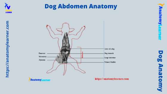
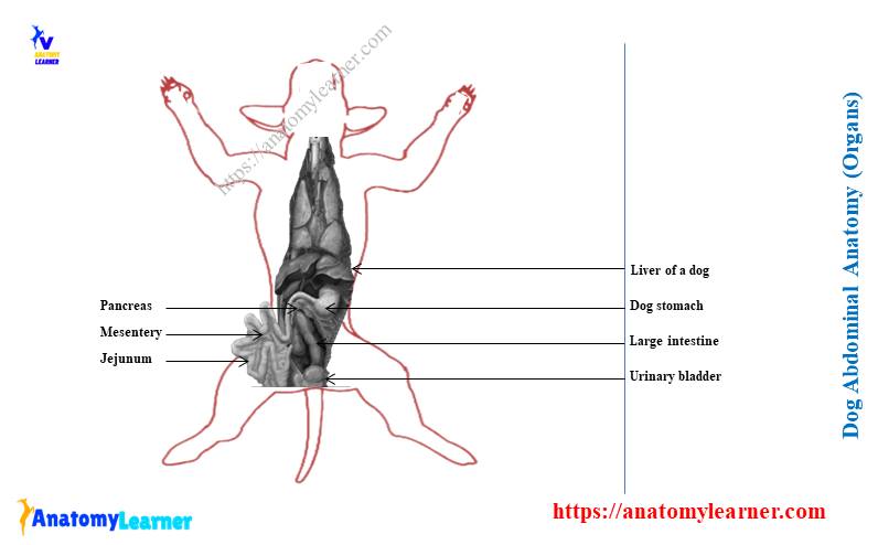
Now, let’s see the male dog abdominal organs from the labeled diagrams (both from the right and left side). The labeled diagram identifies all the essential organs from the male dog’s abdomen.
The significant organs from the dog’s abdomen labeled diagram are the stomach, liver, pancreas, kidney, different segments of the intestine, part of the urinary bladder, and others.
Finally, let’s show the different organs from the female dog abdomen with the labeled diagram. Here, I tried to show the different essential organs in the female dog’s abdomen labeled diagram.
You may find more female or male dog abdomen labeled diagrams here on social media of anatomy learners. Again, you may read the following suggested articles from the anatomy learner to learn more about dog anatomy –
- Dog organs anatomy with the labeled diagram,
- Male and female dog pelvis anatomy with their organs and labeled diagram.
Frequently asked questions on dog abdominal anatomy
Let’s see the dog anatomy learner’s common inquiries on the dog’s abdominal anatomy. All these questions and answers might be helpful for you to learn something more about the dog’s abdomen.
What organs or structures are on the right side of a dog’s abdomen?
I have already enlisted the different abdominal organs from both male and female dogs. You will find the right part of the lung, right part of the heart, right kidney, liver, right part of the stomach, right ureter, greater omentum covering the small intestine, and part of the bladder in the right side of a dog’s abdomen.
Again, you will also see the right uterine horn, descending duodenum, and right ovary on the right side of a dog’s abdomen. The labeled diagram identifies all these organs from the right side of the dog’s abdomen.
You may go to that dog’s abdomen organ labeled diagram and try to identify all these essential organs from the right side of the real sample.
What is on the left side of a dog’s stomach?
You will see the left part of the stomach, left lung, left kidney, left ureter, spleen, greater omentum covering the intestine, and descending colon on the left side of a dog’s abdomen. Again, all these organs from the left side of a dog’s stomach are identified in the labeled diagram.
You may go through that dog’s left side organs labeled diagram and identify all these essential organs from the real sample.
What does abdominal distention look like in dogs?
Usually, the abdomen of a dog is not so distended; it becomes narrower at the caudal third of the abdomen and becomes more narrow in the pelvic region. But, sometimes, you may find an enlarged abdomen in a dog.
Then the belly of the male and female dog goes more significant than the usual structure, and the convexness becomes reduced. This condition may occur due to any disease condition. So, your dog may have some restlessness, panting, pacing, retching, excessive drooling, and other clinical symptoms.
What are abdomen problems in dogs?
Different abdominal problems may occur in the stomach and parts of the intestine. The most common problems of the dog’s stomach and intestine include viral and bacterial infection, non-infection disorders, bloat, and obstruction.
They may cause serious damage if you do not provide the proper management to your dog.
Where is a dog’s abdominal cavity?
A dog’s abdominal cavity is a larger cavity that extends from the diaphragm to the pelvic inlet. You may quickly identify the cranial boundary of the dog’s abdominal cavity by palpating the last rib and costal cartilage.
But, the caudal boundary of the dog’s abdominal cavity is very hard to identify. You may identify only the pelvic inlet by palpation between the thighs. The abdominal cavity of a dog is less voluminous.
Conclusion
I hope you got the basic idea of the dog abdomen anatomy, including the abdominal cavity, muscles, and contents from both males and females. There are four crucial abdominal muscles in the anatomy of a dog’s abdomen – two obliquus, one transverse, and one straight muscle.
This is your first duty to identify the dog’s abdominal cavity boundary. There are different abdominal organs in both the right and left aspects of the dog abdomen. You might learn the exact location (topography and surface) of these essential organs like the stomach, liver, spleen, and different parts of the intestine from the dog abdomen.