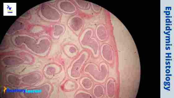
The epididymis is the comma-shaped structure in the male organ system that divides into head, body, and tail. It is made of highly coiled, tortuous ductus tubules and vascular connective tissue. In epididymis histology, you will find pseudostratified columnar epithelium lining and a circularly arranged smooth muscle layer beneath that epithelium.
Here, I will show you all the important identifying features from the actual epididymis histology slide. In addition, I will also provide you with the epididymis histology slide labeled diagram at the end of the article.
If you want to know the basics of the epididymis histology slide, you may read the first two or three parts of this article. But, if you want to know all the histological features of epididymis with their detailed description, please read the article till the end.
Epididymis histology
While studying the ductus epididymis histology slide under the light microscope, you will find many tubules with some characteristic features. The tubules of the ductus epididymis line with the pseudostratified columnar epithelium. Here, you will find two types of cells in the lining epithelium of epididymis – the tall columnar principle cells and the small basal cells.
Again, the tall columnar cells of the ductus epididymis slide contain the microvilli (known as the stereocilia). These stereocilia of the ductus epididymis involve in both secretion and absorption.
Beneath the lining epithelium of the ductus epididymis, you will find a layer of circularly arranged smooth muscle fibers. In addition, there are numerous cells and fibers of the connective tissue present adjacent to the smooth muscle layers.
Okay, let’s enlist the most important histological structures you might identify from the epididymis slide under the light microscope.
- Cross-section of the ductus epididymis
- The longitudinal section of the ductus epididymis
- A lining epithelium of the ductus epididymis tubules (pseudostratified columnar epithelium with stereocilia)
- The basement membrane of the lining epithelium
- A single layer of circularly arranged smooth muscle fiber beneath the lining epithelium of ductus epididymis
- The spermatozoa in the lumen of the ductus epididymis (tubules)
- Interstitial connective tissue (between the tubules)
Now, you might try to identify these histological features from the epididymis slide under the light microscope. I think the epididymis slide image and labeled diagram might help you identify the important structures so quickly.
Epididymis histology slide identification
Fine, in this part of the article, I will enlist some of the important identifying features from the epididymis histology slide. You should write these identification points to identify the epididymis slide under the light microscope at the laboratory.
The epididymis covers with a thick tunica albuginea externally that contains dense irregular connective tissue. Again, you may also find a few smooth muscles cells that are scatteringly distributed in the tunica albuginea of a horse’s epididymis.
- The sample tissue section shows numerous tubules that line with the pseudostratified columnar epithelium.
- Presence of tall columnar cells with stereocilia and basal cells in the lining epithelium of the provided tissue sample.
- There is a thin, smooth muscle fiber layer and loose connective tissue that surrounds each tubule.
- Presence of sperms in the lumen of the tubules.
So, this is a slide of the epididymis.
You know three different parts (head, body, and tail) present in the epididymis. The thin, smooth muscle layer surrounding the tubules may vary in the different regions of the epididymis.
In the head of the epididymis, you will find a thin layer of smooth muscle layer that surrounds each tubule. But, there is a thick, smooth muscle layer present in the outer surface of the tubules. This thick layer of the smooth muscle of the epididymis tail consists of inner circular and outer longitudinal portions.
Features of the epididymis histology with labeled diagram
Grossly, the epididymis of an animal is a comma-shaped structure that possesses a head, body, and tail. The head of the epididymis forms by the highly convoluted continuation of the efferent ductules. So, sometimes you may find the ductules efferentes in the section of the epididymis histology slide.
Again, the body and tail of the epididymis consist of the greatly coiled ductus. But the coil of these ducts gradually decreases towards the tails of the epididymis.
Histologically, the tubules of the ductus epididymis are lined by the pseudostratified columnar epithelium. This epithelium of the epididymis slide consists of two types of cells – tall columnar principal cells and small basal cells.
The tall columnar cells of the epididymis contain long branching microvilli (known as the stereocilia) involving both secretion and absorption. Again, the small basal cells of the epididymis are spherical and located near the base of the epithelium.
In addition, the tubules of the ductus epididymis histology slide surround by the thin, smooth muscle layer. The thickness of this smooth muscle layer gradually increases from head to tail of the epididymis. You will find an inner circular and outer longitudinal smooth muscle layer in the tail region of the epididymis slide.
The smooth muscle of the ductus epididymis also possesses sympathetic nerve fibers. There is rhythmic contraction found on these smooth muscles that help in the expulsion of the spermatozoa from the lumen of the epididymis.
You will also find loose connective tissue cells and fibers adjacent to the ductus epididymis slide’s smooth muscle layer or tubules.
Ductuli efferents histology
Sometimes, you may find the ductules efferentes within the epididymis histology slide (head region). These occur as a small lobule with distinct boundaries of connective tissue. You will find simple columnar cells on the lining epithelium of the ductules efferents containing ciliated and non-ciliated principal cells.
There are also present small basal cells where you will find scatter-distributed mononuclear cells. The principal ciliated cells of the ductules’ efferentes help to move spermatozoa towards the ductus epididymis. Again, the non-ciliated principal cells possess the brush border of the microvilli.
The ratio of ciliated and non-ciliated principal cells of the ductules efferents vary along the ductules. But, the principal ciliated cells of the ductules increase the number towards the ductus epididymis.
There are also three to six layers of loosely arranged myofibroblast and connective tissue around the lining epithelium.
Other information on lining epithelium of epididymis
Generally, there are two types of cells (columnar principal and small spherical basal cells) in the lining epithelium (pseudostratified columnar) of the epididymis slide. These characteristics feature most commonly found in domestic mammals.
The height of the epididymal epithelium of domestic mammals remains the same throughout the year. But, in camel, you will find a change in the epididymal epithelium over the year.
Again, in many species, you will find other cells like apical and clear cells in the lining epithelium of the epididymis slide. There may also present the macrophage and lymphocytes within the lining epithelium of the epididymis slide.
Again, the principal cells of the lining epithelium contain the stereocilia on their apical surface. The length of the stereocilia of these cells becomes shorter towards the tail of the epididymis histology. There you may also find some pinocytic vesicle at the base of the microvilli.
Epididymis anatomy
Anatomically, the epididymis of animals is covered by the thin tunica albuginea. You will find the distinguish three parts in the epididymis – head, body, and neck. The head of the epididymis consists of a dozen of coiled tubules that are grouped into lobules.
The tubules of a lobule unite to form a single tube. Thus it forms the ducts of the epididymis that is full coli. The head of the epididymis forms the body and tail part. Again, the tail of the epididymis terminates in the ductus deferens.
In addition, the tubules and the colis of the ducts of the epididymis are held together by the connective tissue and smooth muscle fiber.
If you want to learn more about the anatomy of an epididymis, you may read another article from anatomy learner.
Functions of epididymis
I will not provide more information on the function of the epididymis. Here, I will provide the basic functions of the epididymis of the animal.
- The proximal part of the epididymis help (head and body) involves the spermatozoa’s maturation process.
- Again, the tail of the epididymis is the main storage place of spermatozoa.
- The epididymal spermatozoa can carry a wide range of potent antigens that easily trigger autoimmune responses.
- It also helps to absorb the fluid from the main organ of a male
The spermatozoa undergo a series of morphologic and functional changes during the passes through the ductus epididymis. The motility of the spermatozoa progressively developed within the ductus epididymis.
A fully matured spermatozoa may be stored in the tail of the epididymis for a long period.
Epididymis histology slide and labeled diagram
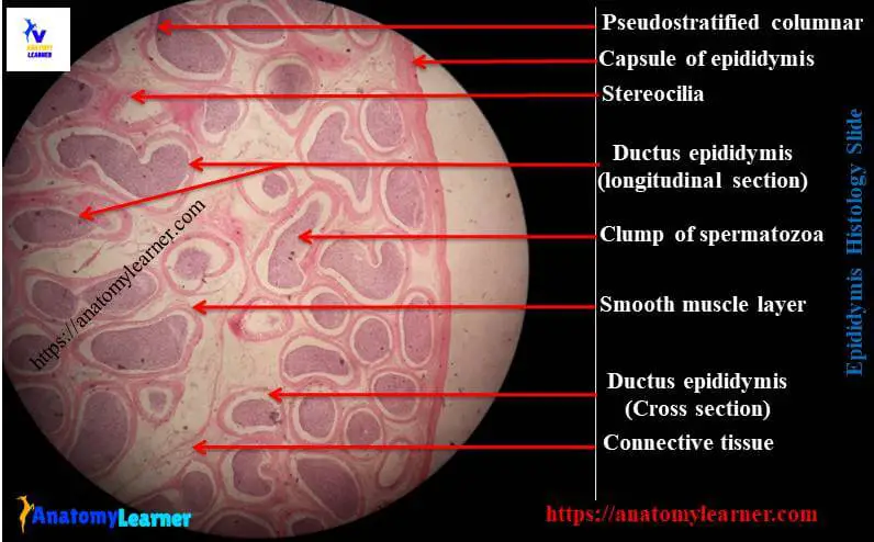
I tried to show you the most important structures in the epididymis histology slide and the labeled diagram. There is a thick connective tissue capsule at the periphery that surrounds the epididymal tubules (known as tunica albuginea).
The pseudostratified columnar epithelium (with a variety of stereocilia) is also shown on the epididymis labeled diagram. A circularly arranged smooth muscle fibers that surround the epididymal tubules are also identified in the labeled diagram.
There is a clump of spermatozoa present in the lumen of the epididymis. But, you may not see the same figure in other slides of the epididymis. You will find more clumps of spermatozoa in the tail region of the epididymis compared to that of the head and body regions.
If you need more microscopic pictures of the epididymis or the labeled diagrams, you may join anatomy learner on social media.
Epididymis macroscopics slide drawing
The following guide might help you to draw the epididymis microscopic slide image. Let’s try to draw the long, convoluted ducts of the epididymis in the section. You might draw the pseudostratified ciliated columnar epithelium in the section. Don’t forget to draw the small spherical basal cell on the epididymis microscopic slide.
You might draw some stereocilia in the apical surface of the principal columnar cells of the epididymis image. Let’s draw a circularly arranged single layer of smooth muscle after the basement membrane of each duct or tubules of the epididymis.
You know clumps of spermatozoa are present in the lumen of the duct, so let’s provide some spermatozoa (clump) in the lumen of the ducts or tubules of the epididymis.
Now, let’s try to draw the loose connective tissue between the epididymis’s ducts or tubules. You might also provide some blood vessels (artery or arterioles) on the connective tissue framework of the epididymis.
I would like to suggest you read the following articles from anatomy learner –
- Histological features of the ureter and the identification points (with actual microscopic slide pictures and labeled diagrams)
- Features of ductus deferens and its identification points (with the actual microscopic images and also labeled diagrams)
- Histological features of seminal vesicle and prostate glands (real microscopics slide images and diagrams)
In addition, you may find more articles (organs of different systems) on the histology section of anatomy learner.
Frequently asked questions on epididymis slides.
Fine, now, I will try to solve the common inquiries on the histology of epididymis slides. I think you will find these answers helpful for you to understand the histological structure of an epididymis.
What type of epithelium is found in the epididymis?
In the epididymis slide, you will find the pseudostratified columnar epithelium that contains principal columnar (ciliated or non-ciliated) and small spherical basal cells. The apical surface of the principal columnar cells possesses some microvilli (brush border). The length of the microvilli gradually decreases towards the tail of the epididymis.
But, you may find other different types of cells within the lining epithelium of the epididymis slide. You may find the apical cells, clear cells, macrophages, and lymphocytes within the basal layer of cells.
The above-mentioned labeled diagram represents the principal columnar (ciliated and non-ciliated) and the small basal cells. If you don’t read the above information, I will hight recommend you read the full article to know the basic histological feature of the epididymis slide.
What is the histology of the epididymis?
I will provide very short histology of the epididymis to identify it under the light microscope quickly. The outer surface of the epididymis covers by a thin layer of connective tissue. In the inner section, you will find different tubules that possess some characteristic features.
The lining epithelium of the tubules is pseudostratified columnar (containing two types of cells – columnar and basal cells). There are numerous microvilli on the apical surface of the principal columnar cells of the lining epithelium.
After the basement membrane of the lining epithelium of the epididymis presents a single layer of smooth muscle fiber. Again, there is interstitial connective tissue present among the tubules of the ductus epididymis.
What is the epididymis structure?
I hope you already got an idea of the structure of epididymis in my previous answer. You should identify these structures from the epididymis slide with the help of the labeled diagrams.
You should identify the thin connective tissue capsule, tubules, lining of the ductus, single smooth muscle layer, spermatozoa, and connective tissue from the epididymis slide.
What are the three types of epididymis?
If you follow this article, you know three different parts of the epididymis (head, body, and tail). But, how do you identify these three parts of epididymis histologically? It is so simple to identify and make differences among the three parts of the epididymis histology slide.
In the head part of the epididymis slide, you will find the ductules efferents, epididymal tubules (along with their lining epithelium), a single layer of the smooth muscle layer, less amount of spermatozoa in the lumen.
All the histological features of the head region of the epididymis slide are almost similar. But the height of the microvilli decreases. Even the smooth muscle layer that surrounds the tubules increase.
Finally, in the third part of the epididymis (tail), you will also find the epididymal tubules that contain lower microvilli on the apical surface of the principal columnar cells. Again, you will find the inner circular and outer longitudinal smooth muscle layer in the third part of the epididymis slide.
You will not find any ductile efferentes in both the body and tail part of the epididymis slides.
What are the structure and functions of the epididymis?
I have already described the structures of the epididymis histology slide in this article. Would you mind reading that portion of the article to know the basics of the structure of an epididymis? The epididymis has a great function to help in the maturation of the spermatozoa. Again, the tail of the epididymis is the place of storage spermatozoa.
Conclusion
In the ductus epididymis histology, you will find the pseudostratified columnar lining epithelium (ciliated) surrounded by the smooth muscle fiber and connective tissue. I hope this simple article might help you understand epididymis histology basics with the actual microscopic images and labeled diagrams.
To identify the three different parts of the epididymis histology slides (head, body, and tail), you might focus on the length of apical microvilli and layers of smooth muscle fiber. Now, you might study the epididymis slide under the light microscope from the laboratory