The dog knee injury is very common in the field. If you want to manage a knee injury, you might have a good piece of knowledge on the dog knee anatomy. Here, I will show you everything on the dog knee, including the bone involvement, ligaments, tendons and their arrangement with a labeled diagram.
You will also get an idea of the anatomy of the menisci of the knee joint. I will describe the detailed anatomy of the straight patellar ligament and cruciate ligament from the dog’s knee joint.
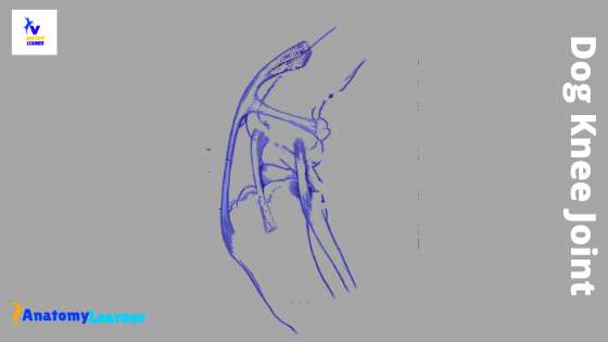
That’s nice; let’s get started to learn everything about dog knee joint anatomy from the hind leg.
Dog knee anatomy
Another name of the dog knee joint is stifle or genual articulation. It is a complex condylar synovial joint found in the hind leg of a dog. In the dog knee anatomy, you will find two main joints – femoropatellar and femorotibial joints.
I will show you the femoropatellar and femorotibial joints separately with their interesting anatomical facts. But, let’s see the basic anatomy of the knee joint from the below mentioned labeled diagram. That might provide you with a summary of the dog knee joint.
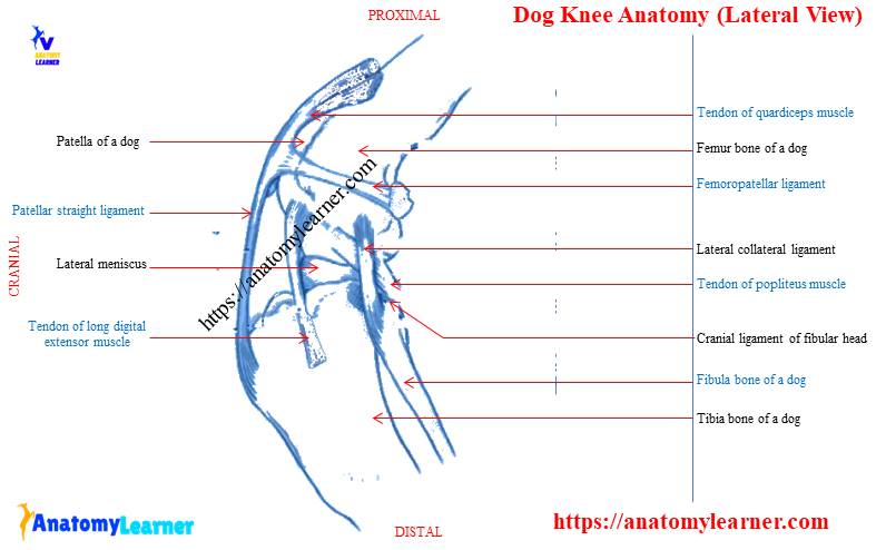
I hope you could understand and identify some of the structures involved with the dog knee joint. Here, you might have a good piece of knowledge on the hindlimb bones of a dog. Again, if you have an idea of the structure of a synovial joint, you will easily understand knee anatomy.
The femoropatellar joint of the dog’s knee is formed by the trochlea of the femur and articular surface of a patella. Again, in the femorotibial joint structure, you will find the condyles of the femur have a connection with a proximal end of the tibia and other. So, make sure you know the different structures or features from a dog’s femur, patella, tibia, and fibula bones.
Bones and joints of dog’s hindlimb
I am not going to describe the detailed anatomical facts of bones and joints from the hindlimb of a dog. I want to help you to memorize the bones and joints from a dog skeleton.
In the hindlimb of a dog, you will get the same bones that you found in a goat or other mammals. So, pelvic bones, femur, tibia, fibula, patella, tarsal, metatarsal, and phalanges are present in the hindlimb of a dog.
Again, in the hindlimb of a dog, you will find the following different joints –
- Sacro-iliac joint or articulation of dog
- Pelvic symphysis or joint of a dog
- Coxal or hip-joint of a dog
- Stifle or knee or genual joint of a dog
- Hock or tarsal or pedal joint of a dog hindlimb and
- Fetlock, pastern, and coffin joints of the hindlimb of a dog
I hope you will identify all of these bones and joints of the hindlimb from the dog leg labelled diagrams.
Details on dog knee anatomy
This joint is formally known as stiffle in most mammals. But in the case of a dog, human, you may term it as a knee joint. This is the largest and more complex joint of the hindlimb of a dog. The dog knee anatomy consists of two joints – femoropatellar and femorotibial.
The main movement of the knee joint is extension and flexion with limited rotation. Now, let’s know what the structures involving the knee joints in a dog are. Okay, I will show you the structures or bones that you will find in the knee joint of a dog.
- In femoropatellar joint – trochlea of the femur and articular surface of the patella
- In femorotibial joint – condyle of femur, the proximal extremity of the tibia, and interposed articular menisci or semilunar cartilage.
Again, you will find other structures (ligaments) like joint capsule, lateral and medial collateral ligaments, straight patellar ligaments, cruciate ligament, and different meniscus ligaments.
Ligaments of knee anatomy
Here, I will provide a little information on ligaments of dog knee anatomy. You will find the joint capsule in common both in the femorpatellar and femorotibial joints of the knee. The joint capsule is a thin but very extensive part of the knee joint of a dog.
In the femorpatellar part of the knee joint, you will find the below-mentioned ligaments. Please try to identify these ligaments from the knee joint ligaments labeled diagram.
- Joint capsule of femoropatellar joint of a dog
- Femoropatellar ligaments – lateral collateral and medial collateral ligaments of dog’s knee
- Patellar straight ligaments of dog’s knee (lateral, middle, and medial patellar ligaments)
Again, in the femorotibial part of the knee joint, you will find the following ligaments –
- Joint capsule of femorotibial joint of a dog
- Lateral and medial collateral ligament of femorotibial joint
- Cruciate ligament of a femorotibial joint of a dog (cranial and caudal parts)
In addition, you will find five or six meniscal ligaments (may vary) that attach the menisci to the tibia and femur bones of a dog.
- Cranial tibial ligament of the medial meniscus
- Caudal tibial ligament of the medial meniscus of dog’s knee
- Cranial tibial ligament of the lateral meniscus
- Caudal tibial ligament of the lateral meniscus of the knee
- Femoral ligament of the lateral meniscus of the knee joint and
- The transverse ligament or intermeniscal ligament of the knee joint of a dog
These meniscal ligaments of the dog knee anatomy are not fixed. You may find less or more meniscal ligament in the knee joint of a dog.
“There are two menisci in the knee joint of a dog (showed in the diagrams)”
The femoropatellar joint of dog knee
The femoropatellar joint locates between the patella and trochlea of the femur. You will find two slightly oblique ridges with a wide and deep groove within them in the trochlea of a dog. The medial ridge is larger, whereas the lateral ridge is narrower and more irregularly curved.
Again, the articular surface of the patella of a dog is much smaller than that of the trochlea of femur. You will find articular cartilage on the trochlea that completely covers the medial ridge.
The joint capsule of the femoropatellar joint
The joint capsule of the femoropatellar or stifle joint of a dog is very thin and largest. There are three different sacs of the joint capsule that communicates with each other in the femoropatellar articulation.
The two sacs are present between the femoral and tibial condyle, and the third is beneath the patella. The patellar part of the joint capsule is very thin and attaches around the margin of the articular surface. But on the femur, the line of attachment is at a varying distance from the articular surface.
Proximally, a sac of femorpatellar joint capsule protrudes deep to the dog’s tendon of the quadriceps femoris muscles. In the large breed of dog, the patellar part of the joint capsule extends from the trochlear ridge to femoral epicondyles laterally. The patellar and femorpatellar parts of the joint capsule join without any sharp boundary at the distal part.
In addition, you will find a large quantity of fat in the fibrous layer of the cranial part of a joint capsule. This is the infrapatellar fat body that increases the thickness of the joint capsule distally.
Femoropatellar ligaments of the knee
The femoropatellar ligaments of dog knee anatomy are two thin bands reinforcing the joint capsule on either side. You will find the lateral collateral ligament of femoropatella easily. But, the medial collateral ligament of femoropatellar joint is indistinct and very hard to separate from the joint capsule.
The lateral collateral ligament arises from the lateral condyle of the femur. It ends at the lateral border of the patella. Again, the thin medial collateral ligament of the dog’s knee arises from the medial epicondyle of the femur and ends on the medial border of the patella.
Patellar straight ligament of dog knee anatomy
You know the patella is the largest sesamoid bone intercalated in the tendon that comes from the quadriceps femoris muscle of a dog. A portion of the tendon from the patella to the tibia tuberosity forms the patellar ligament in the dog’s knee. This is one of the most important ligaments of the dog knee anatomy.
The patellar ligament separates from the synovial membrane of the joint capsule by a large quantity of fat. You will find a small synovial bursa between the patellar ligament’s distal part and tibial tuberosity. Do you know how the patella held in their position in the dog’s knee? The dog’s patella is held in the trochlea of the femur by the thick fascia lata and thinner medial femoral fascia.
The patellar ligament of a dog is very strong bands and divides into three parts –
- The lateral patellar ligament of a femoropatellar joint of a dog
- Secondly, the middle patellar ligament of the dog’s knee and
- The third one is a medial patellar ligament of the femorpatellar joint
You will find an extensive band (lateral patellar ligament) that extends from the lateral part of the cranial surface of the patella to the lateral part of the tibial tuberosity. This lateral patellar ligament receives strong tendons from the biceps femoris muscle and also from fascia lata.
The middle patellar ligament (strongest) extends from the cranial part of the apex of the patella to the distal part of a groove of the tibial tuberosity. In addition, the medial patellar ligament is distinctly weaker than the other. It attaches proximally to the parapatellar fibrocartilage. And it ends on the tibial tuberosity at the medial side of the groove.
There is a lateral and medial cartilage ridge on the crest of the femoral trochlea that helps prevent the patella’s dislocation.
The femorotibial joint of the knee
The femorotibial joint of dog knee anatomy forms with the condyle of the femur, a proximal end of the tibia bone and interposed articular menisci. I will show you the crescentic menisci and very important cruciate ligament from the femorotibial joint with a labeled diagram.
In this joint of dog’s knee, you will find the joint capsule structurally almost similar to that of femorpatellar’s joint capsule. There are two collateral ligaments, two cruciate ligaments, and six meniscal ligaments present in the femorotibial articulation.
Now, let’s see the femorotibial joint diagram of the dog. I hope you will find all the structures that involve in this joint from this diagram.
Collateral ligaments of a femorotibial joint of a dog
The medial collateral ligament of femorotibial articulation attaches proximally to the prominent medial epicondyle of the femur. Distally, it joins to the rough margin area of the medial epicondyle of the tibia bone of the dog.
On the other hand, the lateral collateral ligament of dog’s knee is thinner. It arises from the upper depression of the lateral epicondyle and ends on the head of the fibula bone. Again, it covers the tendon of the origin of the popliteus muscle. Here, you will find a bursa in between the epicondyle and popliteus muscle.
Again, you will find another bursa between the lower part of the ligament and the margin of the lateral condyle of the tibia bone.
The joint capsule of femorotibial articulation
This strong flat band arises from the femur just lateral to the medial head of the gastrocnemius muscle. Distally it extends to the caudal border of the medial border to the tibia bone of a dog.
The joint capsule of femorotibial articulation attaches to the margin of the tibial articular surface. It also attaches to the convex border of the menisci and the cruciate ligaments. The cranial part of the joint capsule is thin and consists of a synovial layer only. But the caudal part is stronger and have a connection with the oblique popliteal ligament.
Here in the joint capsule of the femorotibial joint, you will also find two synovial sacs. But the femorotibial sacs are smaller than that of femorpatellar. Each of these femorotibial sacs divides by the menisci into femoromeniscal and tibiomeniscal parts.
The menisci develop in the fibrous layer of the capsule, and the two parts communicate primarily around their concave, sharp-edge axial borders.
Again, you will find a transverse communication between the lateral and medial border condyloid part of the joint.
Cruciate ligament of dog knee anatomy
The cruciate ligament of the dog knee anatomy is two strong round band-like structures. They locate mainly in the intercondyloid fossa of the femur in between two synovial sacs. Two cruciate ligaments cross each other in knee anatomy.
The cruciate ligaments of the dog knee joint limit cranial and caudal sliding movement of the tibia on the femur bone. The cranial cruciate ligament of dog’s knee runs from the caudomedial part of the lateral condyle of the femur. It across the inercondyloid fossa to the cranial intercondyloid area of tibia (or central fossa on tibial spine).
In addition, the caudal cruciate ligament of the dog knee anatomy runs from the lateral surface of the medial femoral condyle to the lateral edge of the popliteal notch of the tibia. It is slightly thicker and longer than the cranial one. Again, the caudal cruciate ligament lies medial to the cranial one.
The collateral ligaments of the dog knee joints work together with cruciates to limit medial rotation of the tibia on the femur bone. In extension movement, the collateral ligaments are the primary check against the medial and lateral rotation. The lateral rotation of the tibia limits by the collateral ligaments in both flexion and extension.
Menisci of the knee joint of a dog
You already know that the menisci develop in the fibrous layer of the femorotibial’s joint capsule. They are crescentic plates of fibrocartilages that produce congruence in the articular surfaces.
You will find two menisci (lateral and medial) in the dog knee anatomy or stifle joint. Each of these menisci has a proximal concave surface that adapts to the condyle of the femur. Again, the distal surface of each meniscus fits for the corresponding condyle of the tibia bone.
The lateral meniscus is slightly thicker and forms a greater arc than the medial one. In addition, the peripheral border of the meniscus is thick and convex in a dog.
The lateral meniscus does not cover the lateral and caudal parts of the tibial condyle. You will find ligaments that attaché to the tibia, cranial and caudal to the spine. Again, the lateral meniscus has a third attachment by an oblique band that passes from the caudal end to the caudal part of the intercondyloid fossa of the femur.
The medial meniscus of the dog knee anatomy retains its attachment to the joint capsule of the femorotibial. You will find different ligaments that are described in the next part of this article.
Ligaments of menisci of dog knee
I already told you that the meniscal ligaments attach the menisci to the tibia and the femur of a dog. The cranial tibial ligament of the medial meniscus goes from the cranial axial angle of the medial meniscus to the cranial intercondyloid area of the tibia.
You will find a caudal tibial ligament of the medial meniscus that goes from the caudal axial angle medial meniscus to a caudal intercondyloid area of the tibia. This attachment is just cranial to the tibial attachment of the caudal cruciate ligament of the dog’s knee.
The cranial tibial ligament for the lateral meniscus goes to the cranial intercondyloid area of the tibia. It attaches caudally to the transverse ligament and cranial tibial attachment of the medial meniscus.
You will find a caudal tibial ligament for the lateral meniscus that arise from the caudal axial angle of the lateral meniscus. It ends at the popliteal notch of the tibia bone just caudal to the intercondyloid area of the tibia.
Again, the femoral ligament of the lateral meniscus is the only femoral attachment of the meniscus in dog knee anatomy. It passes from the caudal axial angle of the lateral meniscus to the medial femoral condyle.
You will also find intermeniscal ligament in the stifle joint of a dog. They are the small transverse fibrous band in the knee anatomy. Another name of this intermeniscal ligament is a transverse ligament in a dog.
It leaves the caudal side of the cranial tibial ligament of the medial meniscus. Then it goes to the cranial part of the cranial tibial ligament of the lateral meniscus.
Dog knee anatomy diagram
I hope all the diagrams of dog knee anatomy were helpful for you to understand it easily. If you need more diagrams on the dog knee joint, please follow anatomy learner on social media. You will get a notification if I update or publish any new diagrams on the dog knee in future.
Frequently asked questions on a dogs knee
In this frequently asked question section of the article, I will try to answer the most commonly asked questions on dog’s knee joint.
Where is a dog’s knee?
The dog’s knee is located in the hindlimb of a dog between the femur and tibia, fibula bones. Here, you will find two important joints – the femoropatellar and femorotibial joints.
How many ligaments are in a dog’s knee?
You will find two lateral ligaments, one joint capsule, three straight patellar ligaments in the femoropatellar joint of the dog’s knee. Again, several ligaments are found in the femorotibial joint part like – collateral ligaments, cruciate ligaments, and meniscal ligaments.
What ligament do dogs have in their knees?
You may follow my previous answer on the knee joint’s ligament. The most important ligaments of the dog’s knee are collateral ligaments, patellar ligaments, and cruciate ligaments.
Which joint is a dog’s knee?
I hope you have a basic idea of the different joints of the hindlimb of an animal. The stifle joint of a dog is formally known as the knee joint.
Conclusion
So, the dog knee anatomy consists of two joints – femorpatellar and femorotibial joints. The femoropatellar joint form in between the femur and patella of the dog. Again, the femorotibial joint from among femur, tibia and menisci. In association with these bones, you will find different types of ligaments in the dog knee anatomy.
The joint capsule, collateral ligaments are common in both femorpatellar and femorotibial joints of knee anatomy. You will find some special ligaments (patellar straight and cruciate ligaments) in a dog’s knee joint. Would you please use all the labelled diagrams to understand the knee anatomy clearly? If you want to know more about knee joints, let me know.