The bird wing anatomy possesses some unique features. In this article, I will show you the outstanding features of a wing with diagrams. You will get the detailed anatomy of bird wing bones, muscles, joints, and more.
There are different types of feathers present in the wing of a bird. The feathers are the distinctive feature of the member of avian species. Here, I will also show you the external parts of the birds wing with a diagram.
If you are a veterinary student, this article might help you learn the anatomical features of bones, joints, muscles, nerves, and vessels from the birds wing.
Bird wing anatomy
So, first, you might have an idea of the wing of a bird. The wing of a bird is nothing but the modified forelimb in avian species used for flying. You will find some unique large feathers in the external anatomy of a wing of a bird.
You will find almost similar (modified) structures in the bird wing-like forelimb of mammals or other animals. There are the humerus, radius, ulna, fused carpal, metacarpal, and digits present in a bird’s wing.
You will find different muscles in the wing anatomy that provides specific localized control of the movement of bones. I will show you some of these important flight muscles from the wing of a bird.
In addition, you will find some important vessels like the deep brachial artery and its branches deep to the muscles of the arm and shoulder region of a wing. I will also show you the large wing vein (deep ulnar vein) from a bird.
There are also ulnar nerve, axillary nerve, radial nerve, and others present in the wing anatomy of a bird. So, here you will learn the following anatomical features of a wing –
- Unique anatomical features of wing bones of a bird
- The important flight muscles and other muscles from the wing
- The name and formation of joints in the bird wing
- Anatomical features of nerves and vessels of a bird wing.
Nice, let’s start to learn these anatomical features one by one from a bird’s wing.
Bird wing anatomy feathers
As you know, the feathers are the unique features of the member of the avian species. They develop from the epidermal cells in a similar way to grow hair in mammals. Here, I will provide some information on feathers from the birds wing anatomy.
The feathers are made of keratine and create a strong but lightweight covering over a bird’s wing. Several feathers are present in the bird wing-like flight feather, contour feather, and down feather.
The flight feathers are the long rigid feathers attached to the wing of a bird. You will also find this type of feather in the tail region of a bird. They are two types – primaries and secondaries in bird wing.
The primaries attach with the digits and to fused metacarpal bones of the birds wing. Again, the secondaries are shorter than that of primaries and connect with the ulna bone of the wing.
The contour feathers cover the rest of the wings of a bird. It will also cover the outermost layer of the body to produce a smooth outline. You will find the down feathers lie close to the body of a bird.
Bird wing anatomy bones
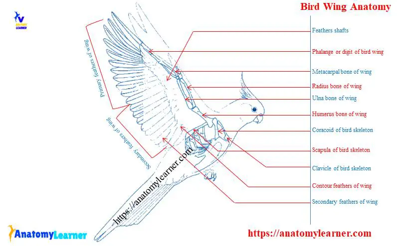
The wing bones consist of humerus, radius-ulna, fused carpals, metacarpal, and digits. Again, a bird’s pectoral girdle comprises three pairs of bones (clavicle, coracoid, and scapula) that support the wing. Now, I will show you the unique features of bird wing anatomy bones.
But, let’s first discuss the bones of the pectoral girdle of a bird. The most strong bone of the pectoral girdle of a bird is the coracoid. It directs caudally and ventrally to articulate with the sternum at the coracoidial sulcus. You will find air sacs in the hollow coracoid bones.
In addition, the clavicles are the slender rod-like bones that fuse ventrally into a flattened plate. This flattened plate is known as hypocleidium that joins with the cranial apex of the sternum by hypocleidum ligament.
You will find a long, flat scapula bone that extends caudally, paralleling the vertebral column. This bone articulates cranially with the coracoid and furculum. It also helps to the formation of the glenoid fossa in a bird.
That’s fine; now, I will discuss the prominent bones of a wing of a bird.
Main wing bones
The humerus is the most prominent wing bone of bird anatomy. It joins proximally with the pectoral girdle at the glenoid fossa. You will find pneumatic foramen at the proximal end of the bird’s humerus. There is a deltoid crest on the ventral surface of the humerus of a bird. The humerus of the bird joins with the radius and ulna bones distally by two condyles.
Other prominent bones of the wing are the radius and ulna. You will find considerably larger ulna bone that radius in the wing of a bird. But the length of these wing’s bones is approximately equal. The radius and ulna bones of the wing articulate with the condyle of the humerus proximally and distally with the carpus bones.
You will find an articulate facet at the proximal end of the radius bone of the bird that joins with the small condyle of the humerus. In addition, there is a large concave articular surface at the proximal end of ulan bone that joins with the more prominent condyle of the humerus. You will also find some nutrient foramen midway along the length of the ulna bone of the birds wing.
There is also a small facet at the distal end of the radius for articulation with the ulna bone. You will find two articular facets for carpal bones on the distal end of the ulna bone.
There are three fused carpometacarpus bones present in a bird’s wing (metacarpal II, III, and IV). You will find a sizeable interosseous space in between the metacarpal III and IV. Again, the metacarpal II is the small projection on the radial side of the carpometacarpus.
Generally, there are three digits present in a bird’s wing (may vary with species). Among these three digits, the third one is the largest and contains two phalanges.
Bird wing muscle anatomy
Here in the bird wing muscle anatomy, I will show you some important muscles from the brachium, antebrachium, and maneus regions. You will find some unique features in a wing’s muscles that help draw the wing’s bones. They also help in flexing and elevating the wing bones. Thus it helps to control the movement of the wing’s bones and flying.
You know there are lots of muscles in the wing of a bird. So, it is pretty hard to describe all the muscles anatomy from the wing in a single article. But, I will try to summarize all the muscles anatomy from different regions of a wing.
So, you will find the summary of the following muscles from the bird wing –
- Muscles of the pectoral girdle and brachium of bird
- List of muscles of forearm or antebrachium of bird and
- The important muscles of the maneus region of the bird wing
Nice, let’s continue this article to know about the muscle anatomy of a wing.
Muscles of pectoral girdle and brachium of bird
There is a flattened superficial latissimus dorsi muscle on the dorsal aspect of the body of a bird. This muscle has two different parts – the cranial and the caudal part.
The cranial part of latissimus dorsi arises from the spinous process of a variable number of cervical and few thoracic vertebrae. Again, it inserts on the caudal surface of the humerus of the bird, between the scapular and humeral head of the triceps brachii. This muscle has a significant role in drawing the wing caudally, flex and elevate the humerus to govern the movement.
The following muscles – scapulohumeral cranialis, caudalis, subscapularis, subcoracoideus, coracobrachialis cranails, and caudalis all have a proximal attachement with the scapula and insert on the proximal end of the humerus. The function of these six muscles provides the specific localized control of the movement of the humerus that affects wings.
The primary force component of power for the downstroke provides by the pectoralis thoracicus muscle. Again, the supracoracoideus muscle provides the upstroke power for the wing of bird anatomy.
You will find a large muscle complex (subcoracoscapularis) muscle that attaches to the scapula, coracoid, and humerus. The coracobrachialis caudalis origins from the lateral surface of the coracoid and inserts on the medial tuberosity of the humerus.
The pars thoracicus muscle helps to draw the wing craniad and at the same time depress the leading edge of the entire wing. In addition, the deltoideus minor muscle of the wing assists in flexing the shoulder and elevating the humerus.
On the other hand, the deltoideus major muscle helps elevate the humerus and wing, thus allowing for movement of the wing caudally.
Other brachium muscles and their functions in wing
You will also find other different muscles in the wing anatomy of a bird. I will provide a list of muscles that helps in upstroke and downstroke movement of the bones of wings at the end of this article. But if you read the whole article, you may make a list by yourself.
You will find topographically similar muscles (triceps brachii) in bird wings as seen in mammals. There are typically two well-defined heads – pars scapularis and pars humeralis.
The pars scapularis muscle flex the shoulder and extend the elbow joint of the wing. Again, the pars humeralis also help in extends the elbow joint and the wing of a bird.
A borad biceps brachii muscle helps to flex the forearm and assists in extending a shoulder of a bird. The brachialis muscle of the wing inserts on the proximal end of the shaft of the ulna bone. It helps to flex the elbow joint of the wing.
Muscles of antebrichum and maneus of wing
There is a large, two-headed extensor metacarpi radialis muscle present at the craniodorsal border of the forearm of a bird. This muscle has a great function to extend the metacarpus and flex the wing’s elbow joint.
The extensor digitorum communis muscle paly a significant role in extending the hand, significantly when the forearm is initially extended. It also helps to maintain the position of the digits during the flying of the bird.
The extensor metacarpi ulnaris is the most caudal muscle of the forearm of a bird. This muscle helps to flex the metacarpus when the wing is extended. There is a flexor digitorum profundus muscle at the ventral aspect of the forearm of a bird that depresses the metacarpus.
At the ventral surface of the ulna bone of the wing, you will find the ulnimetacarpalis ventralis muscle that helps to flex and depress the cranial surface of the metacarpal. The abductor digiti majoris muscle extends the primary digit of the wing with a slight depressing action on the metacarpal.
Again the interosseus dorsalis inserts on the base of the second phalanges of the significant digit of the bird’s wing. It also helps to extend the major digit of the birds wing.
Artery of bird wing
This is also important to know the anatomy of arteries from the birds wing as a veterinary student. The cranial pectoral artery supplies the dorsocranial part of the pectoralis and inserts on humeurs. At eh ventral aspect of the scapulohumeral caudalis muscle of the wing, the axillary artery gives off a deep brachial artery.
The deep brachial artery gives rise to the doral humeral circumflex artery for the proximal muscle of the arm and shoulder joint of the wing. Again, it provides the new branch (ulnar collateral artery) courses with the dorsal brachial artery.
You will also find a deep radial collateral artery that rum parallel to the radial nerve between the proximal end of the triceps brachii muscle of the wing.
The brachial artery runs along the arm with the median, ulnar nerve between the biceps and triceps brachii muscle. At the mid-arm of the bird, the brachial artery divides into the ulnar and radial arteries.
The ulnar artery enters the ventral forearm distal to the cubital fossa. In the cubital region, the ulnar artery of the wing gives the recurrent ulnar artery that runs towards the tip of the elbow joint. At the proximal end of the forearm, the ulnar artery separates from the caudal ramus of the ulnar nerve.
The radial artery of the bird runs to the deep muscles of the flexor aspect of the arm. You will find the following different branches of the radial artery in the wing of a bird.
- Superficial radial artery of bird
- Deep radial artery of bird wing
- The dorsal interosseous artery of the wing
- Small radial recurrent artery of bird wing
Do you know the largest venous channel of the forearm of a bird? Well, the largest venous channel of the wing is a deep ulnar vein. It begins proximal to the wrist joint and runs deep into the flexor carpi ulnaris.
Nerves of the bird wing
Here in this part, I will only discuss the major nerves from the birds wing anatomy. You will find the brachial plexus in a bird that innervate the wing region.
The median, ulnar nerve enters the arm proximoventral to the scapulotriceps muscle. You will find the biceps nerves into the deep surface of the biceps muscle of the bird.
The ulnar nerve innervates the flexor muscles of the forearm, follicle of the feathers, ventral muscle of the metacarpal, and joints of the elbow of the bird.
At the caudal edge of the flexor carpi ulnaris muscle, the ulnar nerve divides into two parts- the caudal and cranial parts. The caudal part of the ulnar nerve is the largest and extends the entire length of the forearm of a bird. Again, the cranial part of the ulnar nerve runs parallel to the ventral surface of the ulna bone.
You will find a median nerve in the wing of a bird that supplies the flexor muscles of the forearm. In the cubital fossa, the median nerve runs just ventral to the radial artery and the biceps muscle tendon.
You will find two different median nerve branches in a bird: a superficial branch and a deep branch.
The superficial branch of the bird’s median nerve runs caudodistal between the two pronator muscles obliquely. It supplies the flexor digitorum external muscle of the wing.
Again, the deep branch of the median nerve runs along the caudal border of the radius bone of the bird. You will also find the axillary and radial nerve in the wing anatomy of a bird. The axillary nerve of bird is the terminal branch of the dorsal end of the brachial plexus.
You will find the radial nerve (largest peripheral) in the wing of a bird that supplies the muscles of the forearm and arm regions.
Frequently asked questions on bird wings.
Great, I will try to cover all the inquiries on bird wing anatomy with their possible answers in this part. If you don’t find your desire question on bird wing, please let me know. You may join with anatomy learners on social media for more labeled diagrams on bird wings for your kind information.
What are the parts of a bird wing?
You will find humerus, radius – ulna, fused carpus, metacarpal, and two or three developed digits in the bird wing. I have already described all the bones from the bird’s wing. Would you please go and read the anatomy of these bones from the wing again?
How do bird wings bend?
What is the top of a bird wing called?
Does a bird wing have bones?
Yes, a bird wing has lots of bones. Mainly, you will find the largest humerus, radius, ulna, carpal, fused metacarpal, and digits in the wing bone of a bird. They possess some unique osteological features than that of the mammals’ forelimb bone.
The birdwing is the modification of the forelimb bone of mammals or other animals. You may find variation in the number of digits in the wing of a bird.
How do birds wings?
Conclusion
I hope this short guide might help you to understand the bird wing anatomy. But this guide is not enough to learn the details anatomy of bones, muscles, joints, vessels, and nerves from a bird’s wing. Please read the anatomical features of bird wing bone, muscles, joints, and vessels separately from the avian anatomy.
Don’t forget to practice all the identification that you have learned from the bird wing anatomy labeled diagram with the actual sample at the laboratory.