The dog larynx anatomy consists of laryngeal cartilage, muscles, nerves, and vessels. Here, I will show the details anatomical features of these components from the dog’s larynx with a diagram.
Quick overview: a dog’s larynx is a musculocartilagenious structure that mainly consists of 5 laryngeal cartilages. There are different intrinsic muscles in the larynx that covers these cartilages. You will also find 5 different transverse segments in the structure of the cavity of a dog larynx.
I will also discuss these 5 transverse segments of the laryngeal cavity. This article will also tell the true prominent vocal cord from the dog’s larynx.
I will also differentiate the dog’s larynx from animals like horses and cows. So, You will know all about the animal’s larynx structure from this article.
Now, let’s start learning the anatomical features of the canine larynx with a labeled diagram.
Dog larynx anatomy
The dog larynx is a relatively short musculocartilagenious organ that attaches to the entrance of the trachea. It serves as the air passageway aids vocalization, and prevents the inspiration of foreign materials.
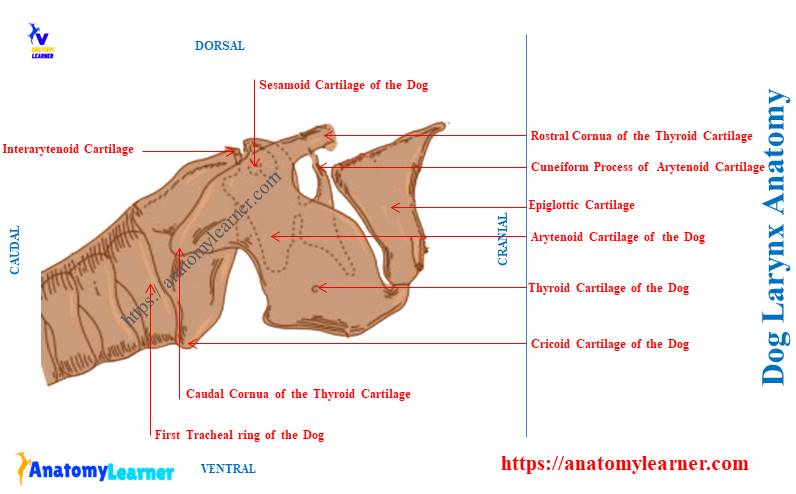
First, I suggest you identify the cartilage, muscles, nerves, veins, and other structures from the dog larynx. Then go for the details description of these components with the labeled diagram.
Let’s identify the below-mentioned cartilages from the dog larynx anatomy –
- Cricoid cartilage,
- Thyroid cartilage of the dog larynx,
- Arytenoid cartilage of the dog,
- Epiglottis of the dog, and
- Cuneiform cartilage of the dog larynx,
All these cartilages from the dog larynx are identified in the labeled diagram. But you might also identify other different structures (discussed in the next section) from each laryngeal cartilage of the dogs.
You will find different muscles in the structure of the canine larynx structure. But the below-mentioned intrinsic muscles are important in the dog larynx structure –
- Thick cricothyroideus muscle of the larynx,
- Cricoarytenoideus lateralis and dorsalis muscles,
- Thyroarytenoideus muscle of the larynx,
- Vocalis muscle of the dog larynx,
- Ventricularis muscle,
- Arytenoideus transverse muscle, and
- Spindle-shaped hypoepiglotticus muscle,
Again, all these abovementioned muscles from the dog larynx are also identified in the labeled diagram. You will find more labeled diagrams on the canine larynx here.
Finally, you might also identify other components from the canine larynx, like –
- 5 segments of the laryngeal cavity (discussed in detail),
- Vocal fold, cord, and ligaments from the canine larynx, and
- Laryngeal nerves and vessels,
Now, I will show the specific anatomical features of every single component of the canine larynx. So, let’s continue the article to learn more about these laryngeal components from dogs.
Where is a dog’s larynx?
The dog’s larynx is directly located back to the tongue’s root, the pharynx’s oral part, and the soft palate. It continues as the elongated musculocartilagenious tube and extends to the trachea.
Let’s see the relationship of the dog’s larynx –
- The roof of the dog’s larynx is related to the pharynx and the beginning of the esophagus,
- Two lateral walls of the larynx are with the omohyoideus and sternomandibularis muscles, and
- The ventral wall of the dog’s larynx is related to the sternothyrohyoideus muscles,
Here, the labeled diagram shows the location of the dog larynx between the pharynx and trachea.
The below-mentioned article might help you to understand the structure of the tongue and mouth cavity of dogs –
- Dog tongue anatomy with labeled diagram – muscles, papillae, glands, veins, and nerves, and
- Dog mouth anatomy – lip, cheek, oral cavity, and salivary glands with the diagram,
What are the cartilages of the larynx dog?
The dog’s larynx has cricoid, thyroid, arytenoid, epiglottis, and cuneiform cartilages. Sometimes you may find the intraarytenoid and sesamoid cartilages in the laryngeal structure of a dog.
Thus, you will find 5 principal cartilages in the dog larynx. Whereas the ruminant typically shows only 4 principal cartilages. They (ruminants) lack the cuneiform cartilage in their larynx.
Among these 5 principal cartilages, you will find the paired and unpaired groups –
- Unpaired cartilages of the dog larynx – cricoid, thyroid, and epiglottis, and
- Paired cartilages of the dog larynx – arytenoid and cuneiform cartilages,
Let’s see the summary of the various cartilages from the dog larynx in Table 1 –
| Dog larynx cartilages | Location | Special features |
| Cricoid cartilage | In front of 1st tracheal ring | Lamina is wide Signet ring shape structure Connect thyroid cartilage and trachea |
| Thyroid cartilage | Ventral to the larynx Cranial to the cricoid | Largest single cartilage Lamina are high but short Have laryngeal prominence ventrally |
| Arytenoid cartilage | In front of cricoid cartilage Form the caudal part of the root of the laryngeal box | Small and irregular Have various processes |
| Epiglottis of the dog | Rostral to laryngeal box | Leaf or quadrilateral plate like Fit into the angle of the thyroid cartilage |
| Cuneiform cartilage | Most rostral to the arytenoid cartilage | Larger and somewhat cresent shaped |
| Intraarytenoid cartilage | Between two arytenoids | Small and flat Lies cranial to cricoid lamina |
| Sesamoid cartilage | Between two arytenoids | Small or dumble – shaped nodules |
What is the anatomy of the larynx in a dog?
To describe the anatomical features of the dog larynx, you might cover the followings –
- Number and features of the laryngeal cartilages (5 – 7),
- Intrinsic muscles of the larynx,
- Formation of the vocal cord in dogs,
- Structure of the laryngeal cavity in the dog, and
- Blood supply and innervation to the dog’s larynx structure,
The dog larynx anatomy is relatively short compared to the cows. Here, the lamina of the cricoid cartilage is wide. You will find a lateral groove in the arch of the cricoid cartilage.
The lamina of the thyroid cartilage is high but short and unites ventrally to the body. There is a ventral prominence and caudal notch at the ventral body of the thyroid cartilage.
The arytenoid cartilage of the dog is relatively small. You will see the intraarytenoid cartilage between two arytenoid cartilages.
The Epiglottis of the dog is a quadrilateral plate-like structure that fits into the angle of the thyroid cartilage. Again, the cuneiform cartilage is a larger, crescent-shaped structure that does not fuse with the epiglottis.
The dog’s larynx shows the more significant and prominent vocal cord. You will also see the extensive laryngeal saccule in the structure of the dog larynx.
The cricothyroid muscle is very thick, whereas the hyoepiglotticus muscle is well-developed and double. Cranial and caudal laryngeal nerves provide the main innervation to the dog’s larynx structure.
Now, let’s know the details of the canine larynx structure with a diagram.
Cricoid cartilage of the dog’s larynx
The cricoid is the only cartilage that forms a complete ring. The more comprehensive dorsal and narrow ventral portion in the dog’s cricoid cartilage structure will be seen.
The anatomy of the dog’s cricoid cartilage shows the following –
- Lamina of the cricoid cartilage – it is the dorsal expanded portion, and
- Arch of the cricoid cartilage – ventral and lateral part of the ring that forms a curved band,
The dorsal lamina of the dog’s cricoid cartilage is a broad, thick, and quadrilateral plate-like structure. You will find a dorsal median ridge on the dorsal surface of the lamina.
This dorsal median ridge on the cricoid cartilage is called the crest or crista mediana. It separates two shallow cavities from which the dorsal cricoarytenoid muscle arises.
The dog’s cricoid cartilage has two pairs of articular surfaces –
- A distinct pair of thyroid articular surfaces, and
- More prominent pairs of arytenoid articular surfaces,
Here, the distinct pairs of the thyroid articular surface are located at the junction of the lamina and arch. You may find it practically about 1 millimeter from the caudal border of the arch.
This articular surface is designed for articulation with the apices of caudal cornua of the thyroid cartilage.
“You should know the borders of the cricoid cartilage arch and the cornua of the thyroid cartilage. I will discuss these topics from cricoid and thyroid cartilages in the next section.”
Again, the arytenoid articular surface locates on the rostral border of the lamina lateral to the median crest. This articular surface is designed for articulating with the arytenoid cartilage.
Both pairs of articular surfaces form the synovial joint with respective cartilage. You may know the details of the synovial joint from the below-mentioned article –
Arch of the cricoid cartilage
Here, I have identified the arches from the dog’s cricoid cartilage. It is the ventral and lateral narrow part of the cricoid cartilage.
Two lateral parts of the cricoid cartilage form the curved band of the ring. Ventrally it becomes narrowest than the dorsal part.
The lateral surface of the arch of a dog’s cricoid cartilage has a groove. This groove is designed for the cricothyroid muscle of the larynx.
You will find two borders in the structure of the arch of the dog’s cricoid cartilage –
- Cranial border of the arch, and
- Caudal border of the arch,
Here, the cranial border of the arch is concave ventrally. It gives attachment to the cricothyroid muscle. The lateral part of the cranial border of the arch is thick and attached to the cricoarytenoideus muscle.
But the cranial border of the dorsal lamina of the cricoid cartilage is thick and slightly concave. Again, the caudal border of the dorsal lamina is thin and irregular.
The caudal border of the arch of the cricoid cartilage attaches to the first tracheal ring. Here, you will find the cricotracheal membrane that binds the caudal border with the tracheal ring.
The internal surface of the arch and lamina of the cricoid cartilage is smooth. The internal surface of the arch and lamina lines with the mucous membrane.
Summary of the cricoid cartilage of the dog
Now, let’s see the summary of the dog’s cricoid cartilage from Table 2 –
| Features | Cricoid Cartilages of the dog |
| Shape | Signet ring shape cartilage |
| Lamina | Wide quadrilateral dorsal lamina |
| Crest in lamina | Dorsal crest |
| Articular surface | 2 – cranial and caudal |
| Cranial articular surface | For articulating with arytenoid cartilage |
| Caudal articular surface | For articulating with thyroid cartilage |
| Arch | Right and left lateral arches Curved band like |
| Borders on arch | Cranial and caudal borders |
| Cranial border of the arch | Concave ventrally Attaches with cricothyroid membrane |
| Caudal border of the arch | Attaches with the first tracheal ring |
You might know the structure of the first tracheal ring from the dog trachea. It is somewhat different than the trachea of the ruminant.
Let’s find the cow tracheal ring anatomy from the below-mentioned article –
Thyroid cartilage of the dog’s larynx
The thyroid is the most prominent cartilage of the dog’s larynx structure. It forms the middle part of the laryngeal box and opens dorsally.
First, let’s identify the below-mentioned structures from the thyroid cartilage of the dog larynx –
- Right and left laminae of the thyroid cartilage,
- Body of the thyroid cartilage,
- Rostral and caudal cornua of the dog’s thyroid cartilage,
- Hyoid articular surface,
- Cricoid articular surface,
- Thyroid fissure,
- Laryngeal prominence on the dog’s larynx,
- Caudal thyroid notch,
- Cricothyroid ligament, and
- Thyrohyoid membrane,
All these structures from the thyroid cartilage of the dog larynx are identified in the labeled diagram. Now, let’s know the details of these structures from the thyroid cartilage of the dog’s larynx.
Body and lamina of the thyroid cartilage
The median thick portion of the thyroid cartilage is the body. You will find right and left lateral wings (lamina) that arise from the body of either side.
The lamina of the thyroid cartilage forms the large part of the lateral wall of the dog’s larynx. They are like rhomboid plates and possess slightly convex lateral surfaces.
You will find an oblique line (known as linea obliqua) that divides the lamina into two distinct areas. The sternothyroideus muscle primarily inserts on this oblique line of the lamina.
Each right and left lamina expands dorsally and forms two transverse thin processes –
- Rostral cornua – articulate with the cartilage of the thyroid cornua of the hyoid bone, and
- Caudal cornua – articulates with the caudolateral aspect of the lamina of the cricoid cartilage,
Thus, the rostral cornua of the thyroid cartilage possess a hyoid articular surface. Similarly, you will find the cricoid articular surface on the caudal cornua.
Both hyoid and cricoid articular surfaces are located on the median side of the respective lamina. You will find the thyroid fissure that separates the rostral cornua from the thyroid lamina.
The cranial laryngeal nerve and artery pass through this thyroid fissure.
What is laryngeal prominence in the dog?
The laryngeal prominence in a dog is the ventral prominent area of the thyroid cartilage. When the right and left lamina of the thyroid cartilage fuses, they form a slight ventral laryngeal prominence.
This dog’s laryngeal prominence is related dorsally to the base of the epiglottis. It attaches to the epiglottis by the elastic ligament.
In the case of dogs, this laryngeal prominence is not visible externally. But, you may feel the laryngeal prominence from the dog’s larynx.
You will find a median deep caudal thyroid notch at the ventral caudal border of the dog’s thyroid cartilage. Here, the cranial border of the thyroid cartilage is slightly convex.
The caudal border of the thyroid cartilage attaches to the ventral arch (va) of the cricoid cartilage. Here, you will find the cricothyroid ligament between the thyroid and the cricoid cartilage.
The cranial border of the thyroid cartilage attaches to the basihyoid and thyrohyoid bones. Within this articulation, you will find the thyrohyoid membrane.
Summary of the thyroid cartilage of the dog larynx
Let’s see the summary of the thyroid cartilage from the dog larynx in Table 3 –
| Features | Thyroid cartilage of the dog |
| Parts | Body and two lateral lamina |
| Cornua in lamina | Rostral and caudal |
| Articular surfaces | Hyoid and cricoid articular surfaces |
| Laryngeal prominence | Elevated area ventral to the lamina |
| Location of thyroid notch | Ventral caudal border of the cartilage |
Epiglottis of the dog larynx anatomy
Epiglottic is the rostral most cartilage in the dog larynx anatomy. It provides the basic structure of the epiglottis that closes the laryngeal opening during deglutition.
The dog epiglottis locates above the body of the thyroid cartilage and curves towards the root of the tongue. It is shaped like a pointed ovate leaf.
You will find two surfaces, two borders, an apex, and a base in the structure of the dog’s epiglottis. First, let’s try to identify the below-mentioned structures from the dog’s epiglottis –
- Laryngeal and lingual surfaces of the dog’s epiglottis,
- Thin, irregular right and left lateral border of the epiglottis,
- Thick stalk or base of the dog’s epiglottis,
- The pointed and curved apex of the epiglottis, and
- Vallecula on the dog’s epiglottis,
The labeled diagram identifies all these structures from the dog’s epiglottis. Now, let’s know the details of the features of the canine epiglottis.
Surfaces and borders of the dog’s epiglottis
The laryngeal surface of the dog epiglottis is the aboral surface (caudal). It is concave in its length and faces dorsocaudally. It also shows the slight convex surfaces transversely.
The lingual surface is the oral surface of the epiglottis. It possesses a convex surface and faces the oral pharynx.
The lingual surface attaches to the middle of the body of the hyoid bone. You will find the short, stour hyoepiglottic muscle between the lingual surface and hyoid bone.
The two median mucosal folds cover this muscle. Within these two median mucosal folds, you will see the deep pocket of the mucosa. And this deep pocket of mucosa is known as the vallecula.
The vallecula is limited laterally to the small fold of stratified squamous epithelium. It runs from the lingual surface of the epiglottis to the lateral wall of the laryngeal part of the pharynx.
But the stratified squamous epithelium can not identify grossly from the vallecula. It would help if you located it (vallecula with lining epithelium) under the light microscope.
For this, you may get an idea of the stratified squamous epithelium from the below-mentioned article –
Base and apex of the canine epiglottis
The base of the canine epiglottis is thick and attaches to the dorsal surface of the thyroid cartilage. A cartilaginous structure projects upward and backward from each side of the epiglottis base.
These projected structures from the dog epiglottis are the cuneiform processes. I will also discuss these cuneiform cartilage from the dog larynx separately.
The apex of the canine epiglottis is pointed and curved ventrally. Nearly all the epiglottic cartilage of the dog covers with the mucous membrane.
The normal position of the epiglottis allows the apex to rest dorsal to the soft palate. Now, let’s summarize the features of the dog epiglottis in Table 4 –
| Features | Epiglottis of the dog |
| Shape | Sharp-pointed spade or ovate leaf |
| Surfaces | Laryngeal and lingual surfaces |
| Apex | Pointed cranially |
| Base | Thicken handle of fibrous tissue |
| Borders | Two lateral borders Thin, irregular, and everted |
| Vallecula | A deep pocket of mucosa on the epiglottis |
Arytenoid cartilage of the dog’s larynx
Arytenoid is the paired irregular cartilage in the dog larynx structure. Thus, you will find them on either side and articulating with the cricoid cartilage’s craniodorsal border.
If you view the dog’s laryngeal cartilage laterally, you will not see the arytenoid. This is due to the larger lamina of the thyroid cartilage.
The morphology of the arytenoid cartilage is different in various animals. In the dog, the arytenoid cartilage embodies the corniculate and cuneiform cartilages.
You will see the following different parts in the dog’s arytenoid cartilage –
- Surfaces (medial, dorsal, and lateral) and borders of the dog arytenoid cartilage,
- Muscular process,
- Corniculate process,
- Vocal process of the arytenoid, and
- Cuneiform process of the dog’s arytenoid cartilage,
First, see all these abovementioned processes and surfaces from the dog arytenoid cartilage labeled diagram.
Location and shape: dog arytenoid cartilage locates on either side, in front of the cricoid, and partly medial to the thyroid cartilage. They are pyramidal and possess 3 surfaces, 3 borders, a base, and an apex.
Base, apex, surfaces, and borders of the dog arytenoid cartilage
The base of the dog’s arytenoid cartilage is concave and primarily faces backward. You will find an oval concave facet on the base of the dog arytenoid cartilage.
Within this facet, the lamina of the cricoid cartilage attaches. Again, the medial angle of the base attaches to the transverse arytenoid ligament.
The apex of the dog’s arytenoid cartilage is curved upward and backward.
The medial surface of the dog arytenoid cartilage is concave-convex and slightly curved. It is also smooth and covered with mucous membranes.
The lateral surface of the dog arytenoid cartilage is concave and separated from the lamina of the thyroid cartilage. You will find the cricoarytenoideus lateralis, vocalis muscle, and laryngeal saccule within this lateral surface and lamina.
The dorsal surface is also concave and covered with the arytenoideus muscle. Again, the articular surface locates on the caudal border of the arytenoid cartilage.
It is slightly oval and possesses a concave surface caudally. This surface faces caudomedially and joins with the arytenoid articular surface of the cricoid cartilage. And thus, the cricoarytenoid articulation is formed between the arytenoid and cricoid cartilages.
Processes of the dog arytenoid cartilage
You will find 4 important processes of the arytenoid cartilage in a dog’s larynx – muscular, vocal, corniculate, and cuneiform. The muscular process is a relatively thick and rounded structure in the dog arytenoid cartilage.
This muscular process directly locates lateral to the articular surface of the arytenoid cartilage. You will find the insertion of the cricoarytenoideus muscle within this muscular process.
The corniculate process of the dog arytenoid cartilage is longer than the muscular process. You will find this corniculate process more caudally that forms the dorsal margin of the laryngeal inlet.
The dog arytenoid cartilage also shows the caudoventral projection. This projection of the dog’s arytenoid cartilage is the vocal process.
This vocal process of the dog is approximately 3 millimeters wide and 5 millimeters long. Within the vocal process, you will find the attachment of the vocal ligament and vocal muscle.
And these vocal muscles and ligaments attach to the vocal process from the thyroid cartilage.
Cuneiform process of the dog larynx
The cuneiform process is the most rostral portion of the dog arytenoid cartilage. This cuneiform process is connected with the primary part of the arytenoid cartilage with a narrow neck.
For this reason, the dog larynx cuneiform process is considered the separate cartilage. You may also call this process the cuneiform cartilage in the dog.
The cuneiform cartilage of the dog larynx is roughly triangular in shape. The dorsal part of the cuneiform cartilage lies on the vestibular fold.
Again, the ventral portion of cuneiform serves as the attachment of the aryepiglottic fold. It also helps in forming the laryngeal inlet.
Within the cuneiform cartilage, you will find the attachment of the ventricular ligament and ventricularis muscle.
Now, let’s see the summary of the dog arytenoid cartilages from Table 5 –
| Features | Arytenoid cartilage of the dog |
| Shape and type | Paired and pyramidal-shaped cartilage |
| Parts | 3 surfaces, 3 borders, apex, and base |
| Base | Concave and backward |
| Apex | Curves upward and backward |
| Articular surface | Caudally Oval and concave |
| Processes | Muscular, vocal, corniculate, cuneifrom |
Sesamoid and intraarytenoid cartilage of dog larynx
The sesamoid cartilage of the dog larynx is located cranial to the cricoid lamina and between the arytenoid cartilage. It is an oval or dumble-shaped nodular cartilage in the dog’s larynx.
It is paired occasionally, and an intersesamoid ligament connects two pairs of sesamoid cartilage. You will find a small contact surface with the dorsal part of the arytenoid cartilage.
You will also find a small, flat, and easily overlooked intraarytenoid cartilage in the dog larynx. It lies cranial to the cricoid lamina and caudodorsal to the transverse arytenoid muscle.
Dog larynx muscle anatomy
There are several intrinsic muscles in the structure of the dog larynx. Here, Table 6 shows the important muscles from the dog larynx anatomy with their location –
| Dog larynx muscles | Location |
| Cricothyroideus muscle | Between thyroid lamina and cricoid cartilage |
| Cricoarytenoideus dorsalis muscle | Cricoid to arytenoid cartilages |
| Cricoarytenoideus lateralis muscle | Lateral and cranial surface of cricoid |
| Thyroarytenoideus muscle | Thyroid to the arytenoid cartilage |
| Vocalis muscle | Medial division of thyroarytenoideus |
| Ventricularis muscle | Cranial division of thyroarytenoideus |
| Arytenodideus transversus muscle | Arise from a muscular process |
| Hyoepiglotticus muscle | Ceratohyoid to mid ventral of epiglottic |
Now, let’s discuss these muscles from the dog larynx with the diagram.
Cricothyroideus muscle of the canine larynx
The cricothyroideus is the thick muscle on the lateral surface of the dog’s larynx. This intrinsic muscle of the canine larynx lies between the thyroid lamina and cricoid cartilage.
You will find the attachment of this muscle with the lateral surface of the cricoid cartilage. Then it passes dorsally and cranially to attach to the caudal margin of the thyroid cartilage.
This muscle also attaches to the vocalis muscle (a medial part of the thyroarytenoideus muscle). Let’s see the function of the cricothyroideus muscle of the canine larynx –
- It helps to pivot the cricoid cartilage on its thyroid articulation. Thus, it tenses the vocal cord of the dogs.
Criocoarytenoideus dorsalis and lateralis muscles
The cricoarytenoideus dorsalis muscle of the dog larynx arises from the dorsolateral (dl) surface of the cricoid cartilage. It runs craniolaterally and inserts into the muscular process of the arytenoid cartilage.
You will also find the connection of this muscle with the thyroarytenoideus muscle. There are 3 heads in the structure of the cricoarytenoideus dorsalis muscle.
It helps to contribute to vocal fold abduction during exercise. Thus, it opens the glottis by abducting the vocal folds.
The cricoarytenoideus lateralis arises from the cranial and lateral aspects of the cricoid cartilage. It runs cranially and inserts on the muscular process of the arytenoid cartilage between crocoarytenoideus dorsalis and vocalis muscles.
This muscle helps to pivot the arytenoid cartilage medially and close the rima glottis of the dogs.
Thyroarytenodieus muscle of the dogs
The thyroarytenoideus is the large muscle that arises along the internal midline of the thyroid cartilage. It passes caudodorsally and inserts on the arytenoid cartilage.
The dog’s thyroarytenoideus muscle gives rise to two other muscles –
- Vocalis muscle of the dog, and
- Ventricualris muscle of the dog,
This muscle also sends fibers to the thyroarytendoideus muscle dorsally and cricoarytenodeus dorsalis caudally. Again, the middle part of the thyroarytenoideus muscle blends with the aponeurotic part of the arytenoideus transversus muscle.
Finally, this muscle also attaches to the muscular process of the arytenoid cartilage deeply. It helps to relax the dog’s vocal cords and constrict the glottis.
Dog vocalis muscle
The dog vocalis muscle is the medial division of the thyroarytenoideus muscle. Another name for this muscle is the thyroarytenoideus aboralis muscle.
This muscle arises from the internal midline of the thyroid cartilage and partly to the thyroarytenoideus. Again, it inserts on the vocal cord of the arytenoid cartilage.
You will find the vocal ligament at the cranial border of the vocalis muscle. This muscle helps to draw the arytenoid cartilage ventrally. Thus, it helps in relaxing the dog’s vocal cords.
Ventricualris muscle of the dogs
It is believed that the ventricualris is the cranial division of the thyroarytenoideus muscle. It arises from the thyroid cartilage to the cuneiform process of the arytenoid cartilage.
Another name for this muscle is thyroarytenodieus oralis. It lies medial to the laryngeal ventricle and aids in dilating the ventricles.
The ventricualris receive some connecting fibers from the cranial dorsal surface of the thyroarytenoideus. This muscle constricts the glottis and dilate the laryngeal ventricle.
Dog’s arytenoideus transverses and hyoepiglotticus muscle
The arytenoideus transversus muscle arises from the muscular process of the arytenoid cartilage. Again, it inserts on the lateral expanded ends and dorsal surface of the interarytenodi cartilage.
This muscle of the dog larynx blends with the vantricualris muscle. The arytenoideus transversus constrict the glottis and adduct the vocal cord.
The hyoepiglottis is the small and spindle-shaped muscle in the dog’s larynx. It originates from the medial surface of the ceratohyoid bone of the dogs.
This muscle runs dorsally after its origin and inserts on the ventral midline of the epiglottis. It helps to draw the epiglottis ventrally.
What is the cavity of the larynx in a dog?
Five transverse segments form the cavity of the larynx in a dog. Let’s see what are these 5 transverse segments in the laryngeal cavity of the dogs –
- Aditus laryngis (laryngeal inlet),
- Laryngeal vestibule,
- Vestibular cleft,
- Cleft of the glottis, and
- Infraglottic cavity,
Let’s discuss these transverse segments of the dog’s laryngeal cavity individually. First, start with the features of the aditus laryngis or laryngeal inlet of the dog.
What is a laryngeal inlet in a dog?
The laryngeal inlet directly lies caudal to the intrapharyngeal ostium. The epiglottis bounds this inlet.
With the help of the oral part of the nasal part of the pharynx, air can enter or exit from the larynx. Most of the air passes through a dog’s mouth and oral portion of the pharynx.
You will find an imperfect triangle in the margin of the dog’s laryngeal opening. The margin of the epiglottic forms the lateral boundary and apex of the laryngeal inlet.
Again, the caudal boundary of the laryngeal inlet is formed by the right and left aryepiglottic folds. The aryepiglottic folds run from the dorsal part of the arytenoid cartilage.
It is closely associated with the corniculate cartilage to the caudolateral angle of the epiglottic cartilage. You will find two prominent tubercles that are separated by a deep notch. Here the dorsocaudal tubercle under the corniculate process is called the corniculate tubercle.
Ventral to this tubercle, two arytenoid cartilage lies together and form the intraarytenoid groove. This groove connects dorsally with the middle of the rostral part of the laryngeal pharynx.
You will also find the cuneiform tubercles that unite with the corniculate tubercle. These are the large cone-shaped projection from the cuneiform process of the arytenoid cartilage.
What is the laryngeal vestibule in a dog?
The laryngeal vestibule is the funnel-shaped structure in the laryngeal cavity of a dog. It opens freely dorsocranially and extends from laryngeal aditus to vestibular folds.
Ventrally it is bounded by the mucosa and dorsally by the concave epiglottis. You will also find the part of the epiglottis on the cranial wall.
The flattened cuneiform process forms the remainder part of the wall. Finally, the dog’s laryngeal vestibule opens caudally into the rima vestibuli.
Vestibular cleft of the dog laryngeal cavity
The vestibular cleft is the part of the laryngeal cavity bilaterally bounded by the vestibular folds. You will also find the mucosa membrane that also covers the vestibular cleft.
Another name for the vestibular cleft of the dog is rima vestibuli.
The ventral boundary (v) of the vestibular cleft is covered by the mucous membrane that comes from the thyroid cartilage. Here, the vestibular fold is the short and wide plica of the mucosa.
You will also find a few elastic fibers in the structure of the vestibular folds. This vestibular cleft runs from the expanded ventral margin of the cuneiform cartilage to the cranial dorsal surface of the thyroid.
Cleft of the glottis
It is part of the dog laryngeal cavity between the vocal fold and arytenoid cartilage. In the structure of the cleft of the dog’s glottis, you will find the following components –
- Vocal folds,
- Arytenoid cartilage, and
- Rima glottides,
You will find two parts in the cleft of the glottis – intramembranous and intracartilagenious. Here, the intramembranous part locates between the vocal folds and is considered part of the rima glottides.
Again, the intracartilagenious part locates between the medial aspect of the arytenoid cartilages.
The rima glottides are a dog’s most important part of the larynx. It is the narrowest part of the laryngeal passage and contains the vocal fold. Again, this structure plays a vital role in the dog’s vocalization.
What is the vocal fold in a dog?
The vocal fold in a dog is the mucosal fold on either side of the arytenoid cartilage. It extends from the vocal process of the arytenoid cartilage to the dorsocaudal part of the thyroid cartilage.
The dog’s vocal fold length is 13 – 15 millimeters, whereas the wide is about 5 – 6 millimeters. You will find the slitlike opening of the laryngeal ventricle between the vocal and vestibular fold.
An elastic vocal ligament strap is attached to this vocal fold structure. It supports the cranial border of the vocal fold.
The vocal ligament of the dog’s vocal fold is 1 – 2 millimeters thick. Finally, the vocal ligament continues with the vocalis muscle of the dogs.
What is the laryngeal ventricle in a dog?
The laryngeal ventricle is a small mucosal sac between the vocal and vestibular folds medially and thyroid lamina laterally. You will find the border of the laryngeal ventricle that attach to these vocal and vestibular folds.
The lumen of the ventricle opens into the laryngeal cavity at the junction of the rima vestibule and rima glottis. Again, the vocal and vestibular folds can vibrate the cavity of the glottis during sound production.
The lateral laryngeal ventricle of the larynx is present only in horses, dogs, and pigs. But you will not find this ventricle in cats and also in ruminants.
Infraglottic cavity of the dog’s larynx
The infraglottic cavity of the dog larynx extends from the rima glottides to the tracheal cavity. This infraglottic cavity is wide dorsally and narrow ventrally.
Dog anatomy laryngeal nerve
The cranial and caudal laryngeal nerves (2) innervate the dog’s larynx. These are two primary branches of the cranial nerves in the dog that are found in the larynx.
Let’s discuss the cranial and caudal laryngeal nerves from the dog larynx structure.
The cranial laryngeal nerve of the dog
The cranial laryngeal nerve arises from the vagus nerve of the dog. It leaves the vagus nerve at the level of its distal ganglion.
At the larynx of the dog, this cranial laryngeal nerve divides into (2) external and internal branches. Here, the external branch of the cranial laryngeal innervates the cricothyrohyoideus muscle.
Again, the internal branch of the cranial laryngeal receives the axon from the mucosa of the larynx. This part of the laryngeal nerve anastomoses with the caudal laryngeal nerve.
You will also find a pharyngeal plexus nerve that innervates the cricothyroideus muscle. Again, the cranial laryngeal nerve innervates taste buds on the epiglottis and luminal mucosa of the larynx.
A cranial laryngeal initiates reflex and closes the cleft of the glottis during swallowing.
The caudal laryngeal nerve of the dog’s larynx
Except for the cricothyroideus muscle, most intrinsic muscles are innervated by the caudal laryngeal nerve. It provides the motor supply to all of the dog’s laryngeal muscles.
The caudal laryngeal is the terminal part of the recurrent laryngeal nerve. This nerve arises in the thorax by leaving the dog’s vagus nerve.
The somatic efferent axon of the caudal laryngeal nerve has its neuronal cell bodies. They exist in the medulla of the cranial roots of the accessory nerve.
This nerve follows the vagus nerve in the thorax. Then it returns to the larynx as the recurrent laryngeal nerve.
How to differentiate the dog’s laryngeal cartilage from the cows?
The cartilages of the larynx show little difference between the dog and cow. Again, the cow larynx does not possess any cuneiform and corniculate cartilage.
The cow larynx has only 4 well-developed cartilage, whereas the dog has 5 – 7 cartilage. Table 7 shows the main difference between the laryngeal cartilage of the dog and cow –
| Features | Dog laryngeal cartilages | Cow laryngeal cartilages |
| Numbers | 5 – 7 | 4 |
| Cricoid lamina | Broad | Narrow |
| Muscular process | Wide; quadrilateral | Small, narrow |
| Thyroid lamina | Rectangular | Quadrilateral |
| Epiglottis | Sharp pointed | Obovate leaf |
| Arytenoid cartilage | Irregular shape | Pyramidal shape |
| Cuneiform cartilage | Present | Absent |
| Corniculate | Wel-developed | Indistinct |
| Sesamoid cartilage | Small nodular | Absent |
| Intraarytenoid cartilage | Present | Absent |
Frequently asked questions on dog larynx anatomy
Now, I will enlist most of the common questions on the canine larynx anatomy that the learners ask. Here, you will get concise answers to these questions related to the dog’s larynx.
But you might read the complete guide to get advanced knowledge on the dog larynx structure. Okay, let’s see the commonly asked questions on the dog larynx structure –
Does a dog have a larynx?
Yes, dogs have a larynx that consists primarily of the cartilage, laryngeal muscles, nerves, and vessels. Thus this is a musculo-cartilagenious structure in the dog’s neck.
This larynx extends from the pharynx to the beginning part of the dog’s trachea.
What are the 4 parts of the dog’s larynx?
You will find 4 main cartilages in the structure of the dog larynx. The 4 main cartilages of the dog larynx are – cricoid, thyroid, epiglottis, and arytenoid.
But, the dog arytenoid possesses different parts and processes. The main processes of the arytenoid cartilage are – muscular, vocal, corniculate, and cuneiform.
The corniculate and cuneiform processes are comparatively larger and attach to the arytenoid. Thus, the different authors defined these processes as single cartilage.
Again, the dog larynx also shows two different cartilages – sesamoid and interarytenoid.
Can you feel a dog’s larynx?
Yes, you can feel the thyroid cartilage of the dog’s larynx on palpation. But it is invisible in the live dogs.
Again, the other cartilages of the larynx can not feel appropriately on palpation.
How many vocal cords does a dog have in their larynx?
There are 2 vocal cords in the dog’s larynx. One vocal cord extends from the cuneiform cartilage to the thyroid cartilage.
Again, other vocal cords (true) are larger and more prominent. It’s located in the dog’s thyroid cartilage.
Conclusion
So, the dog larynx anatomy primarily consists of 5 – 7 cartilages, 8 essential muscles, and 2 nerves. It extends from the caudal part of the oral cavity (pharynx) to the first ring of the dog trachea.
The structure and appearance of the dog’s laryngeal cartilage are slightly different from other animals’ cartilage. You might identify all these cartilages, muscles, and nerves from the dog larynx structure at your anatomy learning laboratory.