The dog tongue anatomy consists primarily of skeletal muscle, mucous membrane, glands, vessels, and nerves. You will find different important anatomical facts on the mucous membrane of a dog tongue. This article will show you the anatomy of dog tongue muscles, mucous membrane, glands, vessels, and nerves in detail.
Again, you will find the detailed anatomical facts of different types of papillae from the dog tongue. I will also differentiate the gross features of the dog tongue from the ruminant at the end of this article.
So, if you are interested in learning the gross anatomical features of the dog tongue, you may continue this article till the end.
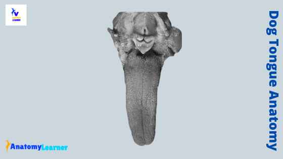
Dog tongue anatomy
You know the dog tongue is a highly movable muscular structure responsible for prehension, mastication, and deglutition. It is also an important organ for taste as it contains taste buds. You will find the dog tongue on the floor of the mouth cavity, between the two rami of the lower jaw.
The dog tongue anatomy includes the features of skeletal muscle, mucous membrane, glands, vessels, and nerves. You will find a very thick root, a long body, and a thin apex in the dog tongue. The body of the dog tongue presents two surfaces – dorsal and ventral.
The muscles of the dog tongue are very complex and divided into two groups (extrinsic and intrinsic). Again, the extrinsic muscles of the dog tongue include styloglossus, hyoglossus, and genioglossus. You will find superficial longitudinal, deep longitudinal, perpendicular, and transverse muscle fibers in the intrinsic group.
Again, the dorsal surface of the dog tongue bears five different types of cornified lingual papillae. The next part of the article will get the detailed anatomy of these papillae and other different parts of the tongue.
There are numerous glands (both serous and mucous) present in the dog tongue. The major blood vessels of the dog tongue are the sublingual artery and lingual vein. Again, the dog tongue is innervated by different nerves, where the most important one is the lingual nerve. Here, I would like to enlist some of the dog tongue’s most important anatomical facts that differ from the ruminant tongue.
Special features of dog tongue
The dog tongue is highly mobile compared to that of the ruminant. You will find the following special features in the dog tongue structure.
- The dorsal surface of the dog tongue represents a median groove.
- You will find two to three valet papillae at each side of the caudal third of the dorsum.
- The tongue of a newborn dog presents marginal papillae.
- A thick fibromuscular cord locates along the mid-ventral surface of the tip of the dog tongue (known as lyssa body).
These are the few exceptional anatomical facts of the dog tongue. But, these are not enough to learn the dog tongue structure. To learn the detailed anatomical facts of the dog tongue, you may continue this article till the end.
Gross anatomy of dog tongue texture
The gross anatomy of a dog tongue includes the forms and structures. Again, the forms of the dog tongue may vary in different species. You will find a longer and narrower shaped tongue in a dog compared to the ruminant. Structurally, the dog tongue comprises skeletal muscle, mucous membrane (including different papillae), glands, nerves, and vessels.
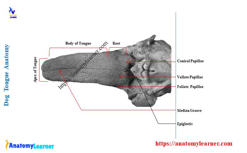
Let’s discuss the different parts of a dog tongue. As I told you before, the dog tongue has three different parts – the root, a long body, and apex. Again, there are two lateral swelling, one median tongue bud, and one median proximal swelling present in the dog tongue.
A row of vallate papillae separates the body and root of the dog tongue. You will find a very thick root attached to the hyoid bone and mandible by hyoglossus and genioglossus muscle. The ventral surface of the root is fixed, and the dorsal surface is free.
You will find an elongated body in a dog tongue that presents two surfaces. The dorsal surface is free and known as the dorsum linguae. Again, this dorsum linguae divides into two lateral halves by a median groove. The dorsal surface of the dog tongue is rough due to the presence of different types of papillae like filiform, fungiform, vallate, foliate, and conical.
On the ventral surface, you will find an unpaired mucosal fold (lingual frenulum) that connects the body of the tongue to the floor of the mouth cavity. Again, the tip of the ventral surface presents a thick fibromuscular cord-shaped structure (lyssa).
There are two margins in the dog tongue separate from the dorsal and ventral surface. These two margins meet rostrally to form the thin apex.
Papillae of the dog tongue
The lingual mucosa of the dog tongue anatomy is thick and heavily cornified on its dorsal surface. But, the ventral surface of the dog tongue is less thick and cornified than that of the dorsal surface. Again, the dorsal surface of the dog tongue possesses five different types of lingual papillae.
Let’s enlist the five different types of papillae from the dog tongue –
- The smallest filiform papillae
- A mushroom-shaped fungiform papillae
- The vallate papillae of the dog tongue
- Two groups of foliate papillae, and
- The conical papillae of the dog tongue
In addition, you will also find the marginal papillae in the tongue of a newborn dog. Now, I will show you all the papillae’s anatomical facts from a dog tongue.
Let’s start with the most numerous and smallest papillae of the dog tongue.
Filiform papillae of dog
The filiform are the numerous and smallest papillae of all the lingual papillae in the dog tongue. These numerous filiform papillae are located on the dorsum of the rostral two-thirds of the tongue body. You will find the filliform papillae with the fungiform papillae in a dog tongue.
The filliform papillae of a dog tongue may extend caudally at the level of vallate papillae. Again, the structure of a filliform papillae is very complex. It consists of a broad base and a dermal core. You will find three different types of filliform papillae in this complex – primary, secondary, and tertiary.
In the filliform papillae complex, the primary papillae are single and located centrally. The dermal core forms the axis of the single primary papillae. Again, the secondary filiform papillae arise from the primary filliform. You will find four to six secondary filiform papillae that surround the base of the primary filliform.
The tertiary filliform papillae of the dog tongue arise from the secondary filliform. You will not find any dermal core in a tertiary filliform papillae structure. They are smaller than the secondary filliform papillae and made up of epithelial tissue.
The surface of the tertiary filliform papillae is well cornified to protect the deeper structures. Again, the cornification of the tertiary filliform papillae is more in the tip of the dog tongue.
The filiform papillae are innervated by the lingual nerve and a branch of a facial nerve.
The fungiform papillae of dog tongue
The fungiform are the mushroom-shaped papillae present on the dorsal surface of the dog tongue. They are located at the rostral two-third of the tongue, along with the rough and numerous filliform papillae. The fungiform papillae may also extend caudally at the level of vallate papillae.
You will not find any fungiform papillae on the median groove of the dog tongue. How will you differentiate the fungiform papillae from the filliform? Well, the fungiform papillae of the dog tongue are shorter, broader, and less numerous than that of the filliform papillae.
They are the second numerous papillae in the dorsum of the dog tongue. You will also find the cornification on the fungiform papillae (lies with stratified squamous epithelium). But, the cornification is much thinner in fungiform papillae than the filliform papillae.
You will find a proximal narrow base and an expanded distal end in each fungiform papillae of the dog tongue. The base of the fungiform papillae may possess spinous cornified projection. Again, the expanded distal end of the fungiform papillae possesses a taste pore that contains the taste bud.
But, some of the fungiform papillae may not contain the taste bud. You will also find the dermal core in the fungiform papillae as you found in the filliform papillae.
The vallate papillae of the dog tongue anatomy
At the caudal third of the dorsum of a dog tongue anatomy, you will find some vallate papillae. They mark the boundary between the filiform and conical papillae. You will find three to six vallate papillae in the dog tongue arranged in V form. But, the number of vallate papillae may vary in a different breeds of dogs.
The V form arrangement of the vallate papillae may be absent if only three or five papillae are present. There are two types of vallate papillae in the dog tongue – simple and complex. The simple vallate papillae are numerous in the dog tongue and contain deep central and outer moats.
The dorsal surface of the simple vallate papillae contains a central depression. From this central depression, a secondary papillae projects. The central moat of the vallate papillae is complete and surrounds the secondary papilla. Again, the outer moat is incomplete and surrounds the vallate papillae.
Do you know how this outer moat of the vallate papillae is formed? The outer moat forms by the modified conical papillae that arrange side by side around the vallate papillae.
The ventral surface of the simple vallate papillae contains the taste bud. But, the number of the taste buds in each simple vallate papillae may vary greatly. You may also find some serous gustatory glands in the base of the vallate papillae of the dog tongue.
The complex vallate papillae may occasionally occur in the dog tongue.
The foliate papillae of the dog tongue
The foliate papillae of the dog tongue locate on the dorsolateral aspect of the caudal third of the tongue. There are two groups of foliate papillae present in the dog tongue. All these papillae are covered with the stratified squamous epithelium (cornified).
You will find eight to thirteen foliate papillae in each group. These papillae remain in the crypts parallel to the papillae and separate. Again, the long axis of the papilla runs obliquely from the side of the tongue towards the dorsum of the tongue in a radiating manner.
Each group of foliate papillae of the dog tongue is bordered dorsally, rostrally, and caudally by conical papillae. But, you will not find the conical papillae in the lateral boundary of the group.
You will find very small and irregular-shaped papillae at each group’s caudal and rostral parts. There is a central primary dermal core in an individual foliate papilla. Again, a secondary dermal papilla arises from the primary dermal papilla.
You will find the taste buds in the secondary dermal papillae and extend the epithelium’s total thickness. There are also taste pores present in the foliate papillae that open in the dorsal surface of the tongue.
Conical papillae of the dog tongue
These are the conical-shaped papillae that are also found in the dog tongue. They are located on the dorsum of the caudal third of the dog tongue. You will find a wide circular base and narrow to a thin apex in each conical papillae of the dog tongue.
The apex is caudally directed and heavily cornified than that of the base. You will find the largest conical papillae just caudal to the palatoglossal arch. But, the number of the conical papillae just caudal to the palatoglossal arch is not numerous.
The dorsal surface of the conical papillae is also lined with the stratified squamous epithelium. Again, some conical papillae are modified to form the wall of the outer moat of the vallate papillae. You will find a secondary dermal papilla in the dermal core of the conical papillae.
The conical papillae of the dog tongue possess mechanical and tactile functions rather than gustatory functions. Again, these conical papillae are innervated by the glossopharyngeal nerve.
The marginal papillae of the tongue
You will find the marginal papillae only in the tongue of a newborn dog. There are no marginal papillae present in the tongue of an older dog.
These papillae of the newborn dog are distributed along the margin of the rostral half of the tongue. You will find the well-developed marginal papillae at the margins of the tongue at the level of premolar. Again, the less developed papillae are present at the margin of the apex of the dog tongue.
In addition, you will not find any marginal papillae at the margins of the caudal half of the tongue. These marginal papillae of the dog tongue are threadlike and narrower at their apices. Again, the marginal papillae are mechanical and tactile rather than gustatory in function.
Dog tongue muscle anatomy
The dog tongue muscle anatomy is very complex and responsible for high movement. You will find two different types of muscles that are responsible for prehension, chewing, and swallowing. The extrinsic muscles of the dog tongue include the styloglossus, hyoglossus, and genioglossus.
Again, you will find superficial longitudinal, deep longitudinal, perpendicular, and transverse muscle fibers in the intrinsic group of muscles in the dog tongue.
Now, I will show you all the extrinsic and intrinsic muscles from the dog tongue with a labeled diagram. I will try to provide the information on the dog tongue’s extrinsic and intrinsic muscles as much as possible.
Fine, let’s discuss the extrinsic muscles of the dog tongue.
Styloglossus muscle of the dog tongue
The styloglossus is the most lateral extrinsic muscle of the dog tongue anatomy. It locates at the caudal third of the dog tongue. You will find a narrow proximal end and wide rostral end in the styloglossus muscle of the tongue.
There are three different heads present in the styloglossus muscle of the dog tongue. These three heads of the styloglossus muscle are –
- A short head of styloglossus muscle
- The rostral head of styloglossus muscle, and
- A long head of styloglossus muscle
The short head of the styloglossus muscle arises from the distal half of the caudal surface of the hyoid bone. Again, this head curves ventrally and rostrally over the epihyoid bone. At the proximal part of the tongue, this muscle inserts its fiber with the muscle fiber of hyoglossus.
The rostral head of the styloglossus muscle arises from the rostrodorsal surface of the proximal half of the stylohyoid bone. In addition, the long head of styloglossus arises from the stylohyoid bone immediately dorsolateral to the origin of the short head.
The long head also curves ventrally and rostrally. So, the fibers of the long head curve ventrally and rostrally along with the ventral surface of the tongue. It crosses the lateral surface of the genioglossus muscle at the ventral surface.
The three heads of the styloglossus muscle draw the tongue caudally.
Hyloglossus muscle of the dog tongue
The hyoglossus is another lateral extrinsic lingual muscle of a dog. You will find this muscle at the root of the tongue and below the styloglossus muscle.
This hyloglossus muscle arises from the ventrolateral surface of the basihyoid and thyrohyoid bones. It will run dorsal to the mylohyoideus muscle and lateral to the genioglossus muscle.
At the root of the tongue, the hyloglossus muscle crosses the medial aspect of the styloglossus muscle. This muscle retracts and depresses the dog’s tongue. Again, this hyoglossus muscle is innervated by the hypoglossal nerve.
The genioglossus muscle of the tongue
The genioglossus muscle of the dog tongue is fan-shaped and lying dorsal to the geniohyoideus muscle. You will find two ends – a narrower and an expanded end. The narrower end of the genioglossus muscle originates from the medial surface of the mandible adjacent to the intermandibular joint.
Again, the expanded end insert to the ventral surface of the dog tongue. You will find three distinct bundles in the genioglossus muscle of the dog tongue. All these muscle bundles comprise three different types of fibers – verticle, oblique, and straight.
The verticle bundle of the muscle fibers locates at the rostral part of the genioglossus muscle. Again, it inserts on the rostral half of the dog tongue just caudal to the lyssa. This verticle bundle arises from the ventromedial surface of the rostral end of the mandible.
The oblique bundle of the muscle fiber lies caudal to the verticle bundle. It is narrow, long, and more oblique than the verticle bundle. These fibers originate from the ventromedial aspect of the mandible and are inserted on the caudal half of the tongue.
In addition, the straight bundle of the genioglossus muscle lies lateral to both the verticle and oblique bundles. This fiber originates from the caudal border of the intermandibular joint with the geniohyoideus muscle.
The genioglossus muscle of the dog tongue helps to depress and protrude the tongue. Again, the genioglossus muscle is innervated by the hypoglossal nerve.
Extrinsic muscle of the dog tongue
You will find the proper lingual muscles in a dog tongue in the extrinsic group. The proper lingual extrinsic muscle of the dog tongue forms the core and possesses four types of fibers.
The fibers of the proper lingual muscles of a dog tongue are –
- A superficial longitudinal fiber of proper lingual muscle
- The deep longitudinal fiber of the proper lingual muscle,
- A transverse fiber of proper lingual muscle, and
- The perpendicular fibers of the proper lingual muscle of the dog tongue
The superficial longitudinal muscle fiber locates immediately deep into the dorsal lingual mucosa. You will find the well-developed superficial longitudinal fibers at the caudal half of the dog tongue.
The deep longitudinal fibers are located in the ventral half of the dog tongue. They are less numerous and organized than that of the superficial longitudinal fibers. You will find compact masses of deep longitudinal fiber at the rostral end of the tongue. These deep longitudinal fibers also surround the lyssa body of the dog tongue.
The transverse and perpendicular fibers occupy a large area in the center of a tongue. These fibers form a network between the superficial and deep longitudinal fibers.
The proper lingual muscle of the dog helps to protrude the tongue, facilitates the local movement, and prevents the tongue from being bitten.
Dog tongue and lyssa anatomy
You will find a special feature at the ventral surface in the dog tongue anatomy. There is a flexible body, a rod-shaped structure that lies on the ventral median surface in the apex of the dog tongue. This special structure is known as the lyssa body of the dog tongue.
This lyssa body of the dog tongue extends from the apex to the level of the rostral part of the verticle fibers of the genioglossus muscle. You will find a dense connective tissue encapsulation on the lyssa body of the dog. Again, you will also find the adipose tissue, striated muscle, and cartilage in the structure of the lyssa body.
The adipose tissue is more prominent in the ventral half of the lyssa body. The lyssa body contains the striated muscle at the middle and dorsal half. Again, the lyssa body of the dog tongue receives the branch of the hypoglossal nerve.
Other features of dog tongue
You might also describe the other anatomical features of the dog tongue. This may include the glands of the tongue, blood vessels, and nerve supply.
You will find some serous and mucous glands in the base of the lingual papillae. I will show you want the important glands present in the dog tongue. There are three important major vessels present in the dog tongue. They are the lingual artery, sublingual artery, and lingual vein. I will also describe these vessels from the dog tongue in a little.
Finally, I will discuss some important nerves like the lingual, glossopharyngeal, and hypoglossal nerves.
Glands of the tongue
You will find numerous salivary glands (serous and mucous) in the dog tongue. These are the gustatory glands that lie at the base of the vallate and foliate papillae of the tongue.
The glands associated with the base of the vallate and foliate papillae of the dog tongue are mainly serous. Again, you will also find the serous glands in the bundle of intrinsic muscles.
In the lingual mucosa (a caudal third of the tongue), you will find the mucoserous types of glands. Again, the lateral margin of the dog tongue consists of a seromucous gland in the submucosa.
Dog tongue blood vessels
The dog tongue is primarily supplied with the paired lingual arteries. They cross the medial surface of the hypoglossus muscle at the root of the tongue. It gives off the muscular branches to the intrinsic and extrinsic muscles of the tongue.
You will also find a sublingual artery that supplies the genioglossus and geniohyoideus muscle of the dog. The most important vein of the dog tongue is a lingual vein.
Dog tongue nerves
The dog tongue is innervated mainly by the lingual, glossopharyngeal, and hypoglossal nerves. The lingual nerve contains the general somatic afferent fibers that convey exteroceptive impulse from the rostral two-thirds of the lingual mucosa.
Again, the lingual branch of the glossopharyngeal nerve innervates the dog’s tongue. It contains special visceral afferent, general visceral afferent and efferent fiber to the caudal one-third of the dog tongue.
The hypoglossal nerve of the dog tongue contains general somatic efferent fiber. It supplies the styloglossus, hyoglossus, genioglossus, and proper lingual muscle of the dog tongue.
Dog tongue anatomy labeled diagram
Now, I will show you again the different structures from the dog tongue anatomy with a labeled diagram. In this tongue-labeled diagram, I tried to show you the different parts of the dog tongue. I will also show you the different types of lingual papillae from the dog tongue.
Again, you will find the median longitudinal groove on the dorsal surface of the dog tongue. The diagram also shows the different types of extrinsic and intrinsic muscles of the dog tongue.
The lyssa body of the dog tongue is shown in the other diagram. If you need more diagrams on the dog tongue, you may find them in a social media of anatomy learners.
Frequently asked questions on tongue
Now, I will try to solve the common inquiries on the dog tongue structure. But, if you read the full article, you may skip this part.
What is special about a dog’s tongue?
This is a very common inquiry about what is special about a dog tongue. Well, first, on the dorsal surface of the dog tongue, you will find a median longitudinal groove. This may be an exceptional feature of the dog tongue compared to the ruminant tongue.
The ventral surface of the dog tongue bears a fibromuscular connective tissue cord-like structure (known as the lyssa body). This is also a special feature of the dog tongue compared to the ruminant.
You will find three to six vallate papillae at the root of the dog tongue that arranges in V form. Again, the dog tongue consists of eight to thirteen foliate papillae at the caudal part.
What are the dogs tongue made of?
I have already described the component of a dog tongue. The dog tongue comprises the striated muscles, mucosa membrane, lingual papillae, glands, blood vessels (lingual artery and vein), and nerves.
How do dog’s tongues fit in their mouth?
What is under a dog’s tongue?
You may also read the other anatomical facts of different organs and structures from the dog and cat. Also, read the following –
Anatomical facts of the dog mouth cavity with labeled diagram
Conclusion
I hope you got the basic idea of the dog tongue anatomy. All the labeled diagrams related to the dog tongue might be helpful for you. The extrinsic and intrinsic muscles are the key component of the dog tongue anatomy. Again, different types of lingual papillae are also important structures of the dog tongue.
The dog tongue’s dorsal median longitudinal groove and the ventral lyssa body are other major anatomical facts. It will be good to learn the detailed anatomy of the glands, blood vessels, and nerves from the dog tongue.