There are three different parts –duodenum, jejunum, and ileum in the small intestine of an animal. You will find some salient microscopic features of each region of the small intestine. In this short article, I will discuss duodenum histology in detail, along with its slide identification points.
I will also show you the fundamental difference in the histological features among the three different parts of a small intestine. So that you may identify the histology slides of the duodenum, jejunum, and ileum of the small intestine easily under a light microscope.
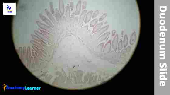
Hi there, welcome back and thanks for learning the typical duodenum histology slide with me. I will try to discuss the different layers of the duodenum slide with a labelled diagram and the actual slide’s picture.
Duodenum histology
Fine, this part of the article might help you to get a basic idea of duodenum histology. Make sure you know the details histology of the general organizational pattern of a tubular organ. You will find four different layers (tunica mucosa, tunica submucosa, tunica muscularis, and serosa) with some salient features in each.
So, what are the salient histological features of the duodenum that you might identify under a light microscope? Well, let me enlist the salient microscopic features from the duodenum of animals.
- Identification of four different layers of a duodenum
- Short leaf-like intestinal villi (lined by simple columnar epithelium and goblet cells)
- Crypt of liberkuhn of duodenum
- Brunner’s gland of the submucosa of a duodenum (very important for identifying duodenum from jejunum and ileum)
- Enteroendocrine cells in the mucosa of the duodenum
You will quickly identify the above mentioned salient features from the duodenum histology slide labeled diagram. This is the actual duodenum slide picture that you will find under a light microscope.
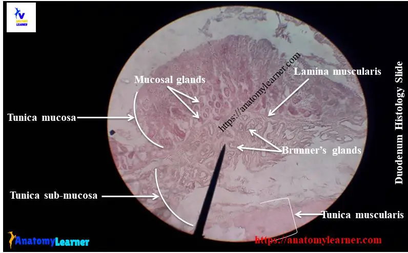
Duodenum histology slide identification
You may ask to identify the different parts of a small intestine histology slides with their proper identifying characteristics at the histology laboratory. Here, I will enlist the essential identifying characteristics of a duodenum histology slide.
- The sample tissue section under the light microscope shows four different layers – tunica mucosa, tunica submucosa, tunica muscularis and tunica serosa.
- There is a short leaf-like (or tongue-like) intestinal villi line with simple columnar epithelium and goblet cells.
- There are minute finger-like projections of the plasma membrane of the simple columnar epithelium.
- The intestinal glands (crypts of liberkuhn) are present in the lamina propria of the tunica mucosa of the provided tissue sample.
- The submucosa of the sample tissue section contains the Brunner’s glands.
So, this is a slide of a duodenum.
You may also add other standard histological features of the duodenum slide. But I think these features are enough to identify the duodenum slide. You might also know the jejunum and ileum histology slides (shown at the end of this article). Thus, you will make differentiate these small intestine histology slides from each other.
Great, I hope you could understand the basic structure of a duodenum slide. Do you want to learn the details histology of the duodenum slide? Fine, you may continue this article to know the histological features of the duodenum slide in more information.
Duodenum histology layers
Thanks for continuing to learn the histological features of different layers of the duodenum in detail. You know there are four different layers in the wall of a duodenum.
- Tunica mucosa layer of a duodenum
- Tunica submucosa layer of a duodenum
- The tunica muscularis of a duodenum and
- Tunica serosa of the duodenum
Let’s discuss the histological features of different layers of the duodenum with labeled diagrams. You will find almost similar histological features in the tunica muscularis, and tunica serosa layers that correspond precisely to the general structure of a tubular organ.
The submucosa layer also has the typical features with some special Brunner’s glands. Okay, first, let’s discuss the tunica mucosa of the duodenum.
Tunica mucosa of a duodenum
The tunica mucosa consists of lining epithelium, a lamina propria with glands, and a lamina muscularis. You will find several specific features in the tunica mucosa layer of a duodenum histology slide.
The surface of the tunica mucosa of the duodenum has the following unique histological characteristics. You should understand these special features first from the mucosa of a duodenum structure.
There are numerous circular folds present in the mucosa of the duodenum.
There are numerous leaf-like or finger-like structures (villi) that project from the surface of the mucosa membrane into the lumen of the duodenum.
Again, the presence of microvilli on the luminal surface of the mucosal lining cells (simple columnar epithelium).
There are numerous depressions or crypts that invade the lamina propria of the duodenum structure.
Fine, let’s discuss the above mention unique structures in detail from the duodenum.
Circular folds of tunica mucosa
You will find numerous mucosal folds in the tunica mucosa of the duodenum. The lining epithelium, lamina propria, and the lamina muscularis form each of these mucosal folds in the duodenum. Sometimes the submucosa also extends in the mucosal folds of the duodenum.
If you explore the gross sample of animal’s duodenum, you will find an extensive and prominent mucosal fold. But, you may not see the first few areas of the duodenum. It is essential to know that these mucosal folds are evident in the middle part of the duodenum and the whole part of the jejunum.
Gradually it becomes fewer and less distinct in the other part of the small intestine (ileum). At the terminal part of the ileum, you will not find any mucosal folds. So, these unique features may be a great point to differentiate the duodenum structure from the other parts of the small intestine.
Do you know what the functions of these mucosal folds in the duodenum are? Great, it may help in two ways –
- Adding the considerable surface area of the mucous membrane of the duodenum.
- Slow down the passage of contents through the duodenum and thus facilitates absorption.
Okay, now let’s discuss the villi and microvilli of the mucosal surface of the duodenum.
The villi and microvilli of tunica mucosa
The villi are the permanent finger-like projection of the lamina propria of the mucosa that extends into the lumen of the duodenum. These villi are the most characteristics features of the duodenum structure.
The villi are covered by the simple columnar epithelium and are also most prominent in the duodenum’s middle part. You will find the loose connective tissue core, blood capillaries, and lymphatic capillaries (lacteal) in each villus of the duodenum.
You will also find the individual strand of smooth muscle fibre in each villus from the lamina muscularis. In addition, there are also lamina propria plasma cells, tissue eosinophils, macrophages, and mast cells present in the core of each villus.
Do you know what the functions of the strand of smooth muscle fibres in villi are? This is very simple – it introduces the movement of the villi and contact with the digested feed particles with the lumen of the duodenum.
Again, you will find numerous microvilli on the apical surface of mucosa or villi. These are the cytoplasmic extensions that cover the apics of the absorptive surface of the duodenum. They are visible under a light microscope as a brush border. You may also call them striated border.
You will find a glycoprotein coat on the surface of the microvilli of the duodenum. The microvilli increase the absorptive luminal surface area of the duodenum.
Crypts of liberkuhn or mucosal gland of the duodenum
The crypts of liberkuhn are the tubular invazi-nation of the epithelium into lamina propria. This is another essential feature of a duodenum histology slide. They are simple tubular mucosal glands of the duodenum that are lined by a variety of cell types.
The main cell type of the duodenal gland is undifferentiated simple columnar cells. These cells multiply, differentiate, and migrate onto the villus, giving rise to the columnar absorbtive cells and goblet cells.
They are pushed towards the tip of the villus by succeeding cells, sloughing off into the duodenum’s lumen. So, you will find continuous cell renewal in the lining of the crypts of the liberkuhn gland.
The mucosal gland of the duodenum opens into the lumen at the base of the villi. You will find the acidophilic granular cells or panteth cells near the bottom of the mucosal glands in horses or ruminants. Again, you may find some enteroendocrine cells in the mucosal gland or crypts of liberkuhn of the duodenum.
The lining epithelium of the duodenum
The simple columnar epithelium is lining the villi and the mucosal surface of a duodenum. You will also find some goblet cells that intersperse among the absorptive columnar cells. The columnar absorptive cells have flattened nuclei located near the base and possess prominent microvilli that form a straight border.
You will find the mitochondria near the nucleus and in the basal region of the columnar absorptive cells of the duodenum. The prominent microvilli of the mucosal surface of the duodenum are regularly arranging. These microvilli significantly increase the absorptive surface of the cell.
Each of the microvilli is a delicate filament that also has a thin wall of a plasma membrane. These filaments extend into the apical part of the cell and are continuous with a plexus of a similar filament. Thus they form a terminal web on the mucosal surface of the duodenum. A fine fibril and mucous cover the surface of the microvillus.
That’s fine, now let’s discuss the histological features of a goblet cell. The goblet cells disperse among the columnar absorptive cell. A goblet cell is a drinking glass that is broad above and has narrow steam attached to the base.
At the extended upper part of the goblet cell, you will find mucin granules. The nucleus of the goblet cell is flattened and located near the base of the cell.
A goblet cell is a pure mucous secreting cell. They also have a prominent Golgi body and rough endoplasmic reticulum. You will find some irregular microvilli at the luminal surface of the goblet cell.
However, the number of goblet cells decreases at the tip of the villi of the duodenum. But the number of goblet cells is higher in the ileum compared to the duodenum.
Other cells of the duodenum
In the duodenum histology, you will also find other different cells in the mucosa. The cells lining the crypts of liberkuhn are predominantly undifferentiated. These cells multiply to give rise to the absorptive columnar cells to goblet cells. You will find the paneth cells near the base of the intestinal gland (mucosal gland of the duodenum). There are also enteroendocrine cells within the mucosal glands.
Let’s discuss the other different types of cells of the duodenum in a little. The undifferentiated cells of the mucosal glands are the columnar cells of the wall of the crypt of liberkuhn. They are similar to the duodenum’s absorptive columnar cells, but the microvilli and the terminal web are not well-developed. You will find the secretory granules in the undifferentiated cell.
The undifferentiated cells of the duodenum are actively proliferated by mitosis and migrate upwards from the crypt and reach a wall of the villi. Thus, they differentiate either the typical absorptive cells or goblet cells.
You may find the acidophilic granular cells at the base of the mucosal gland of the horse and ruminant’s duodenum. Another name of this cell is the paneth cell. You will find a considerable number of rough endoplasmic reticulum and some irregular microvilli in the panteth cells of the duodenum.
You may also find numerous enteroendocrine cells near the lower end of the crypt of the liberkuhn of the duodenum. The granules of the enteroendocrine cells stain with the silver salt; thus, the term is argentaffin. Some of them have a positive reaction with chromaffin, and hence they are called the enterochromaffin cells.
Again, you will find some lymphocytes, fibroblast, leucocytes, macrophages, and mast cells in the lamina propria of the duodenum.
Lamina propria and muscularis of duodenum
In addition, the lamina propria forms the core of the villi and surrounds the mucosal gland of the duodenum. You will find loose connective tissue with a prominent reticular fibre framework in the lamina propria of the duodenum.
A single lymphatic capillary, the lacteal, is located in the centre of the lamina propria within the villus of the duodenum. But, you will not find any lymphatic nodules in the lamina propria of the duodenum histology slide. Again, there are blood vessels, and different types of cells are present in the extensive reticular fibre framework of lamina propria.
The lamina muscularis of the duodenum consists of inner circular and outer longitudinal layers of smooth muscle. But the muscular layer is thin and incomplete in most of the animals except the dog. In a dog, you will find thick and complete lamina muscularis layers.
There are lots of variations in the lamina muscular layers of different animals.
Tunica submucosa of duodenum histology
The submucosa of the duodenum histology slide presents a layer of connective tissue that is denser than that of the lamina propria. You may easily distinguish the duodenum from the jejunum or ileum because of the presence of duodenal glands in the submucosa. There are no glands present in the submucosa of the jejunum and ileum of animals.
The submucosal glands or Brunner’s glands are the compound tubule-alveolar glands. They locate within the connective tissue of the tunica submucosa of the duodenum.
The duct of these submucosal glands passes through the lamina muscularis. It opens into the crypt of liberkuhn of the duodenum.
You will find these brunner’s glands in all domestic mammals, but the distribution and secretion type may vary with different species. At the proximal part of the duodenum of a dog, you may find numerous duodenal glands, but in a horse, you will see less.
These duodenal glands are mucous in dog and ruminant, serous in pig and horse, and seromucous in a cat. You will not find any distinct, solitary lymphatic nodules in the submucosa of the duodenum like the jejunum or ileum.
You may find some nerve plexus in the submucosa of the duodenum. The nerve fibres form this plexus and extend into the villi of the duodenum.
Tunica muscularis and serosa of duodemun
You will find similar histological features in the tunica muscularis layer and the tunica serosa layers of the duodenum, as seen in the general organization pattern of a tubular organ. In all species, you will find inner circular and outer longitudinal smooth muscle layers in the tunica muscularis.
But the thickness of the tunica muscularis layers may vary in different animals and species. You will find the thickest tunica muscular layer in the duodenum of a horse. The two layers of the smooth muscles in the horse duodenum are nearly equal in thickness. You will find the myenteric plexus in between the two layers of smooth muscles of tunica muscularis.
The tunica serosa covers the whole duodenum. You will find a loose connective tissue layer in the duodenum’s tunica serosa covered by the mesothelium. The other features are similar to the general structure of a tubular organ.
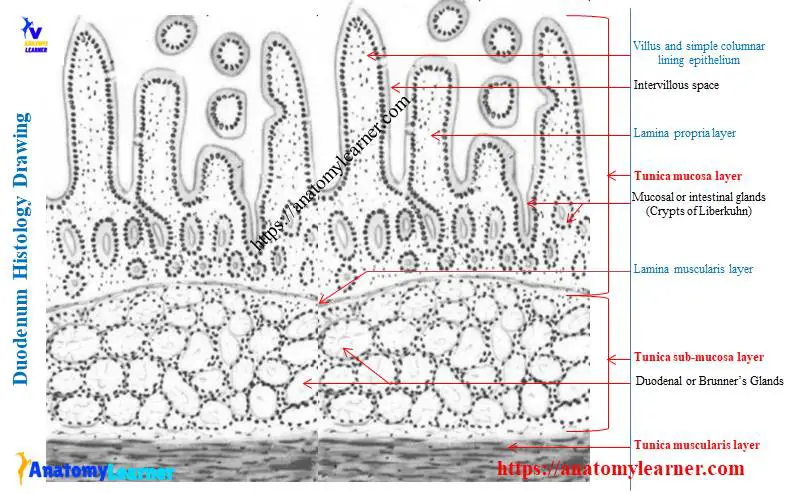
Duodenum histology slide labeled diagram
In this part of the article, I will show you the duodenum histology slide labeled diagram again so that you may summarize it well. The pictures showed the longitudinal section of a duodenum, where you will find almost every structure.
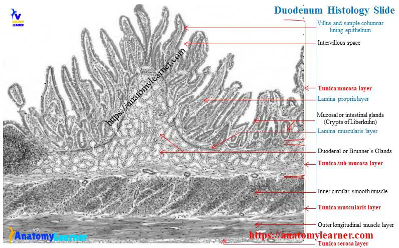
Here, I showed the four different primary layers of the duodenum – the tunica mucosa, tunica submucosa, tunica muscularis, and tunica serosa. Agin, you will find the villi, microvilli, crypts of liberkuhn, brunner’s glands, goblet cells, intervillous spaces, smooth muscle cells, and more.
I will try to show you the lining of the mucosal gland and the brunner’s gland with high magnification. Would you please try to understand the pattern of the smooth muscles of two different layers (inner circular and outer longitudinal) from the labelled diagrams?
Please join the anatomy learner on social media if you need more duodenum histology slides, pictures, or labelled diagrams.
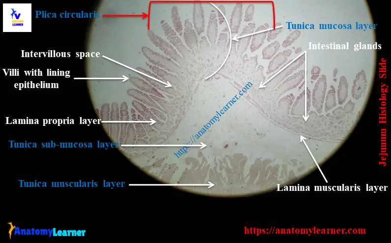
Jejunum histology labeled slide.
The proximal part of the jejunum histology slide shows the significant differences in structure from the other part of the small intestine of the animal. I will show you how you will differentiate the jejunum slide from the duodenum or ileum.
First, look at the jejunum histology slide labelled diagram and determine the following structures or features.
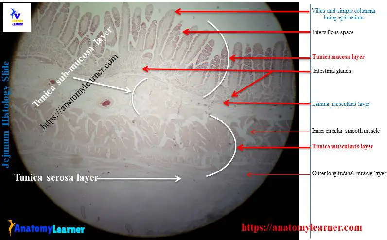
Presence of thicker wall, more prominent and more numerous folds in the jejunum.
There are more extensive and finger-like villi present in the jejunum.
Fewer lymphatic follicles are present in the distal part of the jejunum, but there are no aggregated lymphatic follicles in the proximal portion of the jejunum.
You will not find any glands in the submucosa of the jejunum histology slide.
Have you got the points that make the jejunum different from the duodenum? Okay, now let’s discuss the essential features of the ileum histology slide.
In the jejunum histology slide labeled diagram, I tried to show you the most characteristics features that made the jejunum exception from the other parts of the small intestine.
Ileum histology slide labeled diagram
You may quickly identify the ileum histology slide with the help of the following identifying characteristics. Here, I will enlist only the most important identifying features of the ileum slide. However, if you wish to learn more, please read the complete guide on ileum histology.
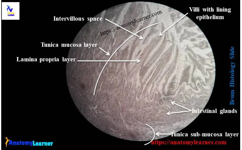
The villi of the ileum are thin and slender.
There are payer’s patches (aggregated lymphoid follicles) in the submucosa.
There is no gland in the submucosa of the ileum.
So, you will identify the ileum histology slide. Could you please summarize the histological features of all three different parts of the small intestine? You will find a great histological variation in the tunica mucosa and submucosa layers of these parts of the small intestine.
Frequently asked questions on duodenum
So, in this part of the article, you will find all the possible answers to the frequently asked questions on the duodenum slide structure.
What type of tissue is found in the duodenum?
Histologically, the duodenum is made of four different layers. They are – tunica mucosa, tunica submucosa, tunica muscularis, and tunica serosa. Again in the tunica mucosa of the duodenum, you will find three distinct layers – lamina epithelium, lamina propria, and lamina muscularis.
I have described all these four layers of the duodenum here in this article. If you want to know these histological features, then please read the full article.
You will find some exceptional features in the tunica mucosa and tunica submucosa layers of the duodenum. In the mucosa, you will find the villi, microvilli, and crypts of liberkuhn. There are brunner’s or duodenal glands in the tunica submucosa layer of the duodenum.
What is the histology of the duodenum?
The histology of the duodenum is similar to the general structural organization of a hollow organ. There are four different layers found in the duodenum of animals.
I have already described all of these four layers (mucosa, submucosa, muscularis, and serosa) in detail. You might read it and will differentiate the duodenum from the other parts of the small intestine.
Here, you will also find a guide to show you the most critical differences in the histological features among three different parts of the small intestine.
What are the 4 parts of the duodenum?
Anatomically, the duodenum of an animal has four parts, and they are – superior part, descending part, the inferior position, and the ascending portion. Histologically, you will find few variations in the four layers of the different parts of the duodenum.
What are the layers of the duodenum?
You will find the four layers (mucosa, submucosa, muscularis, and serosa) in a duodenum if you read this article. The tunica muscular and the tunica serosa have similar histological features as found in the general structure of a hollow organ.
Only the exceptional histological features will find in the tunica mucosa and tunica submucosa layers. Numerous villi, microvilli, and crypts of liberkuhn will be found in the duodenum’s tunica mucosa layer.
Conclusion
I hope you got the essential identifying points that help you to identify the duodenum histology slide. Now, you will also differentiate the duodenum histology slides from jejunum and ileum. I hope all the duodenum slide labeled diagrams were helpful for you.
I always request you to practice these duodenum, jejunum, and ileum slide at your histology learning laboratory. If you need any help related to small intestine histology slides, please let me know. Don’t forget to make a short note by yourself on identifying characteristics of the duodenum slide.