The dog scapula is a large, flat, and triangular bone of the shoulder girdle. In the anatomy of the dog scapula, you might learn the various osteological features from the different surfaces, borders, and angles. This article might help you to learn the details of the dog scapula bone anatomy and muscles that attach to it.
You know the scapula of a dog lies against the cranial part of the lateral thoracic wall in a cranioventral direction. It joins with the wall by the muscles without forming any proper joint. This attachment of the dog scapula with the thoracic wall is known as synsarcosis.
Anatomically, the dog scapula shows two surfaces, three borders, two extremities, and three angles that contain different important osteological features. If you want to learn these features from the dog scapula with the labeled diagram, let’s continue this article till the end.
Dog scapula anatomy
The scapula of a dog is also known as the shoulder blade. First, I would like to identify the different osteological features of the dog scapula anatomy. Let’s see the surfaces, borders, extremities, and angles from the dogs’ scapula.
The scapula of a dog possess –
- Two surfaces – lateral and medial or costal,
- Three borders – dorsal, cranial, and caudal,
- Two extremities – proximal and distal or glenoid, and
- Three angles – cranial, caudal, and ventral or glenoid or lateral,
The dog scapula labeled diagram identifies all these surfaces, borders, extremities, and angles. Now, let’s see the essential osteological features of the lateral and medial surfaces of the dogs scapula.
On the lateral surface of the dog scapula, you will find –
- A lateral prominent ridge or spine (that divides the scapula into two halves),
- The tuberosity of the spine (in the middle),
- An acromion process (pointed terminal structure of spine),
- The supraspinous fossa (upper), and
- An infraspinous fossa (lower depressed area),
Again, on the medial or costal surface of the dog scapula possess –
- A subscapular fossa of the canine scapula (concave), and
- The triangular facies serrate (upper triangular area on both sides),
Again, the proximal and distal extremities of the canine scapula also possess some other important osteological features. Let’s try to identify these osteological features from the proximal and distal extremities of the dog scapula.
The distal extremity shows –
- A narrow band of scapular cartilage (fitted into the pit of the dorsal border),
- Again, the distal extremity of the canine scapula shows –
- The glenoid cavity of the canine scapula,
- Coracoid process of the canine scapula,
- Supraglenoid tubercle of the dog scapula, and
- An infraglenoid tubercle of the canine scapula,
You may also identify all the osteological features from the dog scapula with the help of this video.
Location of dog scapula
The scapula of the dog locates at the cranial part of the lateral wall of the thorax. It slightly curves and slopes laterally, which means it is directed downward and forward.
The cranial angle of the canine scapula lies just ventral to the level of the free end of the spinous process of the first and second thoracic vertebra. Again, the long axis of the scapula extends obliquely from the fourth thoracic spine to the ventral end of the first rib.
You may also tell that the dog scapula extends from a transverse plane cranial to the manubrium (first part of the sternum) to one through the body of the fourth and fifth thoracic vertebra. You will not find any articulation of the scapula with the canine skeleton.
The canine scapula attaches to the thoracic wall and trunk with the help of muscles and synsarcosis. Again, the normal position of the dog scapula may vary due to the variable length of their vertebra.
So, the location and direction of the canine scapula are essential for studying their details anatomy. Before that, make sure you know the borders, extremities, and angles. I will explain how the border, extremity, and angle are formed.
Surface, borders, and angles of scapula
While studying the dog scapula anatomy, you will see two surfaces, two extremities, three borders, and three angles. The surfaces are the flattened areas of the scapula bone in the lateral and medial aspects.
You will find the upper and lower directed portion when you view the canine scapula in the normal position. The upper part of the scapula is the proximal extremity (dorsal border), whereas the lower part is the distal extremity (ventral angle or glenoid angle for scapula).
If you notice the dog scapula bone, the two surfaces meet at three sides (as it’s a triangular outline). Thus it forms three borders – cranial, caudal, and dorsal.
The two surfaces (lateral and medial) of the canine (dog) scapula bone meet at the cranial aspect and form the cranial border. These surfaces again meet at the caudal aspect and form the caudal border.
Finally, these lateral and medial surfaces of the scapula bone meet at the dorsal or distal extremity and form the dorsal border. So, you got the three borders in the canine scapula.
Now, let’s talk about the three angles of the dog scapula. You know there are cranial, caudal, and distal (glenoid) angles in the dogs’ scapula.
How are the angles formed in dog scapula?
When the borders of any bone meet, they form an angle. Here, the cranial and dorsal borders meet at the cranial aspect and form the cranial angle.
When the dorsal and caudal borders of the dogs scapula meet at the caudal aspect, they form the caudal angle. Now, the cranial and caudal borders of this bone meet at the distal extremity.
Thus, the cranial and caudal borders form the distal angle in the scapula. You know there is a glenoid cavity of the distal extremity of the scapula of dogs. So, this angle is sometimes termed the glenoid angle of the scapula.
Special features of the dog scapula
Now, let’s know some of the unique features of the dog’s scapula compared to the other animals’ scapula.
- The spine of the scapula divides it into the nearly equal fossae (whereas in ruminants, it divides the unequal fossae),
- There is a blunt tuber scapulae present (or absent) in the spine (whereas it is large and sharp in ruminants and also in a horse),
- The outline is less triangular compared to the ruminant and horse,
- There is an ill-developed coracoid process in the distal end of the dogs’ scapula, whereas you will find a more developed coracoid process in the horse scapula,
- The subscapular fossa of the dogs scapula possesses some straight muscular lines,
I hope all the above information are enough to learn the basic of dog scapula. But, you may learn more about scapular anatomy from a dog by following this article till the end.
Anatomy of the dog scapula bone
In this article section, I will describe the detailed anatomy of the dog scapula bone with the labeled diagram. Here, you will get the description of the surfaces, borders, and angles of the dogs’ scapula.
Let’s see what you will learn in this section –
- Osteological features of the lateral surface of the dog scapula,
- The anatomy of the medial or costal surface of the dogs’ scapula,
- Osteological features of the three borders (cranial, caudal, and dorsal) from the canine scapula, and
- Osteological features of the angles of the dogs’ scapula,
Okay, let’s start with the surfaces (lateral and medial) of the dogs scapula.
The lateral surface of the dog scapula
The lateral surface of the canine scapula divides into two nearly equal fossae by a prominent ridge. This prominent ridge of the lateral aspect of the scapula is known as the spine.
The spine is the most prominent osteological feature of the lateral aspect of the dogs scapula. It starts proximally at the junction of the cranial and middle third of the dorsal border of the scapula.
At the starting point of the scapular spine, it becomes thick and has low ridges. Again, this structure becomes more expansive in the middle portion. Finally, the spine of the dogs scapula becomes thinner at the distal portion.
You may find two surfaces in the spine of the dogs scapula throughout its length. Again, the free border of the spine possesses a thickened structure in the middle area. This is known as the tuberosity of the scapula (or tuber scapulae, which may be absent).
This tuber scapulae is typically blunt in the canine scapula. But, you may find extensive and sharp tuber scapulae in the horse and ruminants scapula.
The distal end of the dogs scapular spine widens and forms the acromion process. Again, this acromion process is also different than these of the ruminant. In ruminants, you will find a pointed acromion process in the distal end of their spine.
As the acromion process of the dogs scapula is widening, you may easily palpate this structure superficially. There you may find a nutrient foramen at the junction of the distal extension of the spine and the scapular proper.
The deltoid muscle (acromion) arises from the acromion process of the dog’s scapula that extends distally. Again, the omotransversarius muscle also arises from the distal end of the spine adjacent to the acromion process.
Supraspinous and infraspinous fossa of the dog’s scapula
You know the spine divides the dogs scapula into the supraspinous and infraspinous fossa. The upper part of the structure is known as the supraspinous fossa.
Here, the supraspinous fossa is bounded by the cranial surface of the scapular spine and the adjacent lateral surface of the scapula. You will find the widest part of the supraspinous fossa in its middle.
This is due to the arc appearance of the cranial border of the dogs scapula. Here, the cranial border of the scapula extends from the cranial angle proximally to the scapular notch distally as a convex structure.
So, the starting and end parts of the supraspinous fossa are thin. The dog supraspinous fossa is a little concave that holds the muscle tightly.
The supraspinatus muscles arise from the supraspinous fossa (all parts) of the dog. You will learn about the different scapular muscles in the next part of this article.
In the dog scapula anatomy, the infraspinous fossa shows a triangular appearance. This infraspinous fossa is bounded by the caudal surface of the scapular spine and the caudal border of the scapula.
Again, the proximal extremity of the infraspinous fossa is bounded by the thick dorsal border. In contrast, the distal extremity of this fossa connects with the glenoid fossa or angle.
The infraspinatus muscle (dog) arises from the infraspinous fossa of the canine scapula.
Summary of the lateral surface: it shows a prominent spine that divides supraspinous and infraspinous fossa. The spine widens at the distal end and forms the acromion process.
The tuber scapulae are usually blunt or absent in the dogs scapula. Again, you will find the triangular-shaped infraspinous fossa in the dogs scapula.
Medial surface of dog scapula anatomy
The medial surface of the dog scapula is also termed the costal surface. It lies opposite the first five ribs and the adjacent four or five thoracic vertebrae.
You will find two important osteological features in the medial surface of the canine scapula anatomy –
- Two facies serrate, and
- A subscapular fossa,
The facies serrate is the small dorsocranial rectangular area in the medial aspect of the dogs scapula. If you notice the medial surface of the canine scapula, you will see two rectangular areas in the dorsal part – cranial and caudal facies serrate.
From these facies serrate, the thick serratus ventralis muscle arises. You know the thick serratus ventralis muscle of the subscapular fossa insert on the first seven or eight ribs, somewhat ventral to their middle.
Except for these facies serrate, the large remaining part of the medial surface of the canine scapula is the subscapular fossa. The subscapular fossa of the dog is nearly flat and possesses some straight muscular lines.
These straight muscular lines on the subscapular fossa extend from the dorsal extremity to the glenoid angle of the distal extremity. You will see some smooth bone and the concave area between these muscular lines of the subscapular fossa.
But in the horse scapula, you will find a deep subscapular fossa compared with the dogs. Sometimes you may find a large concavity on the subscapular fossa at the distal end.
The subscapaurlis muscle of the dog arises from the whole surface of the subscapular fossa. Again, this muscle also arises from the straight muscular lines.
Summary of the medial surface: the medial surface of the canine scapula possesses two important osteological features –facies serrate and subscapular fossa. The subscapular fossa shows few straight muscular lines.
Dorsal border of the dog scapula
The dorsal border of the dog scapula is sometimes known as the vertebral border or base. This border of the scapula pointed to the vertebral column.
The dorsal border of the canine scapula extends between the cranial and caudal angles. You will see a narrow banded or crescent-shaped scapular cartilage that attaches to the dorsal border of the scapula.
This scapular cartilage acts as a shock absorber and enlarges the area for attachment of the muscles of the scapula. The cartilage becomes calcified and thus more brittle with age.
You may find only a small band like scapular cartilage in the dogs scapula. But, in horse and ruminant scapula, the cartilage is more extensive and extends over the caudal angle that reaches the wither level.
At the four weeks of pregnancy, the ossification center appears in the canine scapula. Again, in the second to fifth months of pregnancy, you may find the coracoid process in the dogs’ scapula.
Rhomboideus muscle attaches to the dorsal border of the dogs’ scapula.
Cranial border of dog scapula anatomy
The cranial border of the dog scapula anatomy shows an arc-shaped appearance. It is thin except at its two extremities. Distally, it forms a concavity which is known as the scapular notch.
This scapular notch of the canine scapula marks the position of the constricted part of the bone. The proximal part of the cranial border becomes rough and thicker. Again, it becomes smoother and thicker at the distal extremity.
The proximal extremity of the cranial border runs into the dorsal border at the cranial angle. In comparison, the distal extremity of the cranial border runs into the distal or glenoid angle.
You will not find the arc-shaped cranial border in all dog species. Sometimes you may find the slender extremities in the dogs’ scapula where the cranial border is nearly straight.
Caudal border of the canine scapula
The caudal border is the thickest of the three borders of the canine scapula. You will see several ridges on the caudal border of the scapula that is responsible for the attachment of the triceps muscle of the forearm.
There is an essential osteological feature present in the caudal border of the dogs’ scapula, and that is the infraglenoid tubercle. This infraglenoid tubercle locates at the distal extremity just dorsal to the ventral or glenoid angle of the scapula.
The infraglenoid tubercle is much thicker than the caudal border and faces the coastal surface. You will find the origin of the triceps (part) and teres minor muscles form this infraglenoid tubercle of the dog scapula.
There are two muscular lines at the distal end of the caudal border – the cranial and caudal lines. The more cranially located line of the caudal border extends nearly to the edge of the glenoid cavity. In contrast, the more caudally located line of the caudal border ends at the infraglenoid tubercle.
The teres minor muscle also originates from the more cranial line and the adjacent caudal border of the scapula. Again, the long head of the triceps originates from the more caudal line and the adjacent caudal border of the scapula.
You will see a smooth and broad part at the middle third of the caudal border of the scapula. The subscapularis muscle curves laterally from the medial side of the middle third of the caudal border.
Again, the teres major muscle arises from the proximal fourth of the caudal border of the dog scapula.
Summary of the borders of dogs’ scapula
The dorsal border of the dog scapula possesses a narrow band like cartilage (scapular cartilage) and forms the synsarcosis with the thoracic wall. Again, the cranial border of the dog scapula is thin and convex (arc appearance).
The distal extremity of the cranial border possesses a vital feature, a scapular notch. Finally, the caudal border of the scapula possesses two important osteological features – an infraglenoid tubercle and two distinct muscular lines.
Cranial and caudal angles of the dog scapula
The cranial angles of the dogs scapula join the thin and slightly concave cranial border at a right angle. You know the thin, convex cranial border meets with the thinner, rough, and slightly convex dorsal border cranially and forms the cranial angle.
At the cranial angle, no muscles are directly attached. But, you know, the cranial border forms the scapular notch at the level of the neck of a dogs scapula. The suprascapular nerve lies on this scapular notch.
The caudal angle of the canine scapula is thickened and palpable through the skin. The adjacent thicker caudal border joins with the thinner, rougher, and slightly convex dorsal border to form the caudal angle (caudally).
The teres major muscle arises from the caudal angle and the adjacent caudal border of the dogs’ scapula.
The ventral angle of dog scapula anatomy
The ventral angle of the dog scapula anatomy is very important as there are different osteological features. You may also call this ventral angle the lateral, articular, or glenoid angle.
The essential feature of the ventral angle, it possesses a glenoid cavity. This glenoid cavity receives the head of the humerus and forms the shoulder joint.
If you notice the ventral angle, the glenoid cavity of the dog scapula is shallow. But, you will find a deep glenoid cavity in the horse scapula. The ruminant scapula’s glenoid cavity is moderately deep compared to the dogs.
The lateral border of the dogs’ glenoid cavity is nearly flattened. Again, the medial border forms a large arc, whereas the caudal border of the scapula forms the smaller convexity.
The lateral border of the glenoid cavity extends out on the articular surface of the supraglenoid tuberosity. You know the supraglenoid tuberosity is the considerable and rough prominence on the ventral angle of the dogs scapula.
The supraglenoid tuberosity is the larger tuberosity that projects cranially from the glenoid cavity. You will see a medial inclination of this supraglenoid tuberosity in the dogs.
A single tendon of the biceps brachii muscle arises from the supraglenoid tuberosity of the dogs’ scapula.
A small beaklike process that arises medial aspect of the scapular tuberosity is the coracoid process. The coracoid process is the remnant of the coracoid bone. From this coracoid process, the coracobrachialis muscle arises.
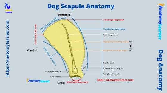
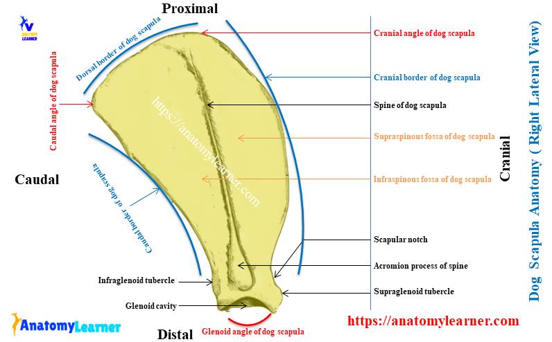
Dog scapula anatomy labeled diagram
You have already got the different labeled diagrams on the canine scapula anatomy. Now, let’s see the other labeled diagrams and practice with the actual sample (dogs scapula).
Here, in the diagram, I tried to show you the different features from the lateral surface of the dogs scapula. Before that, make sure you can identify the surfaces (lateral and medial), borders (dorsal, cranial, and caudal), and angles (cranial, caudal, and glenoid) from the dog scapula.
Here, I have identified these different surfaces, borders, and angles from the dog scapula with a labeled diagram. Now, let’s see the spine of the dogs’ scapula. The labeled diagram also identifies the spine as dividing the lateral surface into two halves.
Here, the broad acromion process of the spine is also identified in the labeled diagram. Again, the supraspinous and infraspinous fossa of the lateral surface of the dogs scapula are also identified in the diagram.
Again, from the medial surface of the canine scapula anatomy, the dorsocranial rectangular facies serrate was also identified. The medial surface also shows a shallow subscapular fossa (identified in the scapula labeled diagram). Again, the labeled diagram also shows a few muscular lines at the medial surface of the dog scapula.
There is the thickest caudal border in the dog scapula (identified). You will see the infraglenoid tubercle that is identified in the labeled diagram.
The ventral angle of the canine scapula identification
From the ventral angle of the canine scapula, I have identified different important osteological features in the labeled diagram. Here, the ventral angle of the dog scapula anatomy shows the glenoid cavity (identified in the diagram).
Again, the cranially located supraglenoid tuberosity from the dogs’ scapula is also identified in this labeled diagram. Medial to the supraglenoid tuberosity of the dog scapula, there is the small coracoid process (shown in the diagram).
You may find more labeled diagrams and videos on the dog’s scapula here on the social media of anatomy learners.
Dog scapula muscles anatomy
In the dog scapula muscles anatomy, I will describe only these muscles closely related to this bone. It will be better if I enlist and describe the muscles from the lateral and medial aspects of the dog scapula.
You may see two major types of muscles (extrinsic and intrinsic) muscles in the thoracic limb of the dogs scapula. The extrinsic muscles are these muscles that originate on the neck and thorax and extend to the scapula or humerus.
You will see the extrinsic muscles like brachiocephalicus, omotransversarius, trapezius, latissimus dorsi, rhomboideus, and serratus ventralis cervicis in the thoracic limb of a dog.
Again, the intrinsic muscles of the dog thoracic limb originate and insert into the bones of the limbs. These intrinsic muscles have no direct connection with the neck or trunk of the dogs.
Let’s know some of the intrinsic muscles of the dogs scapula. It includes supraspinatus, infraspinatus, teres major, deltoideus, and subscapularis.
Now, let’s enlist the muscles that directly relate to the dogs’ scapula from its lateral and medial aspects. In the lateral aspect of the dog scapula, you will see the following essential muscles –
- Trapezius and deltoideus muscles of the dog,
- Rhomboideus muscle,
- Supraspinatus and infraspinatus muscles of the scapula,
- Teres major and minor muscles of the scapula,
- Biceps brachii, and long head of triceps brachii,
- Deltoideus muscle of the dog scapula,
Again, from the medial surface of the dog scapula, you will see the following essential muscles that have a close relationship with this bone –
- Serratus ventralis muscle,
- Subscapularis muscle of the dog scapula,
- Corcobrachialis muscle of the dog,
- Biceps and triceps brachii muscles,
- Rhomboideus and teres major muscles,
Now, you will learn the anatomical features of these lateral and medial muscles from the dog scapula.
Supraspinatus muscle of dog scapula
The supraspinatus is the lateral muscle of the dog’s scapula. It covers the trapezius cervicis and omotransversarius muscles.
This supraspinatus muscle fills the supraspinous fossa and curves over the lateral surface of the neck of the dog’s scapula.
Origins: the supraspinatus muscle of the dog’s scapula originates entirely from the surface of the supraspinous fossa. This muscle also originates from the spine of the scapula and the edge of the neck of this bone. You may also find the origin of the subscapularis muscle from this part of the scapula bone (neck).
Relationship: the strong belly of the supraspinatus muscle curve around the neck of the scapula. So, this muscle may also see from the medial aspect of the dogs’ scapula (partly).
A thick tendinous fold develops from the distal third of the supraspinatus muscle. This tendinous fold extends into the terminal tendon. Again, the distal end of the supraspinatus muscle becomes pennate.
A tendinous sheet originates from the spine of the dog’s scapula. The caudal half of the supraspinatus muscle is covered by this tendinous sheet.
Insertion: the insertion site of this supraspinatus muscle is at the free edge of the major tubercle of the humerus. Here, the entire muscle forms the short and thick tendon that inserts into this major tubercle of the humerus bone.
Action: this supraspinatus muscle of the dogs scapula is important to stabilize and prevent a collapse of the shoulder joint. It helps to extend the shoulder joint and advance the thoracic limb.
Innervation: this muscle is innervated by the suprascapular nerve.
The superficial cervical and suprascapular arteries supply the dog’s supraspinatus muscle.
Dog infraspinatus muscle anatomy
The infraspinatus muscle is another large lateral muscle of the lateral aspect of the dogs scapula. This muscle lies in the infraspinatus fossa of the scapula and extends caudally. Again, the infraspinatus muscle of the dog primarily covers the deltoideus muscle (scapular part of the deltoideus).
Origin: this muscle originates from several parts, especially the infraspinous fossa. It also arises from the scapular spine, the caudal border of the scapula, and from the tendinous sheet that covers the muscle. Again, you may also find the origin of the supraspinatus muscle at the scapular aponeurosis of the scapular part of the deltoideus muscle.
Insertion: the infraspinatus muscle of the dog divides into two parts ventrally. One part of this muscle will insert into the round rough area above the deltoid tuberosity of the humerus. Again, another part of this muscle inserts into the medial aspect of the lateral tuberosity of the humerus bone.
At the shoulder joint, the fleshy infraspinatus muscle becomes a thick tendon and crosses the caudal part of the major tubercle. Here, you will find the infraspinatus bursa.
Action: this muscle help in lateral rotation and abductor of the humerus bone. Again, the infraspinatus muscle help to extend or flex the shoulder joint depending on the position. The tendon of this muscle also acts as the lateral collateral ligament of the shoulder joint.
Innervation: the suprascapular nerve supply to the dogs supraspinatus muscle. Again, you will see the subscapularis and posterior circumflex arteries that supply to the dogs’ infraspinatus muscle.
Dog scapula deltoideus muscle anatomy
You will see a V-shaped deltoideus muscle in the dog scapula anatomy at its lateral aspect. This scapula muscle attaches to the cranial and caudal borders and the ventral end of the infraspinatus muscle.
The dog deltoideus muscle divides into two major parts – scapular and acromion. Here, the scapular part of the deltoideus muscle locates deep in the scapular fascia between the scapular spine and the proximal half of the humerus.
At the shoulder joint, the scapular part of the deltoideus muscle becomes a tendinous sheet that is medially deep to the acromion part. Again, the acromion part of the deltoid muscle possesses an oval, flat belly and crosses the lateral side of the shoulder joint.
The half part of the acromion part of the deltoideus muscle is covered by the aponeurotic part that comprises radiating fibers.
Origin: the scapular part of the deltoideus originates from the caudal border of the scapula and fascia of the infraspinatus muscle. Again, the acromion part of the deltoideus muscle originates from the acromion.
Insertion: the acromion part of the deltoideus muscle join with the tendinous part of the scapular part at the level of lateral tuberosity of the humerus. Again, they insert into the deltoid tuberosity of the humerus and the facia of the triceps brachii muscle.
The action of the deltoideus muscle: this deltoideus muscle helps to flex the shoulder joint. Again, it helps to abduct the humerus bone.
Innervation: this deltoideus muscle of the dog scapula is innervated by the axillary nerve.
Again, you will see the subscapular and posterior circumflex arteries supply to the deltoideus muscle of the dogs scapula.
Teres major muscle of the dogs scapula
The teres major muscle of the dog’s scapula is a fleshy, slender muscle located along the caudal border of the scapularis muscle. This muscle abducts the thoracic limb and flexes the shoulder joint.
Origin: the teres major muscle (dog) originates from the caudal angle and adjacent caudal border of the scapula. This muscle crosses the triceps brachii and coracobrachialis muscles.
Insertion: a short and flat tendon is formed by the teres major muscle of the dogs’ scapula. This short and flat tendon of the teres major insert to the teres major tuberosity of the humerus bone. Again, this short and flat tendon blends with the latissimus dorsi muscle.
The lateral surface of the teres major muscle shows a tendinous sheet. This tendinous sheet is thick distally.
Action: this teres major muscle of the dog’s scapula helps flex the shoulder joint. Again, this muscle helps to draw the humerus bone caudally. Finally, the teres major muscle prevents the lateral rotation of the dogs shoulder joint.
Innervation: the teres major muscle is innervated by the axillary nerve branches. Again, the subscapular artery supply to the teres major muscle of the dogs’ scapula.
Dogs teres minor muscle anatomy
The dog teres minor muscle is the small elongated bundles of the scapula muscle. They are the flexor side muscles of the shoulder joint that lie distocaudally of the scapula. Again, the teres minor muscle is covered by the deltoideus and infraspinatus muscles.
Origin: the teres minor arises from the caudal border of the dogs scapula and primarily from the infraglenoid tubercle. The aponeurosis of the teres minor muscle lies along the head of the triceps brachii muscle.
Insertion: the end part of this teres minor muscle form a short tendon. This tendon of the teres minor muscle inserts on the eminence of the crest of the major tubercle proximal to the deltoid tuberosity.
Action: the dogs teres minor muscle helps in the abduction of the thoracic limb and slight rotation of the arm.
Innervation: the branches of the axillary nerves innervate to the teres minor muscle of the dogs scapula. Again, the scapualris and caudal circumflex arteries supply to the teres minor muscle.
The long head of dogs triceps brachii muscle
In the dog, the triceps brachii muscle consists of four different parts – the long head, lateral head, medial head, and accessory head. Here, I will describe only the long head of the dog’s triceps brachii muscle as it is closely related to the scapula bone.
You may learn the details and anatomical features of the different parts of the triceps brachii muscle from the below-mentioned article –
- Dog thoracic limb muscle anatomy with labeled diagram
The long head of the dog’s triceps brachii is a triangular-shaped muscle that lies on the caudal border of the scapula and apex of the olecranon process. This muscle of the dog assists in extending the elbow joint.
Origin: the long head of the triceps is partly fleshy and partly tendinous. It arises (long head) from the caudal border of the scapula chiefly by the tendon of the infraglenoid tubercle. Again, a small part of the long head arises from the caudal border of the humerus bone.
Insertion: the end part of the long head form an extensive, short, and thick tendon. This extensive tendon of the long head of the triceps brachii inserts into the caudal part of the olecranon tubercle. There is a synovial bursa on the tendinous part of the long head of the triceps brachii muscle.
Action: the primary function of the long head of the triceps brachii is to extend the elbow joint.
Innervation: branches from the radial nerve innervates the long head of the triceps brachii muscle. Again, the posterior circumflex and deep brachial arteries supply to the long head of the triceps brachii muscle.
Dog scapula and biceps brachii muscle anatomy
You might also find the biceps brachii muscle in the dog scapula anatomy. It is an elongated spindle-shaped muscle in front of the humerus and originates from the supraglenoid tubercle of the scapula.
The biceps brachii muscle of the dog covers by the brachiocephalic and superficial pectoral muscle. Again, this muscle helps to flex the elbow joint of the dog.
Origin: the biceps brachii muscle of the dog originates from the proximal part of the supraglenoid tubercle of the scapula. It crosses the shoulder joint in a sharp curve and runs along the cranial surface of the humerus through the intertubercular groove.
Distal to the intertubercular groove, the biceps brachii muscle becomes wide and spindle-shaped. Again, it becomes thick in the middle and runs medial to the cranial surface of the humerus.
Insertion: the tendon of insertion of the biceps brachii divides into two parts. So, the tendons from the dogs’ biceps brachii muscle insert into the radial tuberosity and proximal end of the ulna bone.
The action of the biceps brachii muscle: the main action of the biceps brachii muscle is to flex the elbow joint. Again, this muscle extends and stabilizes the shoulder joint during standing or locomotion.
Innervation: the musculocutaneous branch of the brachial plexus innervates the biceps brachii muscle. Again, the anterior circumflex artery supplies the dogs biceps brachii muscle.
Coracobrachialis muscle of the dog
The coracobrachialis is a small elongated muscle that lies on the medial aspect of the humerus but originates from the coracoid process of the scapula. So, you might learn the anatomy of the dog coracobrachialis muscle here.
Origin of coracobrachialis muscle: it originates from the coracoid process of the scapula by a long and narrow tendon. The tendon runs obliquely over the medial surface of the shoulder joint. It lies in the groove close to the tendon of insertion of the subscapularis muscle.
The muscle runs between the medial and accessory heads of the triceps brachii muscle. It ends on the crest of the minor tubercle and caudal to the crest between the triceps brachii and brachialis muscles.
Insertion: this muscle inserts into the area above the teres tuberosity and cranial surface of the distal third of the humeral shaft.
Action: the coracobrachialis muscle of the dog helps to adduct the thoracic limb and flex the shoulder joint.
Innervation: again, the musculocutaneous nerve from the dogs brachial plexus innervates to the coracobrachialis muscle. The cranial circumflex artery supplies the dog’s coracobrachialis muscle.
Dog trapezius muscle anatomy
Dog trapezius is the extrinsic thoracic limb (dorsum) muscle that is broad, thin, and triangular. It lies deep to the skin and caudal portion of the platysma in the dog’s neck.
You know, the trapezius of the dog arises from the median fibrous raphe of the neck and supraspinatus ligament of the thorax. Again, the origin of this muscle extends from the third cervical vertebra to the ninth thoracic vertebra.
A tendinous band extends dorsally from the spine of the dog’s scapula. This tendinous band divides the dog trapezius muscle into cervical and thoracic parts – pars cervicalis and pars thoracica.
The pars cervicalis of the dogs trapezius muscle arises from the middorsal raphe of the neck. It runs obliquely cranioventrally to the spine of the dogs scapula. Finally, it inserts into the free edge of the scapular spine.
Again, you will see the attachment of the omotransversarius muscle on the spine of dogs scapula (small part).
The pars thoracic of the trapezius originates from the supraspinatus ligament and the spinous process of the third to ninth thoracic vertebrae. Finally, this part of the trapezius muscle inserts into the proximal third of the dog’s scapular spine.
Action: the main action of the dog trapezius muscle is to elevate the thoracic limb and draw it cranially. Again, this muscle (trapezius) rotates the dogs’ scapula.
Innervation: the dorsal branch of accessory nerve supply to the dogs trapezius muscle. The deep cervical branch of the costo cervical and intercostal artery supply to the trapezius muscle.
Omotransversarius muscle of the dog
The dog omotransversarius muscle is a flat, long structure that extends from the atlas to the shoulder and is mainly covered by the brachiocephalicus. This muscle of the dog pulls the ventral angle of the scapula craniodorsally.
Origin of omotransversarius muscle in a dog: it arises from the distal part of the scapular spine. Again, you will also find the origin from the omobrachial facia that covers the acromial part of the deltoideus muscle.
This muscle is separate from the trapezius muscle (cervical part) and passes deep to the cervical part of the cleidocephalicus muscle. You will also find a good relationship between this muscle with the scalenus and intertransversarius cervicalis muscles.
Insertion: finally, the tendon of the omotransversaius muscle inserts into the caudal border of the wing of the dog’s atlas.
Action: the primary function of the omotransversarius muscle is to draw the thoracic limb cranially.
Innervation: the accessories nerve innervates to the omotransversarius muscle of the dog. The inferior cervical and carotid arteries supply to the dog’s omomtransversarius muscle.
Dog rhomboideus muscle anatomy
The dog rhomboideus muscle is a roughly triangular and thick muscle that laterally covers the trapezius. This rhomboideus muscle help to move the shoulder forward and upward.
The dog rhomboideus muscle divides into three parts –
- A cervical part – rhomboideus cervicalis,
- The vertebral part – rhomboideus capitis, and
- A thoracic part – rhomboideus thoracic,
Rhomboideus cervicalis part: it lies dorsolateral on the neck from the second cervical vertebra to the third thoracic vertebra. This part of the rhomboideus muscle arises from the tendinous medial raphe of the dog neck and also from the spinous process of the first three thoracic vertebrae.
The cervical part of the rhomboideus muscle is inserted into the rough medial surface and dorsal border of the dog scapula.
Rhomboideus capitis: it arises from the occipital bone and atlas of the dog. Finally, the tendon from this capitis part inserts into the dorsal border of the scapular cartilage.
The thoracic part of the rhomboideus muscle: this part of the rhomboideus muscle arises from the spinous process of the fourth to seventh thoracic vertebrae. Again, the tendon of the thoracic rhomboideus muscle inserts into the medial surface and partly on the lateral edge of the dorsal border of the scapula.
Here, you will see the latissimus muscle that covers the thoracic rhomboideus muscle.
Action: you will find different functions of the dogs rhomboideus muscle. It elevates the thoracic limb and pulls the thoracic limb and shoulder cranially or caudally. Again, it draws the dog scapula against the trunk.
Innervation: the dorsal branch of the cervical, thoracic nerve innervates to the dogs rhomboideus muscle. The deep cervical and dorsal branches of the costo cervical arteries supply the rhomboideus muscle.
Subscapularis from dog scapula anatomy
In the dog scapula anatomy, you will find this muscle at the medial aspect that fills the subscapularis fossa. It crosses the flexor angle of the dog shoulder joint medially and helps in the adduction of the thoracic limb.
The subscapularis muscle of the dog is a broad and flat structure that overhangs the caudal edge of the scapula. Different tendinous bands divide the subscapularis muscle into several parts.
Origin: the dogs subscapularis muscle primarily arises from the subscapularis fossa. It also arises from the muscular lines on the caudal edge of the scapula. You may also find the origin of the subscapularis muscle on the curved boundary line between facies serrate and subscapular fossa.
This muscle becomes narrower and passes over the shoulder joint medially. At this point, the subscapularis muscle becomes tendinous.
Insertion: the end part of the dogs subscapularis muscle form a short and very thick tendon. This short and thick tendon of the subscapularis muscle inserts on the minor tubercle of the humerus bone.
Action: primarily, this subscapularis muscle helps in the adduction and extension of the dog’s shoulder joint. It draws the humerus cranially during flexion of the shoulder joint. Again, it rotates the humerus medially and prevents lateral rotation of this bone.
The short and thick tendon of this subscapularis muscle act as a medial collateral ligament in the joint.
Innervation: two nerves from the dog’s brachial plexus innervate to the subscapularis muscle, and they are – axillary and subscapularis nerves. Again, the suprascapular and subscapular arteries supply to the dogs’ subscapularis muscle.
Serratus ventralis thoracic muscle of the dog
The serratus ventralis thoracic muscle of the dog is very thick and fan-shaped. This muscle covers the cranial half of the lateral thoracic wall of the dog.
Origin of serratus ventralis thoracic muscle: the fibers of this muscle originate from the roughly triangular area at the craniodorsal part of the medial surface of the dogs’ scapula (facies serrate).
Insertion of serratus ventralis thoracic: it inserts on the first seven ribs, somewhat ventral to the medial aspect of these ribs.
Relationship: the serratus ventralis thoracic muscle attaches to the serratus ventralis cervicis muscle (cranially). You will see the well-defined serration on the thoracic part of the serratus ventralis muscle of the dogs.
This part of the serratus ventralis muscle covers the scanelus muscle. Again, a small part of the serratus ventralis thoracic muscle is covered by the obliques externus abdominal muscle.
The action of serratus ventralis thoracic muscle: this muscle helps in different ways – it supports the trunk of the dog. It carries the dog trunk cranially and caudally and thus helps in inspiration. Again, the serratus ventralis thoracic muscle carries the shoulder cranially and caudally concerning the thoracic limb.
Innervation: the nerve longus thoracic (nerves from brachial plexus) innervate to the serratus ventralis thoracic muscle. Again, some intercostal arteries supply the dogs’ serratus ventralis thoracic muscle.
You may learn the anatomical facts of the muscles from the dog neck region and thoracic limb from the myology section of anatomy learner.
Scapula of horse and pig compare to a dog
Let’s see some of the unique features of the scapula of horse and pig that might help you to identify them from the dogs. The basic structure of the horse and pig scapula is almost similar. You may find a difference in their different osteological features or points.
Let’s see what the peculiar osteological features found in the horse scapula that differ from the dogs –
- The lateral spine of the horse scapula is located further backward and divides the lateral surface into unequal fossae,
- You will not find any acromion process in the scapula of a horse because the spine ends at the distal third part (near the neck),
- There is a well-developed coracoid process at the distal end (glenoid end; medial aspect of the glenoid cavity),
- The tuber scapulae are also more prominent compared to these of the dogs and located further away from the glenoid cavity,
- You will see a deep glenoid cavity that possesses a deep and distinct notch,
- The subscapular fossa is also deep compared to that of the dogs’ scapula,
Again, in the scapula of pig, you will see the following exceptional osteological features –
- The cranial border of the pig scapula forms an arc (supraspinous fossa),
- A prominent scapular spine divides the lateral surface of the pig scapula into almost equal fossae,
- The spine of the pig scapula is wide and further directed backward,
- You will see a well-developed and prominent tuberosity in the spine of the pig scapula (middle),
- The acromion process is rudimentary in the pig scapula,
- There is no glenoid notch in the glenoid cavity in the pig scapula,
These are very short information on the horse and pig scapula. You may learn more about them from another article.
Dog scapula injury
If you are a dog owner, your dog may show some abnormal symptoms when scapular injuries occur. Different types of injuries may occur in the scapular region. This injury may cause the shoulder joint, bones, muscles, ligaments, and cartilage of the scapula.
I will not describe all the things from the dog’s scapular injury. Instead, I would like to provide some essential injuries that may occur in the dog scapula.
The most common injuries in the dog scapula or scapular region –
- Shoulder sprain and frozen shoulder,
- Shoulder dysplasia of the dog,
- Traumatic injury to the dog’s scapular cartilage,
- Ossification on the infraspinatus bursa (bursal ossification),
- Biceps brachii tendon luxation,
- Luxation in the teres major and minor tendons,
As a veterinary student or practitioner, you might know the symptoms and causes of these common injuries in the dogs’ scapula. If you want, you may learn the details about these injuries on the dog’s scapular region from another article by an anatomy learner.
Common inquiries on dog scapula anatomy
Here, I will enlist some common inquiries on the dog scapula anatomy. Again, I will try to solve these common inquiries with concise information. Let’s see the questions commonly asked by the veterinary student, Practioner, and dog owners.
What is the scapula of a dog?
The scapula of a dog is a large, flat, and almost triangular bone in the thoracic limb. This bone helps to form the thoracic girdle in the dog. The dogs’ thoracic girdle is formed mainly by the clavicle and scapula.
The clavicle of the dog is a thin and slightly convex structure located at the tendinous intersection of the brachiocephalic muscle. The medial end of the clavicle attaches to the sternal facia by a distinct ligamentous band.
The clavicle is more closely attached to the clavicular tendon between the cleidocephalicus and cleidobrachialis muscles. Again, you may know more about the scapula of the dogs if you read this entire article from the beginning to the end.
What is the scapula bone in an animal?
The scapula is the flat triangular type of bone in the thoracic limb of the animal’s skeleton. The basic anatomy of the scapula is almost similar in the different animals. But, you may find some peculiar osteological features in the scapula of different animals that makes them unique.
You may easily differentiate the scapula among different animal species by these peculiar osteological features. Here, I have pointed out the peculiar features of the horse and pig scapula along with the description of the dogs’ scapula.
But, you may also learn the osteological features of the ruminant scapula (like ox, goat, and sheep) from the anatomy learner (osteology section).
Where is the dog shoulder blades?
The scapula of the dog is sometimes called the shoulder blades. The dog’s shoulder blades are located at the cranio lateral aspect of the dog’s thorax.
If you notice the direction of the dog’s scapula, it goes downward and forwards. Again, it extends from the third to fourth thoracic spine to the distal end of the first rib.
How do you treat a scapula?
Sometimes the dog shows different symptoms of scapular injuries. The most common injuries of the dog scapula are frozen shoulder, shoulder dysplasia, traumatic injury to scapular cartilage, and luxation of diferent ligaments.
In this condition, you should restrict your dog’s movement and immediately bring them to a professional veterinarian.
Where is the scapula on a dog?
You know the scapula of a dog lies on the cranio lateral aspect of the thoracic wall. The most dorsal part of the dog scapula lies just ventral to the level of the free end of the spinous process of the first thoracic vertebra.
What are the parts of the dog scapula?
You know the dog scapula has two surfaces, three borders, and three angles. The scapular spine on the lateral surface of the dog scapula divides into two major parts – infraspinatus and supraspinous parts or fossae.
Again, the medial surface of the dog scapula shows the subscapular part. The three borders of the dog’s scapula show some distinct, peculiar features. There is an infraglenoid tubercle (special feature) in the distal end of the cranial border of the scapula.
Conclusion
The osteological features of a dog scapula anatomy are somewhat different than these of the ruminant. I think you have got the basic idea of the anatomical features of the dog scapula from this article. The labeled diagrams of dog scapula might help you identify the peculiar osteological features from the sample.
The muscles from the dog scapula anatomy are also crucial for the first-year veterinary student. Here, all the intrinsic and extrinsic muscles from the dog scapula are described with their origin and insertion. Now, at your anatomy learning laboratory, you should compare the anatomical features of the dog scapula with other animals like an ox, horse, and goat.