The dog sacrum anatomy consists of three fused sacral vertebrae. It forms the basic last part of the canine spine.
Here, I will share the osteological features of the dog sacrum anatomy with the labeled diagram.
Quick overview: three fused sacral vertebrae from the dog sacrum, and thus it becomes small in length. It is a four-sided, wedge-shaped bone in the spine that possesses enlarged wings. This canine sacrum shows two intervertebral foramina and a notched median sacral crest.
You will also find the main differentiation between the canine and horse sacrum bones. Thus you will quickly identify the dog’s sacrum bone from other species.
Okay, let’s get started to learn the anatomical features of the canine sacrum bone.
Dog sacrum bone
First, let’s know and identify the below-mentioned osteological features from the dog sacrum bone –
- Dorsal convex and ventral concave surfaces of canine sacrum bone,
- Bodies of the canine sacrum bone,
- The cranial base of the sacrum bone,
- Caudal apex of the sacrum bone,
- Wing of the dog sacrum bone,
- Cranial and caudal articular processes of the canine sacrum,
- Median sacral crest with notches,
- Intermediate sacral crests on the dorsal surface,
- Two dorsal sacral foramina of the sacrum bone,
- Sacral canal of the sacrum bones,
- Ventral or pelvic sacral foramina (two),
- Latera; sacral crest or modified transverse processes of the dog’s sacral vertebrae, and
- Pelvic inlet and outlet of the canine sacrum vertebra,
All these above-mentioned osteological features are identified in the dog sacral vertebrae labeled diagram. This might help you to understand the appearance and structure of the dog sacrum bone.
This canine sacrum is different from the features of a typical vertebra. You may know the details of the animal’s typical vertebra from the below-mentioned article –
This article might help you to understand the basic difference between the typical true vertebra and sacrum.
Unique features of the canine sacrum bone
Here, I will enlist some of the unique features of the dog sacrum anatomy. Let’s find these features and try to compare them with other animal’s sacrum bones –
- Only three sacral vertebrae fuse to form the sacral bones in the dog’s spine,
- It is a four-sided (quadrilateral) and wedge-shape bone in the last part of the canine spine,
- This bone possesses two dorsal and two pelvic sacral foramina,
- The articular process on the lateral high wings is larger,
- Again, the dorsal median sacral crest shows notches, and
- The dorsolateral aspect of the canine sacrum shows intermediate and lateral sacral crests,
You may also learn the other features of the canine sacrum from the next section of this article.
What is a sacrum on a dog?
Answer: sacrum is the atypical vertebra of the dog spine that is formed by the fusion of the bodies and processes of three sacral vertebrae. It articulates with the pelvic bone dorsally and forms the sacroiliac articulation.
Again, you will find the articulation of this sacrum bone with the last lumbar vertebra cranially. The caudal part of the dog sacrum bone articulates with the first caudal vertebra. Thus, it forms the sacrococcygeal articulation in the dog between the last sacral and first caudal vertebrae.
This bone also contributes to forming the pelvic inlet and outlet in dogs. You will learn the details of the pelvic inlet and outlet from the dog sacral vertebrae in the last section of the article.
How many bones are in the dog sacrum?
Answer: there are 3 sacral vertebral bones in the structure of a dog sacrum. These three bones become fused and form a single vertebral bone.
Here, the body of the first segment (first sacral vertebra) is larger than the other two segments. The arches of these three sacral vertebrae form the lateral bony mass.
The number of sacral vertebrae in the sacrum is different in various animals. Here, table 1 shows the number of sacral vertebrae that fuse to form the sacrum in various animals –
| Species | Fused vertebrae in the sacrum |
| Dog | 3 |
| Ox | 5 |
| Horse | 5 |
| Sheep | 4 |
| Pig | 4 |
Dog sacrum anatomy
In the dog sacrum anatomy, you might describe the below-mentioned features –
- Surfaces of the sacrum bone with their osteological features,
- Borders with their osteological features, and
- The features of the apex and base of the canine sacrum bone,
The shape of the canine sacrum bone is quadrilateral, whereas it is triangular in ox. But, you will find similar surfaces, borders, and other features in the canine sacrum, like the oxes.
The canine sacrum shows two surfaces – dorsal and pelvic; 2 lateral borders, a base, and an apex. Let’s discuss these surfaces, borders, base, and apex of the dog sacral vertebrae.
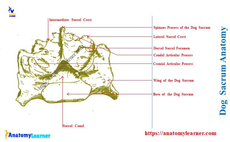
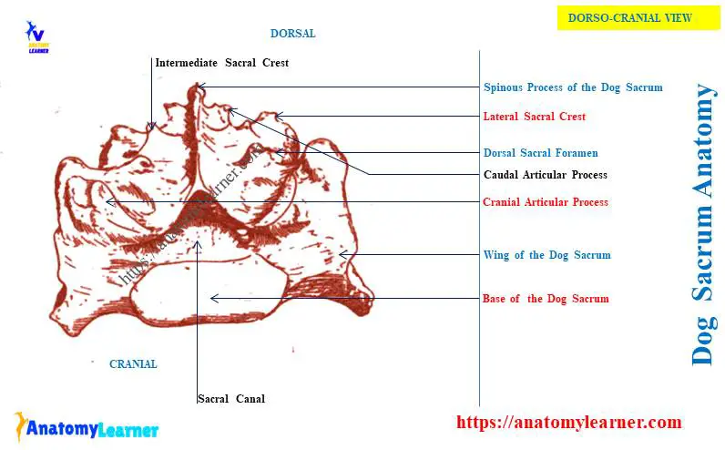
Dorsal surface of the canine sacrum bone
The dorsal surface of the canine sacrum bone is more or less convex and presents the median sacral crest. This is one of the important features on the dorsal surface of this sacral vertebrae.
And you know, this median sacral crest of the dog is formed by the fusion of three spinous processes. But you may also find two notches on the median sacral crest of a dog. These notches indicate the fusion of the three spinous processes.
In the canine sacrum diagram, I already showed two dorsal foramina. The dorsal branch of sacral spinal nerves and spinal vessels pass through these dorsal foramina.
Just medial to these dorsal sacral foramina, you will find bony projections. The fusion of mammillary and articular processes of the adjacent sacral vertebrae forms these projections.
The projections on the dorsal surface of the sacrum are known as the intermediate sacral crests. You will also find the cranial and caudal articular processes of the sacral vertebrae on the dorsal surface of the bone.
Here, the cranial articular process of the dog sacral vertebrae is larger. It faces dorsomedially and articulates with the caudal articular process of the seventh lumbar vertebra.
Again, the caudal articular process of the canine sacrum bone is small. It articulates with the cranial articular process of the first caudal vertebra.
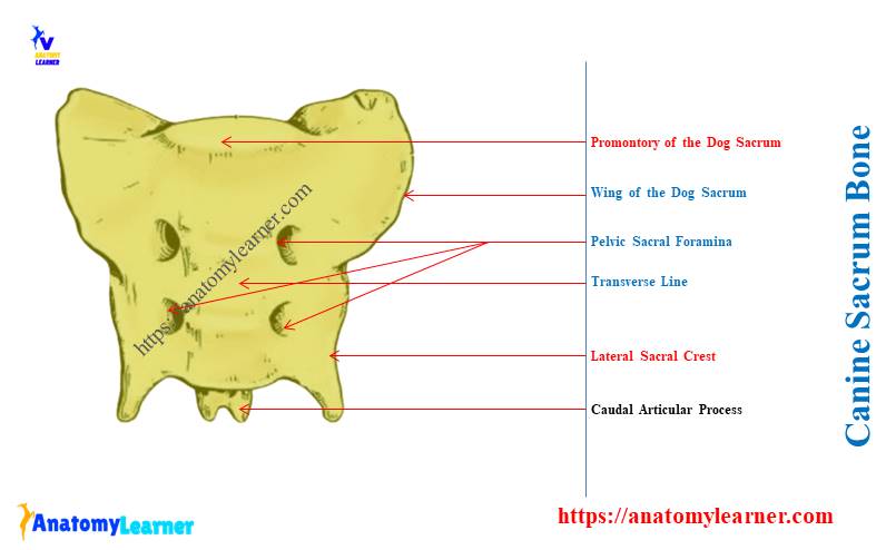
The pelvic surface of the dog sacral vertebrae
The ventral aspect of the dog sacral vertebrae is the pelvic surface which is more or less concave. You will find two important features on the pelvic surface of the canine sacrum structure –
- Two transverse lines (between the bodies of two adjacent sacral vertebrae), and
- Two pairs of pelvic sacral foramina,
Here, the pelvic sacral foramina of the canine sacrum bone are larger than these of the dorsal foramina. Again, the blood vessels and ventral branches of sacral spinal nerves pass through these pelvic foramina.
The transverse process of the first sacral vertebrae is highly modified from the wing. Again, the transverse processes of the second and third sacral vertebrae form a narrow, thin crest. And this is the lateral crest of the canine sacrum bone.
The lateral sacral crest terminates caudally as the flattened and pointed process. Finally, it articulates with the transverse process of the first caudal vertebra.
Let’s know the features of the wing of the dog’s sacral vertebrae with the diagram.
Wing of the dog sacral vertebrae
The wing is the enlarged lateral part of the dog’s sacral vertebrae. And you know, this wing is formed by modifying the transverse process of the first sacral vertebra.
The height of the wing of a dog sacral vertebra is larger compared to the ox and horse. You will find a large and rough articular surface on the wing of a dog’s sacrum. It articulates with the medial surface of the ilium bone (part of the pelvic bone) of the dogs.
You may know more about the anatomical features of the dog’s pelvic bone from the below-mentioned article –
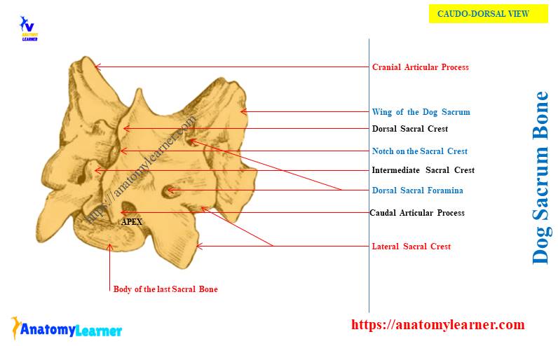
Base and apex of the dog sacral vertebrae
The base of the canine sacrum faces cranially and articulates with the preceding vertebra. It possesses a slightly convex articular surface and a wider sacral canal.
Here, the sacral canal is formed by the fusion of vertebral rings of three sacral vertebrae. You will find the opening of this sacral canal as a triangular opening. Again, it becomes more trinagular at the caudal end.
You will see a transverse ridge at the cranioventral aspect of the base. This is the promontory of the canine sacrum anatomy.
Again, the caudal part of the canine sacrum forms the apex (shown in the diagram). The apex of the sacrum articulates with the first caudal vertebrae of the spine.
Sometimes, you may see the fusion of the first caudal vertebra with the apex of the canine sacrum.
How does the dog sacrum contribute to the pelvic inlet and outlet?
The promontory and the ilia form the dorsal boundary of the pelvic inlet. Again, the apex of the canine sacrum forms part of the dorsal boundary of the pelvic outlet.
What parts are in the sacrum of a dog?
Answer: you will find different parts in the structure of a dog sacrum. The most important parts of the canine sacrum are –
- The base of the sacrum,
- Dorsal and pelvic surfaces,
- Right and left lateral borders of the sacrum,
- Median sacral crest,
- Lateral and intermediate sacral crest (right and left),
- Notches on the median sacral crest,
- Two dorsal and two larger ventral sacral foramina,
- Ventral transverse lines (two) and single longitudinal line,
- Wing of the sacrum, and
- The apex of the sacrum,
All these structures from the canine sacrum have been described in this article’s previous section.
How to differentiate the dog sacrum from the horse and cow’s sacrum?
Answer: you will find a great variation in the number of sacral vertebrae in dogs, horses, and cows. Again, the spinous process of the sacrum of these species also shows a great variation.
You may also find the unique features in the structure of wings in horses, dogs, and ox sacrum bones. Let’s see the major difference among the sacrum bones of various animals from Table 2 –
| Features | Dog sacrum | Horse sacrum | Ox sacrum |
| Sacral bones | 3 | 5 | 5 |
| Spinous process | Fused | Separated | Fused |
| Median sacral crest | Present | Absent | Present |
| Wing | Very high | Laterally extended Pointed Possess – Large oval surface | Curved ventrally Compressed |
| Sacral tuberosity | Absent | Prominent | Absent |
| Transverse process | Prominent | Developed | Rudiment |
The ventral surface of the horse sacrum is less concave compared to these of the ox and dog sacrum. Again, it shows the distinct supraspinous processes due to their incomplete fusion.
The wings of the horse sacrum also show unique features to the dogs. You will find the prismatic shape wing in the horse, which is also pointed at the end.
The cranial aspect of the wings possesses a large oval articular surface. This is one of the unique features of the horse sacrum compared to the dogs. This articular surface articulates with the articular surface on the caudal aspect of the transverse process of the last lumbar vertebra.
Conclusion
So, the dog sacrum anatomy shows the fusion of three sacral bones that possess the features of an atypical vertebra. The external appearance and confirmation of the canine sacrum bone are different from other animals like cows and horses.
Now, you might identify the features from the dog sacrum bone practically from the actual sample.