Dog salivary gland includes these oral glands that pour their secretions into the mouth cavity. You will see both the major and minor salivary glands in the dog’s oral cavity.
The major salivary glands of a dog include parotid, mandibular, sublingual, and zygomatic. Again, the minor salivary glands comprise the molar, buccal, lingual, and palatine.
Within this short article, I will show you the details anatomy of the dog salivary gland so that you may locate their exact location from the surface approach. Again, I will also discuss the infection and causes of swollen of the dog’s salivary glands.
In addition, you will find a clear concept of the salivary mucocele of a dog at the end of this article. So, if you want to know the details of anatomical facts and some common problems of the salivary glands of a dog, let’s continue this article.
Dog salivary gland
As I told you, the dog salivary gland divides into two main groups – major and minor salivary glands. Here, I will only focus on the major salivary glands of the dog.
You know there are four major salivary glands in the oral cavity of a dog. But, you will find three major salivary glands in the ruminant: cows, sheep, and goats. The zygomatic salivary glands are the extra pair of major salivary glands in the dog.
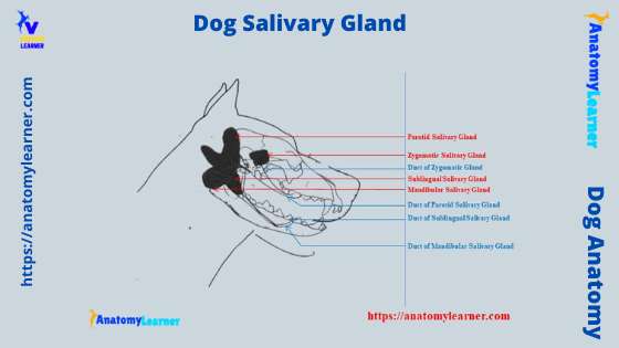
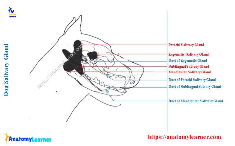
Okay, first try to identify the following four major salivary glands with the help of a labeled diagram –
- Parotid salivary glands of a dog,
- Mandibular salivary glands of the dog,
- Sublingual salivary glands (total four in number) in a dog, and
- The zygomatic salivary glands of a dog,
All these salivary glands are identified in the labeled diagram.
Now, let’s know some of the particular anatomical features of the major salivary glands of the dog’s oral cavity. These anatomical features of the salivary glands are compared with the ruminant (goat, cow, and sheep) salivary glands.
Peculiar features of dog salivary glands
- The parotid gland of a dog is a V-shaped or triangular in outline,
- You will find a notch on the dorsal border of the dog’s parotid salivary gland,
- Opposite the third premolar tooth, the duct of the parotid gland opens,
- The mandibular is an ovoid salivary gland that is larger than the parotid gland,
- You will see two sublingual glands on either side of the mandible,
- The major sublingual duct is closely related to the dorsal aspect of the mandibular ducts and opens on a small sublingual caruncle,
- You will see an extra pair of zygomatic salivary glands in the dog that locates the anterior part of the pterygopalatine fossa.
The major ducts of the zygomatic salivary glands of the dog open at the caudal surface of the last superior check tooth. You will learn more about the anatomy of these salivary glands (including location, shape, size, number of ducts, opening sites of these ducts, and others) in the next part of this article.
Dog salivary gland location
As a veterinary practitioner or a student, this is very important to know the exact location of the dog salivary gland. So, in this part, I will try to show you the exact location of four major salivary glands in a dog.
Let’s see the parotid glands from the diagram; you will see a V-shaped outline of this gland (when viewing from the lateral surface). This parotid gland of the dog lies at the junction of the head and neck.
This gland shows mainly two parts – the base and the apex. Here, the base is directed dorsally, whereas the apex of the parotid gland is directed ventrally.
It again molded around the proximal part of the auricular cartilage of the dog’s ear. In this part, the parotid gland can be rolled on palpation. It also occupies the depression between the masseter and wing of the atlas vertebra.
Topography location of dog’s parotid salivary gland
At the caudal part of the dog’s parotid salivary gland, you will see the mastoid part of the sternocephalicus and the cervical part of the cleidocephalicus muscle. Again, the rostral part of this gland is bounded by the masseter muscle and temporomandibular articulation.
You may also see the parotid lymph node at the rostral part of the parotid gland. Again, you know this parotid gland overlying the basal part of the auricular cartilage.
Caudal to this auricular cartilage, this parotid gland relates medially to the facial nerve and maxillary vein. The ventral part of this gland is thicker, whereas the dorsal part is thinner in a dog.
So, in a live dog, you should palpate through the base of the ear and along the caudal border of the vertical part of the ramus of the mandible to find the parotid gland.
Location of mandibular salivary gland
A dog’s mandibular gland is located close to the mandible’s angle and partially covers the parotid gland. The salivary gland labeled diagram shows an ovoid mandibular gland slightly smaller than the parotid gland.
You will also find this gland subcutaneously, just caudal to the monostomatic salivary gland between the lignofacial and maxillary veins (at the angle of the mandible). The mandibular and monostomatic salivary glands have great clinical importance in a dog.
These glands may undergo cystic changes (renula), which require surgical removal. You may know details about the dog renula in details from the other article by anatomy learner.
You may find a strong fibrous capsule over the dog’s mandibular salivary gland, and thus it may be easily palpable by the surface approach. But, this gland may confuse the dog’s mandibular lymph nodes under finger exploring.
Let’s see the relationship of the dog mandibular gland with other structures. Rostrally, you will see mandibular lymph nodes, sublingual gland, masseter muscle, and digastric muscle.
You will find the digastricus, external carotid artery, and medial retropharyngeal lymph node at the medial aspect of the mandibular salivary gland. In the caudal aspect, these glands have contact with different muscles of the neck region.
Location of the sublingual salivary gland of a dog
You will see the compact narrow sublingual salivary gland in a dog that continues forward from the mandibular gland. The sublingual gland locates medial to the mandible, under the mucosa the underside of the tongue.
Let me clarify the location of the dog’s mandibular salivary gland; from the mandibular gland, it goes forward. This gland passes between the digastricus and pterygoid muscle and reaches the lateral of the root of the tongue.
Again, the gland passes over the mandibular ducts (between the digastricus and pterygoid muscle). The sublingual salivary gland’s duct accompanies the mandibular duct at the sublingual caruncle.
Both the ducts of the sublingual and mandibular glands raise the sublingual fold near the body of the mandible. You may see the slitlike opening of the mandibular and sublingual salivary glands ducts on the lateroventral surface of the lingual caruncles.
The mandibular duct is larger and more rostral than the sublingual salivary ducts. You may easily perform the cannulation on the mandibular salivary gland of the dog. In contrast, it isn’t easy to cannulate the ducts of the dog’s sublingual gland.
A dog has two types of sublingual salivary glands – monostomatic and polystomatic salivary glands. These sublingual salivary glands are the smallest of the four pairs of the major salivary glands in a dog.
You may see a common clinical condition in a dog’s sublingual salivary glands, which is salivary mucocele. This is a condition where the accumulation of the mucoid saliva leaked from the damaged ducts or glands.
Zygomatic salivary glands of the dog
Among the domestic mammals, you will only find the zygomatic salivary glands in dogs and cats. Another name for the zygomatic salivary glands of the dog is the orbital gland.
In other mammals, the caudal condensation of the unilocular dorsal buccal gland insteat of zygomatic glands.
This zygomatic salivary gland is globular to pyramidal shaped, a base directed dorsally and caudally. Again, the gland lies against the ventral part of the periorbital surface.
You will see a poorly developed capsule that surrounds by soft fat. So, you may easily identify these zygomatic salivary glands of the dog more than the other glands.
Again, the apex of the dog’s zygomatic gland lies lateral to the part of the maxilla at the level of the last superior molar teeth.
Summary of zygomatic gland location: located in the ventral part of the orbital cavity and covered by the zygomatic arch. This gland also relates to a maxillary artery, nerve, and medial pterygoid muscle medially.
If there is any eyeball protrusion or eye disease, the swelling may occur in the zygomatic salivary gland. Again, any facial trauma may cause leakage of the salivary from the zygomatic salivary gland that leads to the salivary mucocele.
So, this zygomatic salivary glands of the dog also have clinical significance. Thus, you should know the anatomical facts of the different salivary glands of the dog in detail.
Summary of the location of different major salivary glands
Here, I will summarize the location of the dog’s different major salivary glands. These might help you learn quickly about the surface anatomy of the dog’s salivary glands.
Parotid salivary gland – between the base of the ear and the caudal border of the verticle part of the ramus of the mandible (or mandibular glands); you may easily palpate this gland.
Mandibular salivary gland – caudal to the angle of the mandible; covers partially by the parotid gland dorsally. You may also palpate this gland with the surface approach.
Sublingual salivary gland – medial to the mandible and lateral to the root of the tongue; under the mucosa of the root of the tongue. You can not palpate this sublingual gland by surface approach.
Zygomatic salivary gland – close to the eyeball within the orbital cavity of the dog. This zygomatic gland can not palpate with the surface approach.
I hope you can identify all of the salivary glands from the dog’s mouth cavity. The saliva production from these salivary glands of the dog is continuous, but different factors may increase the quantity.
The sight, smell of the feed, fear, pain, irritant gases, and other different chemicals may cause increased salivation in a dog. Again, excessive salivation may occur just before vomiting and may be a great warning for the owners.
Functions of the dog’s salivary gland
The main function of the dog salivary gland is to secrete saliva with different roles. Here, I will enlist some of the important roles of the dog saliva.
The saliva that secretes from the salivary gland lubricate the feed particle, so making the mastication and swallowing easier for the dog,
The saliva’s most important function is maintaining the thermoregulation of the dog’s body. Evaporation of saliva from the tongue during panting or the fur of the dog’s body during grooming causes the closing of the blood of underlying capillaries. Thus this process reduces the core body temperature of the dog.
Again, the parotid gland of the dog secretes amylase that helps with carbohydrate digestion.
These are the very few functions of the saliva that are secrete from the different salivary glands of a dog. Again, all the salivary glands of the dog are clinically important (told before) as there you may find different diseased conditions.
Dog parotid salivary gland anatomy
The dog parotid salivary gland anatomy is a dark flesh-colored coarse lobulated organ that divides into superficial and deep parts. In an ideal dog, you will find approximately seven grams in weight, six centimeters in length, and a thickness is about one centimeter.
The superficial part of the parotid salivary gland possess two limbs of the V. You will see the thin edge on the two limb and concave area on them.
Again, the deep part of the dog’s parotid salivary gland is wedge-shaped and lies ventral to the external acoustic meatus. It extends medially to the tympanic bulla and the wall of the nasal pharynx.
The dog’s parotid salivary gland possesses three borders and three angles. The borders of the parotid glands are dorsal, rostral, and caudal. Again, you will see dorsocaudal, dorsorostral, and ventral angles in the dog’s parotid salivary gland.
Again, these parotid glands show two main surfaces – superficial and deep. The superficial surface is nearly flat transversely and convex slightly longitudinally. Again, the deep part of the parotid gland relates to the facial nerve, maxillary, and superficial temporal arteries.
The maxillary vein is related to the dog’s parotid gland at a more ventral and superficial level than the nerves and arteries.
Angles and borders of the parotid glands
At the rostral border of the dog’s parotid salivary gland, you will see the palpebral, auriculotemporal, and dorsal buccal branches of the facial nerves. You will also see the parotid lymph nodes that lie to the rostral border of the rostral limb of the parotid salivary gland.
Again, the rostral auricular artery, vein, and transverse facial artery may find deep into the rostral border of the parotid glands of the dog.
The dorsal border of the dog’s parotid salivary gland is closely related to the cartilaginous part of the external acoustic meatus. But, you will not see any large nerves or vessels on the dorsal border of the parotid salivary gland.
The caudal border of the gland is convex (circled) and possesses some structures. You will find some branches of auricular arteries that run over the caudal border of the parotid gland.
The dorsorostral angle of the parotid salivary gland temporal muscle and zygomatic arch. Again, the dorsocaudal angle of this gland relates to the dog’s auricular cartilage and sternocephalicus muscle.
Finally, the ventral angle partially covers the mandibular gland. And, you will find a relationship between the maxillary vein with the ventral angle of the parotid gland.
Parotid ducts of the dog
Two or more small ducts of the parotid gland leave from the rostral border of the gland and unite with each other on the masseter muscle. Again, all the ducts closely unite to the lateral surface of the masseter muscle by superficial fascia.
Here, you will see a moderate diameter of the dog’s parotid gland, which may palpate on the masseter muscle. The parotid duct again runs straight rostrally to the check, parallel to fibers of the masseter muscle of the dog.
Now, the parotid duct opens into the buccal cavity at the rostral end of the small parotid papilla. This small parotid papilla is opposite the caudal part of the upper fourth premolar tooth.
The parotid duct of a dog makes a right-angle bend just before opening at the parotid papilla. So, it is easier to perform cannulation by grasping the mucosa just caudal to the opening of the parotid duct.
Nerve and blood supply for parotid gland
The dog’s parotid salivary gland receives its main blood supply from the parotid artery. You know the parotid artery is the branch of the external carotid artery of a dog. There are also different branches from the auricular, transverse facial, and rostral auricular arteries supply to the parotid gland.
Again, the superficial temporal and caudal auricular veins drain blood from the dog’s parotid salivary gland. In addition, the lymphatics from the dog’s parotid salivary gland drain into the parotid and medial retropharyngeal lymph nodes.
From the auriculotemporal nerve, the dog’s parotid gland receives parasympathetic fibers. You know, the auriculotemporal nerve is the branch of a mandibular nerve from the trigeminal nerve.
The postganglionic sympathetic axons from the cervical cranial ganglion are also supplied to the arteries distributed to the parotid gland.
Accessory parotid salivary gland of a dog
You may also find the accessory parotid salivary gland in a dog on one or both sides. The size of the accessory parotid salivary glands of the dog may vary from single lobule to small granular masses.
The accessory parotid gland of the dog lies dorsal to the parotid ducts and may be placed at any level along these ducts. You will see the smaller ducts in the accessory parotid salivary gland. These smaller ducts open into the main parotid duct.
The thin areolar connective tissue also covers the accessory parotid gland. This facia of the gland blend with the fascia of the head, neck, and ear.
Dog mandibular salivary gland anatomy
The dog mandibular salivary gland anatomy includes the location, shape, size, duct system, blood, and nerve supplies. I have already described the location and shape of the dog’s mandibular salivary gland. Now, you will learn the other anatomical features from the dog’s mandibular gland.
A dog’s mandibular gland is a mixed gland with an oval body. It is slightly heavier than the parotid salivary gland (weighing approximately 8 – 9 grams). The color of the mandibular gland is light buff.
This mandibular gland has a connection with the monostomatic part of the sublingual gland. Mainly the ventral part of the rostral pole of the mandibular gland has a connection with the sublingual gland.
You will see a heavy and specialized connective tissue capsule that covers the mandibular gland. This connective tissue capsule is derived from the deep cervical fascia and buccopharyngeal fascia.
In a dog’s mandibular gland, you will see two extremities or poles and two defined surfaces. The two extremities of the dog’s mandibular gland are –
- Rostral pole or extremity and
- Caudal pole or extremity,
Again, you will see superficial and deep surfaces in the mandibular salivary gland.
Extremities and surfaces of the mandibular gland
The rostral extremity of the mandibular gland is truncated. It relates to the caudal part of the monostomatic sublingual gland of the dog. Again, the caudal extremity from an even ventrally unites the superficial and deep surface at an acute angle.
The superficial part of the mandibular gland is slightly rounded. You will see a dorsal groove on the superficial surface of this gland on which the maxillary vein runs.
Again, the parotid gland overlaps the mandibular gland at its rostrodorsal aspect. You will also find the mandibular lymph nodes and linguofacial vein at the ventral surface of the mandibular gland.
But, the deep surface of the mandibular salivary gland has different subsurfaces. There is a relationship with the sternocephalicus muscle at the dorsocaudal aspect. Again, there are retropharyngeal lymph nodes and larynx at the medial aspect.
You will also find the digastricus muscle and stylohyoid muscle at the rostral aspect of the deep surface of the dog’s mandibular salivary gland.
Duct of the mandibular salivary gland
You know the sublingual gland forms an impression at the ventromedial part of the mandibular gland. The mandibular duct exists near this impression (medial surface of the gland).
First, this mandibular duct runs rostromedially. Then, it lies on the medial surface of the monostomatic sublingual gland adjacent to the major sublingual duct.
Again, the mandibular and sublingual ducts (monostomatic) run between the masseter muscle and mandible laterally. Now, the mandibular duct reaches the lateral part of the pharyngeal mucosa and arches rostrally.
Then, it passes with the monostomatic sublingual ducts between the intermuscular septum of styloglossus and mylohyoideus muscles. Finally, the mandibular duct courses with the main sublingual duct.
The mandibular duct of the dog opens on a small sublingual caruncle with the main sublingual duct.
Blood and nerve supplies to the mandibular gland
The mandibular gland of a dog is supplied with the branch of the facial artery. You will find this artery that enters the mandibular gland where the duct leave from it. You will also find one or two branches of the caudal auricular arteries at the deep surface of the mandibular gland of the dog.
The lingual vein is the main vein that drains the blood from the dog’s mandibular gland. Again, you will find the facial and maxillary veins in the structure of the dog’s mandibular gland.
The mandibular gland of a dog supplies the mandibular nerve, which is the branch of the trigeminal nerve. Again, the preganglionic axon from the mandibular nerve joins with the lingual nerve.
You will find the mandibular ganglion in the anatomy of the mandibular salivary gland. The sympathetic postganglionic axon from the cervical cranial ganglion is also supplied to the dog’s mandibular gland.
Dog sublingual salivary gland anatomy
The dog sublingual salivary gland anatomy also consists of location, size, shape, structure, blood, and nerve supply. You already know the exact location of the dog’s sublingual salivary gland. Now, I will discuss the other different forms and structures of the sublingual glands.
You know there are two types of sublingual glands in a dog – monostomatic and polystomatic sublingual glands. Let’s see the major difference between the dog’s monostomatic and polystomatic sublingual glands.
The monostomatic sublingual salivary gland will see two or more lobulated masses. They are nearly flat and possess a truncated base and an apex. The base of this monostomatic sublingual gland closely relates to the blunt rostral end of the mandibular gland.
You will find the slightly darker-colored connective tissue capsule that covers the monostomatic sublingual salivary gland. The apex or tapered extremity of this gland extends rostromedially between the caudomedial border of the masseter muscle and digastricus muscle.
Finally, the monostomatic part of the sublingual gland reaches the lateral surface of the styloglossus muscle, just medial to the body of the mandible. The secretion of the monostomatic sublingual gland enters the oral cavity through the opening on the sublingual caruncle.
The polystomatic sublingual salivary gland has more than six small lobules. Thye lies deep in the mucosa on each side of the body of the tongue. Again, they open into the oral cavity adjacent to the tongue by several ducts.
So, the ducts of the polystomatic sublingual salivary gland secrete its secretion directly into the oral cavity without passing through the major sublingual duct.
A duct of the sublingual salivary gland
The duct of the major sublingual salivary gland lies ventral to this gland. It closely relates to the dorsal aspect of the mandibular duct throughout its courses.
At the caudal margin of the mylohyoid muscle, you will see two branches of the ducts that pass the medial and dorsal aspects of this muscle. The lingual branch of the trigeminal nerve crosses the sublingual ducts.
The ducts of the sublingual salivary gland again run between the genioglossus and mylohyoid muscles. Finally, the major ducts open on the small sublingual caruncle. This sublingual caruncle is lateral to the rostral end of the frenulum (caudal to intermandibular articulation).
Blood and nerve supply to the sublingual gland
The monostomatic part of the sublingual salivary gland is supplied with the glandular branch of the facial artery. Again, the sublingual artery (branch of the lingual artery) supplies the polystomatic part of the dog’s sublingual gland.
Both parts of the sublingual salivary gland supplies with the lingual nerve and sympathetic fibers. You will also find the sublingual ganglion in the anatomy of the sublingual salivary gland of the dogs.
Dog zygomatic salivary gland anatomy
These are the orbital gland only found in dogs and cats. You will see some globular to pyramidal shapes at the ventromedial aspect of the zygomatic arch and periorbital cavity.
In the anatomy of a dog zygomatic salivary gland, you will see a poorly developed connective tissue capsule. Again, there is the soft tissue surrounding this connective tissue capsule.
The lobules of the dog’s zygomatic salivary glands are very distinct. They are light to dark blue color in a dog. This zygomatic gland of the dog shows a base and a blunt apex.
The base of the zygomatic salivary gland lies against the zygomatic arch. In comparison, the blunt apex of the zygomatic or orbital gland lies lateral to the part of the maxilla. Here, the maxilla bone contains the root of the last superior molar tooth.
From this part, you will see one major zygomatic duct and two or more minor ducts to the caudal part of the vestibule.
A duct of the zygomatic salivary gland
The main duct of the dog’s zygomatic gland opens on a small papilla lateral to the caudal part of the upper first molar tooth. A small ridge connects ducts of zygomatic and parotid salivary glands.
You may also find the small two or more accessory ducts that open caudal to the main duct of the zygomatic salivary gland. Cannulation of the zygomatic ducts is easier to perform.
The first branch of the infraorbital artery supplies to the zygomatic salivary gland. Before supplying to the zygomatic gland, the branch of the infraorbital artery enters the infraorbital canal.
The main vein of the zygomatic salivary gland is a deep facial vein. Again, the parasympathetic preganglionic axon in the glossopharyngeal nerve continues in the tympanic nerve into the tympanic plexus.
These parasympathetic nerve fibers leave the tympanic cavity and synapse with the otic ganglion. In addition, a branch of the mandibular nerve supply to the dog’s zygomatic salivary gland.
Dog salivary gland or lymph node
I am sure you will identify all the salivary glands from a dissected dog and a surface approach. But, sometimes, you may be confused with the dog lymph node while identifying the salivary glands by the surface approach.
So, it is also important to know the exact location of the different lymph nodes, especially parotid and mandibular lymph nodes. I will show you the location of the parotid, mandibular salivary lymph node, and some other important lymph nodes.
But, first, let’s know what the lymph nodes are. Well, they are the structural and functional unit of the lymphatic system. You will always see them in the course of the lymph vessels.
The dog’s lymph node acts as a lymph filter and as a germinal center for lymphocytes. In the structure of a lymph node, you will find the connective tissue capsule that contains elastic fiber and smooth muscle cells.
Again, the internal structure of a dog lymph node contains the septa and trabeculae that are also made with dense regular connective tissue. You will see a poorly defined cortex and medulla in the internal part of a lymph node.
Each lymph node of the dog consists of a convex surface and a small concave area. This small concave area of the dog lymph node is the hilus.
Now, let’s see the exact location of the lymph node that might help you to be different from the dog salivary gland. First, I will show you the exact location of the dog’s parotid and mandibular lymph nodes.
Parotid lymph node of a dog
You know there is three main lymph center in the head of a dog – parotideum lympho center, mandibular lympho center, and retropharyngeal lympho center. The parotid lymph node is part of the parotideum lympho center of the dog.
This parotid lymph node is located at the rostral base of the dog’s ear. It is bean-shaped, and you may easily differentiate this lymph node from the parotid gland.
You will see this parotid lymph node deep to the rostrodorsal border of the parotid salivary gland on the caudal part of the zygomatic arch and adjacent masseter muscle.
The size, length, width, and thickness of the parotid lymph node may vary in different aged groups of the dogs. You may palpate this parotid gland when they become enlarged in any adverse condition.
An afferent lymph vessel comes from the cutaneous area of the caudal half of the dorsum of the muzzle. The main drainage area of the parotid lymph node is the eyelid, external ear, temporomandibular articulation, and parotid salivary gland.
Again, this parotid lymph node drains the lymph from the temporal, masseter, and zygomatic muscles area. I hope you can differentiate the location of the dog’s parotid lymph node and salivary gland.
Mandibular lymph node of the dog
The mandibular and buccal lymph nodes are part of the mandibular lymph node of the dogs. These mandibular lymph nodes form a group of two or three nodes. All these mandibular lymph nodes are flattened and three-sided structures that lie ventral to the angle of the mandible.
You will see the linguofacial vein at the medial aspect of the mandibular lymph nodes of the dog. The afferent lymph vessel to the mandibular lymph nodes come from all the parts of the head.
This lymph node of the dog drainage lymph from the cranium and the temporomandibular articulation. The efferent lymph vessels of the dog’s mandibular lymph node lie medial to the retropharyngeal lymph node.
You will see the buccal lymph nodes at the dorsal, ventral, and rostral angles of the facial vein and dorsal to the buccinator’s muscle.
Now, I will provide a little information on the other different lymph nodes of the dog.
Other lymph nodes of the dog
A medial retropharyngeal lymph node is an elongated and transversely compressed structure. This is one of the large lymph nodes in the dog’s head and neck. This medial retropharyngeal lymph node shows a more pointed caudal end.
This lymph node lies ventral to the wing of the atlas. Cranially, you will see the digastricus muscle, dorsally longus colli muscle, and ventromedially larynx and pharynx.
Again, the cleiodocephalicus and sternocephalicus muscles cover the medial retropharyngeal lymph node laterally.
The lateral retropharyngeal lymph node of the dog lies at the dorsal border of the horizontal part of the cartilaginous external acoustic meatus. The caudal part of the parotid salivary gland covers the lateral retropharyngeal lymph node.
Again, you will see superficial cervical lymph nodes, deep cervical lymph nodes, cranial deep cervical lymph nodes, and caudal deep cervical lymph nodes in the cervical region of the dog. You may find the details guide on these cervical lymph nodes in another article by anatomy learner.
I have discussed only the parotid and mandibular lymph nodes because they are related to the parotid and mandibular salivary gland, respectively.
Dog salivary gland infection symptoms
The most common clinical condition of the dog’s salivary gland is the salivary mucocele. You know that salivary mucocele is the condition where mucoid saliva accumulates that leaked from a damaged duct or glands.
All the salivary glands may be affected, but the sublingual salivary gland is most frequently affected. Extravasated saliva most commonly collects in the subcutaneous tissues of the intermandibular, sublingual tissue (ranula).
The mandibular salivary gland may be swollen due to different adverse conditions. Again, the zygomatic salivary gland may be swell due to different diseased conditions. This condition may cause the protrusion of the eyeball and bulging of the oral mucosa.
How salivary mucocele develop
You know a dog’s salivary glands produce saliva that passes through the duct. If there is any damage to the salivary gland or duct, the saliva may leak and accumulate in the surrounding tissues. Thus, it becomes swollen, and this condition is known as the salivary mucocele or sialocele.
The dog has several salivary glands, but the sublingual, mandibular, and zygomatic may lead to the salivary mucocele. The most common site for the salivary mucocele in a dog is under the lower part of the mandible and the tongue.
Sometimes you may find the salivary mucocele on the cheek and below the eye of the dogs. Again, you may find the salivary mucocele in a dog’s face (very rare).
Causes and symptoms of salivary mucoceles
The main causes of the dog’s salivary mucoceles are damaged glands or ducts, blockage of the ducts, poor oral hygiene, and chronic illness. If there is a salivary mucocele in the lower part of the dog’s mandible, you will see the following symptoms –
Large hard swelling of the affected tissue or area,
Trouble eating, swallowing, and breathing,
Prevent the mouth from closing properly, and
Redness and dry mouth,
If the saliva accumulates under the tongue (sublingual tissue), it becomes larger and causes trouble keeping feed particles in the oral cavity and swallowing.
Mandibular salivary gland dog swollen
Sometimes, the dog owner may find a swollen mandibular gland or swollen area near this gland. This is due to the presence of infection on the mandibular gland. So, this swollen mandibular salivary gland infection may cause an accumulation of saliva on the gland or tissue nearby.
Perhaps the ducts of the mandibular salivary gland have blockage or damage. Thus, the saliva may accumulate or leak from the duct or gland and produce swollen area.
The common inquiries on dog salivary glands
Now, in this part, I will try to solve the common inquiries asked by the learners who want to learn about dog salivary gland anatomy. Again, this is also important for those who want to know about the remedies for swollen salivary glands and the possible complications of the salivary mucoceles.
How do you treat a dog’s swollen salivary glands?
Before the remedies, you might confirm the salivary mucoceles or swollen salivary gland. This may be performed so easily by CT scan, MRI, and examining the fluid or saliva.
If you find the salivary mucoceles occur due to the damaged duct, then you may create a new opening by surgical approach. Again, if you find the blockage in the salivary ducts, you should perform the cannulation.
I have already mentioned that cannulation is easier for the dog’s parotid, mandibular, and zygomatic salivary glands. But, you might have details anatomical knowledge of the location of the salivary glands and the opening of their ducts to perform this cannulation.
You will find the anatomical facts about the salivary glands and ducts of the dog in this article (please see the starting part).
Will salivary mucocele dog go away?
Yeah, if you provide the proper management, then the salivary mucoceles of the dog may go away. But, some infections of the salivary gland may return. The complications of the salivary mucoceles are not so common in the dog.
But, the possible complications of the dog salivary gland mucoceles may include – the abscess of the salivary gland return of the infection. Again, you may find the spread of the infection that may lead to cellulitis and other problems.
What causes enlarged salivary glands in dogs?
The enlarged salivary glands in the dogs may be due to accumulation of the saliva, blockage from salivary duct stone, unhygienic oral cavity, low amount of water in the dog’s body, and chronic illness.
Sometimes the choke, trauma, and sudden hyperextension may also lead to an enlarged salivary gland in the dog. If the salivary mucoceles of the dog become more enlarged, a dog may face problems with eating, drinking, swallowing, and breathing.
Salivary mucoceles under the dog’s lower jaw may also lead to the same problems.
How do you unblock a dog’s salivary glands?
If there is a simple block on the ducts of the dog’s salivary glands, you may perform the cannulation. And you know, the cannulation is easier to perform, especially when you find and confirm the block occurs in the parotid, mandibular, or zygomatic salivary gland ducts.
But, if you find the damage to the salivary glands, then surgical intervention is required. But, this is not a good decision to remove the salivary glands by surgical approach.
Do swollen salivary glands hurt dogs?
Yeah, the swollen salivary glands may hurt the dogs’ a lots. If the swollen salivary glands occur under the lower part of the dog’s jaw, this may cause trouble in eating, swallowing, and breathing. Again, if the swollen salivary gland occurs in the tongue of the dog, that may also cause the same troubles.
In addition, the zygomatic salivary gland mucoceles in the dogs may produce exophthalmos (protrusion of the eyeball). The swollen salivary gland under the tongue leads to the ranula in the dog.
Home remedies for dog salivary gland problem
Some wise measures may reduce the infection of the dog salivary gland. However, the salivary glands’ infection is very uncommon in most dog species. But, it would help if you took care to prevent the dog’s salivary mucoceles.
You should provide the good oral hygiene to the dog that might help to heal and prevent infection from being spread,
Provide more drinking water and sugar-free lemon, which will increase the flow of the saliva and reduce the swollen salivary gland,
You may use the warm compressor on the inflamed salivary gland of the dog and message the infected dog,
These are very common remedies for dog salivary gland problems. But, you might always bring your dog to the veterinarian when the salivary mucoceles symptoms develop.
These were the little information about the anatomical facts of the salivary glands of the dogs with the major infection. You may also read the following suggested articles if you want –
Histological features of the dog salivary glands with the labeled diagram
You may check the latest labeled diagrams on the dog salivary glands (anatomy and histology) here on the social media of anatomy learners.
Conclusion
I think you got the basic idea of the anatomy of the dog salivary gland and salivary mucoceles. The main purpose of this article was to inform you of the exact location of the dog’s salivary glands so that you may identify them perfectly. I hope you can identify these salivary glands from the dog perfectly.
The parotid salivary gland of the dogs is a roughly triangular structure around the proximal part of the auricular cartilage. Again, the mandibular gland of the dog is an oval structure that lies between the linguofacial and maxillary veins just caudal to the angle of the mandible.
In addition, you will see the zygomatic salivary gland at the ventromedial aspect of the zygomatic arch. The dog’s sublingual salivary glands are lateral to the tongue’s root. All the opening of these salivary glands of a dog should know to perform the cannulation if any blockage occurs.
The salivary gland mucoceles in a dog and swollen mandibular, parotid, and zygomatic glands are not so common. Some preventive and home care may reduce the infection of the dog’s salivary mucoceles.