The dog tail anatomy includes the anatomical facts of bone, muscle, artery, and nerve. As a veterinary practitioner, you might have a good piece of knowledge on the anatomy of a dog tail for further surgical intervention. Here, I will show you all the anatomical facts of the dog tail (including bones, muscles, arteries, veins, and nerves) with the labeled diagram.
You will also get information on types of dog tails and some common injuries on the dog tail at the end of this article. I will also try to solve some of the common inquires on the anatomical facts of a dog tail to acquire basic knowledge.
Dog tail anatomy
First, you should know the base of the dog tail anatomy. Well, the caudal vertebrae from the base of a dog tail. You will find fifteen to twenty-five caudal vertebrae (vary in different breeds) in the tail of a dog. The caudal vertebrae of the dog tail gradually reduce from first to last. Again, some last caudal vertebrae of the dog tail become cylindrical rods in their appearance.
In addition, you will find the distinct intervertebral disc in a few joints of the caudal vertebrae of a dog tail. The muscles of the dog tail arise from the vertebral column lie on the lateral, ventral, and dorsal aspects. These muscles cover the individual caudal vertebrae and intervertebral discs of the dog tail.
You will find some levator muscles in the dog tail, like the medial dorsal sacrococcygeal and lateral dorsal sacrococcygeal. The depressor muscles of the dog tail include the medial ventral sacrococcygeal and lateral ventral sacrococcygeal.
Again, the intertransverse is the lateral flexor muscle of the dog tail. In the dog tail anatomy, you will also find the coccygeal, iliocaudal, and pubocaudal muscles. There are also superficial and deep coccygeal fascia present in the dog tail muscle.
The median coccygeal artery and vein are important structures in the dog tail. You will also find other arteries and veins in the dog tail structure like – dorsal coccygeal, dorsolateral coccygeal, and dorsoventral coccygeal.
Externally, the dog tail is covered with thick skin that contains hairs. The hairs of the dog skin vary from breed and individual.
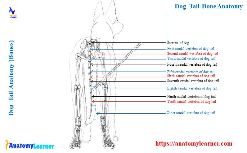
Dog tail bone anatomy
You know the bones of the dog tail anatomy. These are the caudal vertebrae that are also referred to as the coccygeal vertebrae.
They possess a great variation in their structure compared to that of other regions’ vertebrae.
The first few caudal vertebrae of the dog tail anatomy consist of the typical osteological features. But, the most caudal ones are gradually reduced to simple cylindrical rods by losing their characteristic features. You will find the major changes in the arches and processes of the caudal vertebrae of a dog tail.
Again, the major variants are found in the spinous and transverse processes of the caudal vertebrae of the dog tail. There are also mammillary processes, hemal processes, and hemal arches present in the bone of a dog tail.
The hemal process protects the median coccygeal artery of the dog tail. Again, the heaml process forms the hemal arch on certain caudal vertebrae of a dog tail.
How many bones are in a dogs tail
This is very important to know how many bones are in a dogs tail. You may find fifteen to twenty-five bones (15 – 25) in a dog tail. But, this number of bones are not fixed in dog tail. The average number of bones is about twenty in a dog tail.
You may also find a great variation in the number of bones in the dog tail in the different breeds. There may present only four to six (4 – 6) bones in the tail of some dog breed.
The cranial bones of the dog tail almost possess the characteristics of a typical vertebra. At the same time, the caudal bones are gradually reduced into simple rods.
The first caudal bone of the dog tail – body and arch
The first caudal bone of the dog tail anatomy represents most of the osteological features of a typical vertebra. It possesses a body, arch (pedicle and laminae), and the processes (articular, transverse, and spinous). The body of the first caudal vertebra of the dog tail is as wide as it is long. You will find a slightly convex cranial and caudal portion in the first caudal bone.
But, what are the features of other caudal bones of a dog tail? Well, the length of the body become increases up to the middle of the tail. Then the rest caudal bone becomes progressively shorter in their length.
But, the wide of the vertebral body gradually decrease in the dog tail. Again, the last bone of the dog tail is small and ends as a tapering process.
The vertebral arch is also well-defined in the first bone of the dog tail. A smaller vertebral canal is present in the first caudal bone enclosed by the arches.
The vertebral canal (lumen of the individual bone) becomes gradually smaller in the middle of the tail. Then, you will find a small groove that continues with the vertebral canal.
“You know the caudal vertebral canal of the dog tail contains the caudal spinal nerve. But, the spinal-cord usually ends at the joint between the last two lumbar vertebrae in dog.”
The processes of the first caudal vertebrae
You will find the cranial and caudal articular process in the first caudal bone of the dog tail. But, the articular function of the cranial articular process may be lost. Again, the caudal articular process projects from the arches’ caudal border and is frequently asymmetric.
The caudal articular process of the other bone of the dog tail become gradually disappears in a craniocaudal sequence. You will also get the mammillary process in each cranial articular process. When the cranial articular process vanishes in the caudal bone of the dog tail, the mammillary process will also have vanished.
The pedicle and the laminae of the first caudal bone are fused to form the small spinous process. It is thin cranially and thick caudally. The spinous process may disappear in the middle of the dog tail bone.
Again, you will find a well-developed transverse process in the first caudal bone of the dog tail. But, what happens in other caudal bones of a dog tail. The first few caudal bones may possess a well-developed and typical transverse process (first five or six).
But, they become reduced in size from the middle of the tail and disappear in the last few bones. You will also find two other structures – the hemal arch and hemal process in the caudal bone of the dog tail.
A few caudal bones show the paramedian process to protect the median caudal vessels at the ventral surface. These paramedian processes are known as the hemal processes. You will find this hemal process at the ventral surface of the fifth to tenth caudal vertebrae of a dog tail. Again, these hemal processes continue and form the hemal arches.
A second and third caudal bones of the dog tail
The osteological features of the dog tail’s second and third caudal bones are almost similar. You will find the typical osteological features in the dog tail’s second and third caudal bones. Though, there is some modification present in their structure.
The body of the dog tail’s second and third caudal bones becomes long compared to the first caudal bone. Again, the wide of these caudal bones decreases compare to that of the first caudal bone of the dog tail.
These two bones represent the cranial and caudal articular processes. Still, now, the articular processes have their articular functions. You will also find the mammillary process at the cranial articular process of these bones.
The transverse process of these caudal bones is still now well-developed and directed backward. Again, the spinous process of the second and third caudal bone decreases.
But, the lumen of these vertebrae (caudal spinal canal) become reduced compared to that of the first caudal vertebrae of the dog tail.
The fourth and fifth caudal bones of a dog tail
In the fourth caudal vertebrae of a dog, you will find a cylindrical body with a shorter arch. You will also find a narrow vertebral canal in the fourth caudal bone. The cranial and caudal articular processes become smaller than the third caudal bone. Again, the mammillary process is small and sometimes may disappear.
In addition, the spinous and transverse processes of the fourth caudal bone are shorter. What are the most characteristic features of the fourth caudal bone of the dog tail? Well, you will find the hemal process at the ventral surface of the fourth caudal vertebrae.
In the fifth caudal bone, you will find a long cylindrical body and a short arch. The lumen of the fifth caudal vertebra is narrower than the others. Again, the spinous and transverse processes are also short in the fifth caudal bone.
The mammillary process may be present or absent, but the ventral hemal process is distinguished.
Other caudal bones
From these caudal bones, you will not find any typical osteological features. These bones become a typical cylindrical shape. The processes are disappearing in the sixth and seventh caudal vertebrae.
You will find the most outstanding osteological feature at the ventral surface of these caudal bones. The ventral surface of each bone contains the hemal process that protects the median caudal artery.
The rest of the caudal bones of the dog tail also shows the typical cylindrical bodies without distinguished arches. It is also very hard to identify the articular, transverse, and spinous processes from the last caudal vertebrae of a dog tail.
You will find some short cylindrical bones at the end of the dog tail. Again, the last bone of the dog tail is very short and possesses a tapered end. You will not find any lumen (vertebral canal) on the last caudal bones of the dog tail.
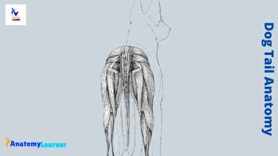
Dog tail muscle anatomy
The muscles enclose the caudal bones of a dog tail. You will find four different muscles in the dog tail muscle anatomy. Here, I will describe all the tail muscles from a dog tail with a labeled diagram.
First, let’s enlist the types of muscles in the dog tail. You will find the following muscles in a dog tail –
- Levator muscles of the dog tail – include medial dorsal sacrococcygeal muscle and lateral dorsal sacrococcygeal muscle.
- Depressors muscles of the dog tail – include medial ventral sacrococcygeal muscle and lateral ventral sacrococcygeal muscle.
- The tail’s lateral flexor muscle includes intertransverse dorsal, caudal muscle, and intertransverse ventral caudal muscle.
- Other muscles of the dog tail – include coccygeal muscle, iliocaudal muscle, and pubocaudal muscle.
Again, you will also find other muscles in the dog tail like – rectococcygeus muscle, sphincter ani extraneous, sphincter ani internus, and more. I will describe all these tail muscles with the labeled diagram.
The medial dorsal sacrococcygeal muscle is the dog tail’s short levator consisting of short, individual segments. On the other hand, the lateral dorsal sacrococcygeal muscle is the long levator of the tail. It is the direct continuation of the longissimus of the back on the tail.
The medial ventral sacrococcygeal muscle is the short depressor of the dog tail that covers a ventral aspect of the vertebral column. In contrast, the lateral ventral sacrococcygeal muscle is the long depressor of the tail that consists of numerous individual parts.
You will also find the intertransverse muscle that flexes the dog tail laterally. The coccygeus medial muscle of the dog tail consists of iliocadualis and pubocaudalis.
Medial dorsal sacrococcygeal muscle
This is the short levator muscle composed of relatively short, individual segments. And it is the direct continuation on the dog tail of the multifidus muscle. The medial dorsal sacrococcygeal muscle of the dog tail lies next to the median plane on the sacrum and caudal vertebrae. Again, it extends from the seventh lumbar to the last caudal vertebra.
The medial dorsal sacrococcygeal muscle segment runs between the spinous process of the cranial vertebrae and on the mammillary process. These muscles are composed of deep, short muscle masses and a large, superficial long mass. Again, there is a small tendon in the large, superficial muscle mass.
You may isolate the individual segment of the medial dorsal sacrococcygeal muscle at the root of a dog tail. At the tip of the dog tail, the muscle segment becomes small, short, and more homogenous.
The medial dorsal sacrococcygeal muscle helps in the extension of the tail and lateral flexion.
Lateral dorsal sacrococcygeal muscle of dog tail
This is the long levator muscle of the dog tail that is flat and segmental. The segmental muscle strand of the lateral dorsal sacrococcygeal becomes larger towards its dorsal border. Again, this muscle is the continuation of the longissimus muscle on the tail of a dog.
In addition, at the caudal part of the lumbar region, this muscle lies between the longissimus muscle and multifidus lumborum muscle. You will find a fleshy origin of the lateral dorsal sacrococcygeal muscle from the aponeurosis of the longissimus muscle.
On the other hand, you will also get a tendinous origin from the mamillary processes of the first to sixth lumbar vertebrae, sacrum, and last eight caudal vertebrae. Two distinct longitudinal divisions are present in the lateral dorsal sacrococcygeal muscle that partly cover one another.
The segments of the lateral dorsal sacrococcygeal muscle are arranged into the flat bundle by accumulating the successive tendons. They embed in the deep fascia of the tail and taper towards the tip of the tail.
Do you know the action of this muscle on dog tail? This muscle (lateral dorsal sacrococcygeal) helps in extension or lifting the tail. It also helps in the possible movement of the tail on its one side.
The medial ventral sacrococcygeal muscle of tail
The medial ventral sacrococcygeal or short depressor muscle of the dog tail is cord-like. It consists of a segmental, short individual portion that extends from the last sacral vertebrae to the last caudal vertebrae of the dog tail anatomy. Again, it lies against the caudal vertebrae’s ventral surface, with the opposite side’s muscle.
Here, it will form a deep furrow for the median coccygeal vessels (medial coccygeal artery). The muscle bundle is very large at the pelvic outlet, and the segmentation may become indistinct.
But, you may find the separated segments of this medial ventral sacrococcygeal muscle at the distal portion. All the fibers of each segment arise essentially from the ventral surface of the caudal vertebrae.
The action of the medial ventral sacrococcygeal muscle is to flex the dog tail. Again, it has a small lateral movement action of the tail.
Lateral ventral sacrococcygeal muscle of the dog tail anatomy
The lateral ventral sacrococcygeal muscle of the dog tail anatomy is the long depressor. It is the very large muscle in the dog tail. You will find numerous long, individual masses and long tendons in the structure of the lateral ventral sacrococcygeal muscle of the dog tail.
The first segment of the muscle arises from the ventral surface of the last lumbar vertebrae and the sacrum. In contrast, the remaining segments arise from the ventral surface and the root of the transverse process of the caudal vertebrae.
You will find the individual long tendon that arises from the different segments of the muscle. The individual long tendon embeds in the thick and deep fascia. Again, this long tendon attaches to the ventrolateral tubercle of the proximal end of the sixth caudal vertebrae.
The action of the lateral ventral sacrococcygeal muscle of the dog tail is the same as the action of the medial ventral sacrococcygeal muscle. So, it will also flex the dog tail. Again, it will show action on lateral movement of the dog tail.
Intertransversarius muscles of the dog tail
The intertransversarius muscle of the dog tail flexes the tail laterally. They are located on the lateral aspect of the caudal vertebrae between the dog tail’s long elevator and long depressor. Again, they occupy the space between the transverse process of the caudal vertebrae.
You will find two divisions in the intertranversarius muscle of the dog tail. The two divisions of the intertransversarius muscle are – dorsal intertransversarius muscle and the ventral intertransversarius muscle of the tail.
Now, I will show you some of the anatomical facts of the dorsal and ventral intertransversarius muscles from the dog tail.
Dorsal intertrnasversarius muscle of dog tail
This is the intertransversarius dorsal, caudal muscle of the dog tail. It lies between the sacrum and middle of the dog tail.
You will find the short, individual segments in the structure of the dorsal intertransversarius muscle of a dog tail. The first portion of the individual muscle is well developed.
The first portion of this muscle arises from the long and dorsal sacroiliac ligament. It also arises and on the lateral part of the third sacral vertebrae. It forms a large, round muscle belly that ends on the transverse process of the fifth caudal vertebrae.
The deep part of the dorsal intertransverse muscle lies on the dorsal surface of the transverse process and becomes gradually independent. These muscle segments become so small at the caudal half of the dog tail.
You will find a long, flat tendon on the superficial parts of the first large segment of the intertransverse dorsal muscle. It extends to the caudal fascia and the transverse process of the sixth caudal vertebrae of the dog tail.
This muscle possesses lateral flexion action of the dog tail. But, this action will combine the intertansversarius dorsal and ventral muscle of the dog tail.
Intertransversarius ventrales caudae muscle
This is the second part of the intertransversarius muscles that lies ventral to the transverse process of the dog tail bones. It also consist of different individual segments, which contain a round belly. The intertransversarius ventralis caudal muscle is smaller than that of the dorsal caudal muscle of the dog tail.
Moreover, you will find a more constant size and well-developed segments compared to that of the intertransverse dorsal muscle of the dog tail. This muscle covers by the long tendon of the long depressor of the dog tail ventrally.
Again, the action of the intertransverse ventral muscle is similar to the dorsal caudal muscle. So, it will help in flexing the dog tail.
The coccygeus muscle of the dog tail anatomy
You will find the two types of coccygeus muscles in the dog tail anatomy. The lateral coccygeus muscle is thick and extends to the dog tail’s lateral surface. Again, the medial coccygeus muscle is a broad triangle muscle that inserts between the levators and depressor of the dog tail.
The lateral coccygeus muscle of the dog tail arises from the narrow tendon on the ischiatic spine cranial to the internal obturator muscle. It crosses the medial aspect of the sacrotuberous ligament and runs on the lateral surface of the dog tail.
The medial coccygeus muscle lies medial and cranial to the lateral coccygeus muscle of the tail. This muscle originates from the medial edge of the shaft of the ilium, on the dorsal surface of the ramus of the dog pubis, and the entire pelvic symphysis.
You will find two divisions of the medial coccygeus muscle based on their origin. The division of the medial coccygeus muscle of the dog tail is – iliocaudalis and pubocaudalis muscles.
The iliocaudalis muscle of the dog tail constitutes the iliac portion of the levator muscle of the caudal region. This muscle originates from the medial aspect of the ilial shaft. The pubocaudalis muscle of the dog tail constitutes the pubic portion of the levator muscle and originates from the pelvic floor along with the pubic symphysis.
The fascia of the dog tail
You will find two types of fascia in the dog tail. The superficial fascia of the dog tail is indistinct. But, the deep fascia is thick and provides thick connective tissue masses for special attachment of the long tendon of the sacrococcygeal dorsalis lateralis and sacrococcygeal ventralis lateralis muscles.
Median caudal artery of the dog
In the dog tail anatomy, you will also find a major structure: the median caudal artery. The medial caudal artery of the dog tail continues the median sacral artery at the first caudal vertebrae. This artery lies between the right and left median ventral sacrocaudal muscles.
It passes through the fourth, fifth, and sixth hemal arches and then between the successive hemal process of the dog tail. You may find some of the segmental arteries that arise from the opposite to the bodies of caudal vertebrae. The segmental arteries of the dog tail run caudolaterally to the vertebrae.
You will find only two caudal arteries and the median artery in the dog tail that reaches the tail’s end. These arteries may anastomose with each other at the end of the tail. Again, you may find the paired ventral arteries on the structure of a dog tail.
The nerve of the dog tail
The dog’s spinal cord becomes tappers at its distal end and forms the cauda equine. Most of the cauda equina lies caudal to the lumbar vertebrae in a dog. You will find some filament at the distal end of the cauda equina of the dog. This structure is known as the filum terminale.
These filum terminale extends to the caudal vertebrae from cauda equina. You may find the filum terminale up to the dog tail’s fifth or sixth caudal vertebrae.
Types of dog tail
There are different types of dog tails found in different dog breeds. Here, I will not provide a detailed description of the types of dog tails. Rather, I will provide only the different tail types from different dog breeds.
You may find the bobbed, docked, curly, snap, sickle, otter, whip, and saber tail types in the different dog breeds. The bobbed tail is a very short tail found in some dog breeds. Again, the curly tail is short but possesses a single ring at the end. The sickle-shaped tail possesses a lower curve at its end part.
You may know the details of the different tail types from the different dog breeds.
Frequently asked questions on dog tail anatomy.
In this part of the article, I will try to solve the common inquires on the dog tail anatomy, including muscles and bones. All these questions were collected from the anatomy learners who love to learn the dog tail structure.
If you think these questions are not enough for you, please let me know the question related to dog tail. Again, if you need a dog tail bone or muscles diagram, you may join anatomy learner on social media (get more labeled pictures on dog tail).
Is a dog’s tail bone or cartilage?
The dog tail is formed by the bones, muscle, cartilage, ligaments, blood vessels, and filum terminale. The base of the dog tail is forming with the bones. These are the caudal vertebrae bone that varies with the different breed of the dog.
But, typically, you may find more than fifteen caudal vertebrae (bones) in the dog tail structure. Again, I showed the different types of muscles from the dog tail anatomy in this article. If you wish to know these muscles, please read the article from the beginning.
What is the anatomy of a dog’s tail?
If you want to know the anatomical facts of the dog tail, you should know the numbers of bones, types of muscle, different arteries and veins, and the nerve fibers. There are fifteen to twenty-five (depending on the breed or individual) present in the dog tail. The first few caudal vertebrae of the dog tail possess the typical osteological features.
But, the bones at the last portion of the dog tail lack the typical osteological features of a vertebra. Most of the caudal bone of the dog tail becomes cylindrical rod-shaped.
The major muscles of the dog tail are divided into the levator, depressor, and lateral group. The levator group will find the lateral dorsal sacrococcygeal and medial dorsal sacrococcygeal muscles. Again, in the depressor group, you will also find the medial ventral and lateral ventral sacrococcygeal muscles.
Do dogs feel pain in their tails?
Is a dog’s tail an extension of its spine?
If you are interested, you may read the following suggested article from anatomy learner –
- The anatomical features of dog leg with the labeled diagram
You may also find more articles on dog anatomical facts in the anatomy learners’ dog and cat anatomy learning section.
Conclusion
I hope you learn the basic idea of the dog tail anatomy. The most important anatomical facts of the dog tails include the characteristics of the caudal vertebrae bone, different types of muscles, nerves, and blood vessels. The most important blood vessel of the dog’s tail is the median sacral artery.
You know, this vein of the dog tail has great clinical importance. Again, the medial sacral artery of the dog tail locates in between the levator and depressor muscles. So, every structure of the dog tail anatomy is important to know as a veterinary student.