The dog skeleton anatomy consists of bones, cartilages, and ligaments. You will find two different parts of the dog skeleton – axial and appendicular. Here, I will show you all the bones from the axial and appendicular skeleton with their special osteological features.
Again, I will provide more labeled diagrams for each dog skeleton bone. This article will provide a clear conception of the dog paw and foot skeleton anatomy. In addition, I will try to solve the common inquiries on the dog bone anatomy at the end of the article.
So, if you are interested to learn the basics of the dog skeleton bones and differentiate them from the other skeletons like goat or horse, you may continue the article till the end.
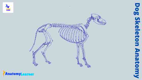
Dog skeleton anatomy
The bones of the dog skeleton anatomy serve to support and protect the visceral organs. Again, all the bones of the dog skeleton provide lavers for muscular action. You will find a total of three hundred and twenty-one (321) bones in the dog skeleton.
The axial skeleton contains one hundred and thirty-four (134) bones. On the other hand, you will find one hundred and eighty-six (186) bones in the appendicular skeleton. In addition, you will also find one heterotropic bone in the dog skeletal system.
You know there are different types of bone-like – long, short, flat, sesamoid, irregular, and pneumatic bone present in the animal skeleton. In the dog skeletal system, you will also find these different types of bone.
I have already described the different types of bone from the animal skeletal system previously. If you wish to memorize these bones, you may read the full article from anatomy learner.
Okay, what should you do now to start learning the anatomical features of the bones from the dog skeletal system? Well, first, you might know all the names of the bones and identify them from the dog skeleton labeled diagram.
Fine, let’s identify the different segments and bones from the dog skeletal system –
- The segments and bones of the thoracic limb – scapula, humerus, radius – ulna, carpal, metacarpal, and phalanx
- The segments and bones of the pelvic limb – hip, femur, tibia -fibula, tarsal, and metatarsal
- Identify single and paired bones from the dog skull
- The bones of the dog vertebrae, sternum, and ribs
I hope the dog skeleton labeled diagram might help you quickly identify all the bones. Now, move to the next part of the article to know the anatomical facts of bones.
Bones of dog thoracic limb
You know the appendicular skeleton of a dog consists of thoracic and pelvic limbs bones. Again, the axial skeleton of a dog consists of skull bones, vertebrae, sternum, and ribs.
You will find thoracic girdle, arm (brachium), forearm (antebrchium), and forepaw (manus) segments in each thoracic limb of a dog. The thoracic girdle of the dog forms by the scapula and clavicle. Again, the arm of a dog represents the humerus bone.
The forearm or antebrachium of a dog consists of radius and ulna bone. In addition, you will find the carpals, metacarpals, phalanges of the digits, and palmar sesamoid bones in the forepaw or manus of a dog.
Clavicle of dog
The clavicle of a dog is a small, thin, irregular triangular bony of the cartilaginous plate. You will not find any direct articulation of the clavicle to the dog skeleton.
It locates at the tendinous insertion of the brachiocephalic muscle. Again, the medial end of the clavicle attaches to the sternal fascia by a distinct ligamentous band.
You will find a slightly concave area in both longitudinally and transversely of the dog clavicle. Again, the dog clavicle is closer to the clavicular tendon between the cleidocephalicus and cleidobrachialis muscles.
Dog scapula anatomy
The anatomy of the dog scapula is somewhat different from that of the ruminant scapula. First, you may know the details of an idea scapula of an animal (example – cow or goat scapula). Then it will be better to compare with the dog scapula anatomy.
Fine, let’s enlist some important osteological features of a scapula from dog skeleton anatomy. But, make sure you have a good piece of knowledge on the ideal structure of animal scapula. If you want, you may read this article (cow scapula) to get a basic idea of the structures of the animal scapula.
These are the essential osteological features of dog scapula that differs from other animal’s scapula –
- The spine of the dog scapula locates in the middle and divides the lateral surface into two halves.
- You will find blunt tuber scapulae.
- There is no coracoid process in the dog scapula
- You will find a short and blunt acromion process that extends to the level of the glenoid cavity.
- There is a concave subscapular cavity that contains few rough lines
That’s fine; let’s know the details of dog scapula anatomy with the labeled diagram.
Structure of dog scapula
The dog scapula is the large, flat bone of the shoulder joint. You will find two surfaces, three borders, and three angles in a dog scapula.
The lateral surface of the dog scapula divides into two halves by the spine. This spine of the dog scapula is the most prominent osteological feature. At the distal end of the dog, the scapular spine possesses a pointed structure (acromion process).
Again, the spine of the dog scapula forms the supraspinous and infraspinous fossa at the lateral surface. The supraspinous fossa is the widest in the middle of a dog. In addition, the infraspinous fossa of the dog scapula is triangular in shape and well defined.
The caudal surface of the dog scapula possesses a triangular area (facies serrate) dorsally and a subscapular fossa ventrally. It is nearly flat and usually presents three relatively straight muscular lines.
The cranial border of the dog scapula is thin, strongly convex, and possesses a scapular notch distally (concave structure). A dorsal border is present in the dog scapula that extends between the cranial and caudal angles. Here in this dorsal border, you will find the scapular cartilage.
The caudal border of the dog scapula is the thickest of the three borders. This caudal border possesses the infraglenoid tubercle.
The caudal angle forms by the thick caudal border with a thinner convex dorsal border. Again, the cranial and dorsal border forms the cranial angle of the dog scapula. The ventral angle is known as the lateral or glenoid angle. This is the expanded part of the distal part of a dog scapula.
Here in the glenoid angle, you will find a very shallow glenoid cavity. Again, other important structures like the supraglenoid tubercle and coracoid process are present in the distal end of the dog scapula.
Dog humerus anatomy
The humerus is a relatively very long, slender, and slight spiral twist bone in the arm of a dog. Anatomically, you will find the head, neck, body, and condyle in the dog humerus anatomy.
To make it simple, you may first learn the basic anatomy of an animal humerus bone. Then you may compare the osteological features of the dog humerus anatomy with another animal humerus.
Let’s enlist some important osteological features of the dog humerus bone that make it exceptional from another animal humerus.
- The dog humerus bone is comparatively more prolonged and less twisted (compared to the cow humerus)
- You will find a more rounded and convex head in a dog humerus
- The musculospiral groove of the dog humerus bone is not so prominent
- You will find the deltoid tuberosity is in the form of a ridge
- There is an exceptional osteological feature present at the distal end of the dog humerus (supratrochlear foramen), where the radial and olecranon fossa is communicated
So, the above-mentioned osteological features are more important to know while studying the anatomy of the dog humerus bone. Now, you will know the detailed anatomical facts of the dog humerus with the labeled diagram.
Details anatomical features of dog humerus
The dog humerus proximally articulates with the scapula and distally joins with the radius – ulna bones. I will show you the different osteological features from the dog humerus’s head, neck, body, and condyles.
The head of the dog humerus is oval and located at the proximal extremity. You will also find some other structures like – intertubercular groove, greater tubercle, and lesser tubercle in the proximal extremity of the dog humerus.
The articular surface of the head continues distally by the intertubercular groove. Again, the greater tubercle is the large cranio-lateral projection of the proximal extremity of the dog humerus. You will find a flattened lesser tubercle at the proximal medial part of the humerus.
The neck of the dog humerus is distinct, only caudally and laterally. Again, the body is long, laterally compressed and possesses four surfaces.
Surfaces of dog humerus body
The lateral surface of the humeral body is marked by the tricipital line (anconeal line). This tricipital line start at the head of the dog humerus caudal to greater tubercle ends to the elongated deltoid tuberosity. The musculospiral groove is smooth, flat to convex, and located at the lateral surface of the dog humerus.
The medial surface is rounded and bounded by the crest of the lesser tubercle caudally. Again, it ends distally in an inconspicuous eminence and the tuberosity for the teres major.
The cranial surface of the dog humerus body is narrow and begins proximally at the crest of the greater tubercle. In addition, the caudal surface is smooth and begins at the neck of the humerus.
The caudal surface extends distally and continues with the lateral supracondylar crest. You will find some distally directed nutrient foramen at the caudal border of the dog humerus.
Condyles of dog humerus
The humeral condyle is the rounded structure at the distal end of the dog humerus. You will find different critical osteological features at the humeral condyle of a dog.
The condyle of the dog humerus contains large articular surface (capitulum), trochlea, olecranon fossa, radial fossa, supratrochlear foramen, lateral epicondyle, and medial epicondyle.
The olecranon fossa is a deep excavation of the caudal part of the dog humeral condyle. Opposite to the olecranon fossa, you will find the radial fossa on the cranial surface of the condyle.
Again, the radial and olecranon fossae communicate through the supratrochlear foramen. You will find two epicondyles (lateral and medial) at the distal end of the dog humerus.
The lateral epicondyle lies atcaudoproximal to the lateral articular margin of the capitulum. On the other hand, the medial epicondyle is a prominence on the medial aspect of the condyle just proximal to the medial border of the articular surface of the trochlea.
Radius and ulna bone of dog skeleton
The radius is the main weight-bearing bone of the forearm of the dog. It is smaller than the ulna bone of a dog.
These radius and ulna bones are completely separated in the dog skeleton anatomy. I will show you the osteological features of these two bones of the dog’s forearm separately.
But, let’s know some exceptional features of a dog skeleton’s radius and ulan bones that make them different from ruminant.
- The radius and ulna are two separate bones and are in contact with each other by their ends
- You will find a narrow interosseous space that extends throughout the length of the bones
- There are pair of small tubercles present at the cranial part of the olecranon process of the dog ulna bone
- The proximal end of the dog radius presents only one articular facet for the lateral condyle of the humerus.
- Again, the medial condyle of the dog humerus articulates with a facet on the semilunar the notch of the dog ulna.
These are short but essential osteological features from the dog radius and ulna bones. Now, you may learn more about the anatomy of radius ulna bone from dog. Or, if you want, you may learn the basic anatomical features of radius and ulan bones from a cow with more images and video.
Dog radius bone
The radius bone of a dog articulate proximally with the humerus and form the elbow joint. You will find a head, neck (proximally), body, and trochlea (distally).
The head of the dog radius bone is irregularly oval in outline. You will find a concave articular fovea that articulates with the capitulum of the humerus. The articular circumference of the head is caudal, smooth, that articulate with the radial notch of the ulna bone.
The neck is the constricted part of the radius bone that articulates the head to the body. You will find a small radial tuberosity at the distal end of the neck on its medial aspect.
The body of the dog radius bone is compressed and present two surfaces and two borders. You will find a convex cranial surface in the dog radius bone.
Again, the caudal surface divides into two flat to concave areas by a verticle interosseous border. The medial and lateral border of the dog radius bone do not possess any specific osteological features. Both the lateral and medial borders of the dog radius bone are smooth and rounded side to side.
The distal extremity of the dog radius possesses the trochlea (most massive part of the bone). You will find a small concave area at the trochlear’s lateral surface that helps form the ulnar notch. Medially, you will find a wedge-shaped projection (styloid process) that extends distally to the main carpal articular surface.
Dog ulna bone anatomy
The ulna of a dog is well developed but diminishes in size distally. You will find two extremities and a body in the dog ulna bone. The proximal extremity is the olecranon process, and the distal extremity is the styloid process.
The body of the dog ulna bone is three-sided and laterally compressed. Again, the proximal end of the body is grooved cranially and enlarged and rounded caudally.
The proximal extremity of the dog ulna includes the olecranon process. It articulates with the humerus and radius bone of the dog.
You will find some essential osteological features in the olecranon process of the ulna. This includes – olecranon tuber, the anconeal process, and the proximal part of the trochlear notch.
The trochlear notch is a smooth, verticle, half-moon-shaped concavity that faces cranially. A sharp-edge, slightly hooked structure (anconeal process) is present at the proximal end of the trochlear notch.
Forepaw of dog skeleton anatomy (manus)
The forepaw of the dog skeleton anatomy consists of carpals, metacarpals, phalanges bones of the digits, and some palmar sesamoid bones. Before going to the details, let’s know the summary of the forepaw skeleton of a dog.
There are seven bones present in the carpus of a dog. These seven carpal bones of the dog leg arrange into two rows (three at the proximal row and four at the distal row).
The bones of the proximal row are radial and intermediate fused carpal, ulnar carpal, and accessory carpal bone. Again, the bones of the distal row are the first carpal, second carpal, third carpal, and fourth carpal bone.
You will find five metacarpal bones in the dog forepaw anatomy. The first metacarpal is the shortest. The third and fourth metacarpal bones of the dog are the largest. Again, the dog’s second and fifth metacarpal bones are almost equal in length. The body of the dog metacarpal is craniocaudal compressed.
There are four developed and one underdeveloped digit present in the forepaw of the dog skeleton. The first digit of the dog is underdeveloped and contains only two phalanges. Again, the other four digits possess three phalanges each. You will find the maximum length in the third and fourth digits of the dog forepaw.
The first phalanx of the dog paw contains four surfaces. The second phalanx is short and concave on its proximal end. Again, the third phalanx resembles the shape of a claw.
The proximal group contains nine caudal and five cranial sesamoid bones. Again, the distal sesamoid bone of the dog forepaw is cartilaginous.
Carpal bones of dog
You know there are two rows of carpal bones in the dog forepaw. Let’s discuss the anatomy of the dog’s proximal row of carpal bone.
The ulnar carpal is the lateral bone of the proximal carpal. It is smaller than the intermediate carpal bone of the dog. This ulnar carpal articulates with the ulna and radius bone proximally and distally with the fourth and fifth metacarpal bones.
The intermediate carpal bone locates on the medial aspect of the proximal row. This is the largest carpal of the forepaw of a dog.
It represents a fusion of primitive radial carpal bone with the central and intermediate carpal bones.
You will find a sizeable articular area at the proximal surface of the dog intermediate carpal bone. Again, the distal surface of the intermediate carpal articulates with all four distal carpal bones.
The accessory carpal bone locates at the palmar side of the ulnar carpal. You will find a slight saddle-shaped articular surface for the ulnar carpal at the base of the accessory carpal. Again, the free end of the accessory carpal is thickened and overhangs slightly.
The first carpal of the dog forepaw is the smallest and somewhat flattened. It articulates proximally with the intermediate carpal and distally with the first metacarpal bone. The second carpal bone is small, wedge-shaped, and compressed distally.
Again, the third carpal bone is larger than the second carpal and possesses a sizeable palmar projection. This palmar projection is articulate with the three middle metacarpal bones. The most prominent carpal bone of the distal row is the fourth one. It is wedge-shaped and presents a sizeable caudal enlargement.
Metacarpal bones of a dog
There are typically five metacarpal bones present in the forepaw of a dog. Each metacarpal bones of the dog are cylindrical shaped and enlarged at both ends. You will find a base (proximal), body (middle), and head (distal) in a dog metacarpal bone.
The first metacarpal bone of the dog forepaw is small and more slender than that of the other metacarpals. Again, the metacarpal II, III, IV, and V are the main metacarpal that irregular rod shape with a uniform diameter.
The metacarpal II and V are shorter and four-sided than the metacarpal III and IV. There are sesamoid fossae present at the palmar aspect of the dog metacarpal (between head and body). You will also find the sharp-edge sagittal crest at the palmar aspect of the dog metacarpal.
Phalanges of dog forepaw
You will find five digits in the forepaw of a dog skeleton. Each leading digit (except first) consists of a proximal phalanx, a middle phalanx, a distal phalanx, and two large palmar sesamoid bones.
There are two phalanges in the first digit and three phalanges present in the other four digits of the dog forepaw.
The first phalanx is medium length rod-shaped with enlarged extremities. You will find a concave articular surface at the proximal end.
The middle phalanx is rod-shaped and one-third shorter than the proximal phalanx of the dog forepaw. Each middle phalanges possesses a proximal base, medium body, and a distal head. The size of the distal phalanx in all digits is almost the same.
You will find an enlarged extremity at the proximal end of the distal phalanx. It possesses a shallow, concave articular area to contact the middle phalanx.
Sometimes, you will find a laterally compressed cone structure at the distal part of the distal phalanx of the dog. This is the ungula process that is shielded by a claw.
Bones of dog pelvic limb
The pelvic limb of the dog skeleton anatomy consists of the pelvic girdle, thigh, leg, and hind paw or pes. Again, the pelvic girdle of the dog comprises of ilium, ischium, pubis, and acetabular bone. The thigh of the dog anatomy represents the femur bone associated with patella sesamoid bones.
In addition, the leg of a dog consists of tibia and fibula bones. The hind paw or pes includes the tarsals, metatarsals, phalanges of the digits, and their associated sesamoid bones.
So, the segments of the dog pelvic limb with their bones are enlisted below –
- The pelvic girdle – ilium, ischium, pubis bone (os coxae or hip bone)
- A thigh of dog – femur bone
- The leg of a dog – tibia and fibular bones
- Again, the pes or hind paw – tarsals, metatarsals, phalanges, and sesamoid bones.
Now, I will show you all the bones from a dog’s different pelvic limb segments with their specific osteological features.
Hip bone anatomy from the dog skeleton
The hip bone of the dog skeleton consists of three well-defined bones (ilium, ischium, and pubis). You will find some structural differences in these hip bones of a dog skeleton compared to that of the cow hip. It will be better if you read the full article on the general osteological features of the cow hip bone. This will help you make the difference between a dog’s hip from ruminant.
Okay, first, let’s know some of the essential osteological features of the dog hip bones. Then we will learn the details of the ilium, ischium, and pubis bones in details.
- The ilium of the right and left sides are almost parallel to each other
- You will find a concave area in the gluteal surface of the ilium bone with an indistinct gluteal line
- The crest of the ilium bone is strongly convex
- Again, the ischium of the dog hip is twisted compared to the cow ischium
- The ischial tuberosity of the dog hip is flat
- Both the superior and inferior ischiatic spine are blunt in dog hip bone
- You will find a broad notch in the acetabular cavity of the dog hip
I hope you will find all the above osteological features from the dog hip bone diagram. Let’s know the detailed anatomical features of all hip bones from a dog.
Dog ilium bone
The dog ilium is the most significant and cranial part of the hip bone. You will find the laterally concave wing and a narrow, more irregular body. The body of the dog ilium bone is expanded on its caudal end.
First, you should identify some of the essential osteological features from the dog ilium bone. Here, I will enlist these structures from the dog ilium bone.
- The tuber coxae and tuber sacrale (iliac crest)
- A caudal and cranial dorsal iliac spine
- The cranial and caudal ventral iliac spines
- The concave greater ischiatic and lesser ischiatic notches
- A gluteal, sacropelvic, and articular surface of the dog ilium bone
- The iliac tuberosity and an arcuate line
I think you will identify all these structures so quickly from the dog ilium bone labeled diagram. The iliac crest consists of the tuber coxae and tuber sacrale. These two structures form the cranial border of the ilium.
The gluteal surface of the iliac wing is concave and faces dorsolaterally. Again, the sacropelvic surface (medial surface of the wing) articulates with the wing of the sacrum. You will find the arcuate line in the ventromedial border of the ilium bone. That extends from the auricular surface to the iliopubic eminence.
Dog ischium anatomy
The ischium bone of the dog forms the caudal third of the hip bone. This bone helps to form the acetabulum, obturator foramen, and pelvic symphysis.
The most important osteological features of the dog ischium bone are the body, arch, tuber ischii, and tuberosity.
Fine, let’s know the structures that you might identify from the dog ischium bone. Okay, let’s enlist the important osteological features from the dog ischium bone.
- The body of the ischium
- A greater and lesser ischiatic spine and notch
- The ischiatic symphysis
- A deep ischial arch and ischiatic tuberosity
The body of the ischium is the cranial part of the bone that lies lateral to the obturator foramen. You will find a thick dorsal border that continues with the ilium bone and forms a slightly concave ischiatic spine.
The medial border of the ischium forms the ischiatic symphysis with the opposite medial border of the ischium. Again, the caudomedial border of the ischium bone forms the deep ischial arch. You will find an ischiatic tuberosity at the caudolateral aspect of the ischial arch.
Dog pubis bone
The pubis of the dog hip bone is dorsoventrally compressed. It extends from the ilium and ischium laterally to the pubic symphysis medially. Again, the caudal border of the pubic bone is bounded by the cranial border of the obturator foramen.
You will find two-part in the dog pubis bone – the body and two rami. The body of the dog pubis bone is flat triangular that helps form the acetabular cavity and obturator foramen.
Again, the cranial ramus fuse with the ilium bone and help form the acetabulum. On the other hand, the caudal ramus forms the medial border of the obturator foramen.
You will find the iliopubic eminence on the cranial border of the cranial ramus of the dog pubis bone. The iliopubic eminence serves as the attachment for the prepubic tendon.
The two-part of the dog pubis bones join ventrally at the pubic symphysis. You will find the ventral pubic tubercle on the cranioventral surface of the pubis adjacent to the pubic symphysis.
The femur of the dog skeleton anatomy
The femur is the longer and heavier bone in the dog skeleton anatomy. It articulates with the hip bone proximally and with the tibia bone distally. For description, you may divide the dog femur into proximal extremity, a body, and the distal extremity.
The proximal extremity of the dog femur consists of a head, neck, and two trochanters (greater and lesser). Again, the body of the dog femur is long and possesses some interesting osteological features. In addition, the distal extremity of the femur is quadrangular and consists of condyles (lateral and medial) and the trochlea (cranially).
Let’s know some peculiar osteological features of the dog femur bone. Here, I will enlist some of the specific osteological features of the dog femur bone.
- The greater trochanter is at a lower level than that of the head
- You will find a tuberculous lesser trochanter in the dog femur
- The trochanteric ridges are of equal shape and size
- There is no third trochanter in the dog femur
- You will not find any defined supracondyloid fossa in the dog femur
- There is a facet (for sesamoid bone) present at the postero-lateral aspect of the distal end of each femur of the dog
These are very common and exception osteological features of the dog femur anatomy. I hope you will get all the osteological characteristics from the dog femur labeled diagram.
The proximal extremity of dog femur
I think you already know the main structures that find in the proximal extremity of the dog femur bone. Here, I would like to enlist the structures from the proximal extremity of the dog femur that you should practically identify.
So, the structures are –
- Smooth and spherical head with small fovea capitis femoris
- Constricted smaller neck
- Larger or greater trochanter (caudolaterally) and lesser trochanter (caudally)
- The trochanteric fossa and intertrochanteric crest
You will find a smooth and spherical head at the medial part of the dog femur. Again, a small, indistinct circular pit (fovea capitis femoris) is present in the medial part of the femoral head.
The neck is a constricted part below the head, and it connects the head with the other part of the proximal extremity. You will find a prominent tubercle (known as the greater trochanter) at the proximal extremity of the dog femur on its lateral aspect.
A trochanteric fossa is present between the femoral neck and the greater trochanter. Again, you will find a distinct, pyramidal-shaped eminence that projects from the caudomedial surface of the proximal extremity of the dog femur. This is the lesser trochanter and connects the greater trochanter by an intertrochanteric crest.
Body of the dog femur
The body of the dog femur is more prolonged and nearly cylindrical. You will find very ill-defined four surfaces (lateral, medial, cranial, and caudal).
The caudal surface of the dog femur is almost flat. You will not find any distinct third trochanter in the body of the dog femur bone.
Again, the caudal surface possesses some fine lines (rough surface). The distal part of the caudal surface is the popliteal surface of the femur. This surface contains two tuberosities – the lateral and medial supracondyloid tuberosities.
The distal extremity of the dog femur
The distal extremity of the dog femur is quadrangular and possess the three main articular surface. You will find an articular surface on the lateral and medial condyles. Again, an articular surface is present in the cranial part of the trochlea.
So, what are the structures that you need to identify from the distal extremity of the dog femur? Fine, let’s enlist the structures from the distal part of the dog femur bone.
- The lateral and medial condyle with their articular surfaces
- An intercondyloid fossa (deep)
- The lateral and medial epicondyles and
- A cranial trochlea with the articular surface for the patella bone
The lateral condyle is convex in both the sagittal and transverse planes. Again, the medial condyle of the dog femur is small and convex in both the sagittal and transverse planes.
You will find a deep fossa (intercondyloid fossa) between the dog femur’s lateral and medial condyles (caudally). In addition, the trochlea of the femur possesses a cranial smooth, articular groove or surface for the patella.
The patella is the large sesamoid bone in the dog skeleton. The dog patella possesses a base, an apex, and articular surfaces like the patella of a cow or goat.
Tibia and fibula bones of dog skeleton
The tibia is a long, thick bone and part of the dog leg. It articulates with the femur proximally and tarsus bones distally. Again, the tibia articulates with the fibula bone both proximally and distally.
If you want to learn the anatomical facts of the tibia and fibula bone in detail, you may read the previous article from the anatomy learner. You will find more tibia bone labeled diagrams and videos in that article.
Okay, the tibia bone of a dog skeleton also possesses the proximal extremity, a long body, and distal extremity. You might learn and identify all the structures from the dog tibia bone’s proximal extremity, body, and distal extremity.
Before that, let’s know some special osteological features of the dog tibia and fibula bones –
- The shaft of the dog tibia along the length is convex medially at the upper part and again convex laterally at the lower part.
- You will find a very prominent tibial crest in the dog tibia bone
- The upper part of the body is prismatic, and the lower part is cylindrical. Again, the cylindrical part possesses facets laterally for articulation with the fibula bone
- The fibula is a long and thin bone that extends the whole length of the dog tibia
- You will find a proximal flat end that articulates with the lateral condyle of the tibia
- The distal end of the dog fibula is thick
These are concise information on the anatomical facts of dog tibia and fibula bones. Let’s head to the next part of the tibia and fibula anatomy.
Tibia of a dog
The dog tibia is long, thick bone and the same length as the femur. It divides into the proximal extremity, a long, double curve body, and the distal extremity. The proximal extremity of the dog tibia is relatively flat and triangular.
You will find two condyles (lateral and medial) at the proximal end of the dog tibia that provides a proximal articular surface. Again, you will also find an intercondylar eminence in between the condyles for attachment of the ligament.
There are also lateral and medial intercondylar tubercles on the intercondylar eminence of the dog tibia. The popliteal notch locates caudally in between the lateral and medial condyles. A prominent tibia tuberosity is present in the proximal end of the dog tibia bone.
The body of the dog tibia is triangular and possesses three surfaces and three borders. You will find a smooth, comprehensive, and concave lateral surface. Again, the medial surface is also comprehensive and nearly flat proximally. There is some popliteal line present at the caudal surface of the dog tibia.
The cranial border originates from the tibial tuberosity and extends to form the tibial crest (prominent). Again, the lateral border of the tibia is flat and closely attaches to the fibula bone.
The distal end of the dog tibia is quadrangular and more massive. You will find medial and lateral malleolus at the distal end of the tibia bone anatomy.
Dog fibula anatomy
The dog fibula is a thin, slender and somewhat twisted bone of the dog skeleton anatomy. You will find the enlarged part at both ends of the dog fibula. The proximal part of the body separates from the tibia by a considerable interosseous space. But, the distal part is flattened and closely applies to the tibia bone.
The proximal extremity of the dog fibula contains the head and neck. It is flattened transversely, being expanded beyond the plane through the border of both cranially and caudally. You will find a short neck on the distal end that bends with the body of the fibula.
The body of the dog tibia is slender and irregular. The proximal half of the fibula may be twisted. You will find the interosseous spaces and borders throughout the length of the fibula bone of a dog.
Again, the distal end of the dog fibula also contains the lateral malleolus. Medially, you will find the articular surface on the lateral malleolus of the fibula bone.
Hindpaw or pes of dog skeleton anatomy
The hind paw of the dog skeleton consists of the tarsus, metatarsus, phalanges, and associate sesamoid bones. There are seven tarsal bones present in the dog hind paw anatomy. These are tibial tarsal, fibular tarsal, central tarsal, first tarsal, second, third, and fourth.
You will find five metatarsal bones in the dog skeleton. The first one is ill-developed and is in the form of a flat cone. Again, the rest metatarsal bones of the dog hind paw are comparatively more prolonged than those of the metacarpal bones.
In addition, the phalanges and sesamoid bones of the dog hind paw are so similar to those of the forepaw. The first digits are usually absent in dogs. But, sometimes, you may find the first digit in some breed. This first digit of the hind paw is known as the dewclaw.
Tarsal bones of a dog
The tibial tarsal (talus) is the second-largest tarsus in the dog that consists of a body, neck, and head. The body forms the proximal half of the talus bone. Again, the head of the talus is transversely elongated at the distal extremity.
The calcaneus (fibular tarsal bone) is the largest and longest bone in the dog tarsus. You will find the central tarsus of the dog hind paw that lies in the medial part of the tarsus between the proximal and distal rows.
The first tarsal bone of a dog hind paw varies significantly in the different breeds. The second tarsal (smallest tarsus) articulates with the central tarsus proximally and the third tarsal distally. Again, the third tarsal bone of a dog is three times larger and two times longer than the second tarsal bone. In addition, the fourth tarsus is as long as the combined dimensions of the central and third tarsal.
The metatarsal bone of a dog
The metatarsal of the dog resembles the corresponding metacarpal bones. You will find an ill-developed metatarsal bone in the dog hind paw anatomy. The other metatarsal of the dog is similar in structure.
Each of the metatarsal bones of the dog hind paw possesses an irregular proximal base, a triangular body, and a distal head. You will also find the deep, transverse sesamoid fossae at the planter part of the metatarsal bones of the dog hind paw.
Vertebrae of the dog skeleton anatomy
The vertebrae of the dog skeleton anatomy consist of approximately fifty irregular bones. These vertebrae are part of the axial skeleton of the dog. The vertebrae of the dog skeleton arrange into five regions – cervical, thoracic, lumbar, sacral, and caudal or coccygeal.
“You know the axial skeleton of the dog consists of the bones of skull, vertebrae (five regions), sternum, and ribs. The osteological features of the axial skeleton are described previously in details. So, here, I will provide only the summary of osteological features of the bones from dog axial skeleton.”
A typical vertebra of a dog consists of a body, vertebral arch (left and right pedicle and laminae), and different processes. The processes of the typical vertebra are – transverse, spinous, articular, accessory, and mammillary. If you want, you may read the full osteological features of a typical vertebra of an animal from an anatomy learner.
The essential osteological features of the dog vertebrae are –
- You will find seven cervicals, thirteen thoracic, seven lumbar, three sacral, and eighteen to twenty-five caudal vertebrae in the vertebral dog column
- The foramen transversarium is present in the dog atlas at its posterior aspect
- The odontoid process is rod-shaped and pointed
- The spinous processes are comparatively short. They are broad ventrally and narrow dorsally in the lumbar region of the dog skeleton.
- You will find a short sacrum in a dog which consists of three bones
- The caudal vertebrae of the vertebral dog column are well-developed
So, these are some of the specific osteological features of the dog vertebrae. But, this information is not enough to learn the anatomy of the dog vertebrae.
Cervical vertebrae of dog skeleton
The cervical vertebrae of the vertebral dog column are also seven in number, whereas the first two vertebrae differ significantly in their structure. The first cervical vertebrae are atypical and possess ventral arch or body, wing, dorsal arch, cranial concave articular surface, ventral tubercle, and foramina on the wing.
The axis is the second cervical vertebrae of the vertebral dog column that contains the odontoid process. You will find a slight variation in the structure of the dog skeleton’s third, fourth, and fifth cervical vertebrae.
But, the dog’s sixth and seventh cervical vertebrae possess some specific osteological features that make them an exception from the other cervical bones. If you want to learn the details anatomical facts of the cervical vertebrae of the dog skeleton, you may read the previous article from the anatomy learner.
Dog thoracic vertebrae
There are thirteen thoracic vertebrae present in the vertebral dog column. The first nine thoracic vertebrae are almost similar in their structure, but the last four thoracic vertebrae possess some differences from each other.
The bodies of the thoracic vertebrae are wide and compressed dorsoventrally significantly at each end of the region. You will find the facets on the caudal surface of the thoracic vertebral body for the head of the ribs (except the last two vertebrae).
The transverse process of the dog thoracic vertebrae resembles those of the horse. You will find the mammillary processes on the transverse process of the dog thoracic vertebrae. There is also an accessory process present in the last three thoracic vertebrae of the dog.
The spinous processes of the first three thoracic vertebrae are equal in length. But, the length becomes gradually decreases in the dog thoracic vertebrae.
The lumbar vertebrae of a dog
The lumbar vertebrae of the dog are seven in number and possess a longer body than those of the thoracic vertebrae. They gradually increase in width throughout the series and length through the first five vertebrae.
The spinous process of the dog lumbar vertebrae is broad ventrally, narrower dorsally. You will find the highest and more massive spinous process in the mid lumbar region of the dog skeleton.
The transverse process of the dog lumbar vertebrae directs cranially and ventrally. Their length increases to the fifth and sixth lumbar vertebrae. They form no joint with each other or with the sacrum bone of the dog.
The accessory process is well-developed in the first three lumbar vertebrae of the dog. You will find an extensive cranial articular process compressed laterally and bears mammillary processes.
Dog sacrum anatomy
The dog sacrum is formed by the fusion of the three sacral vertebrae. Again, the dog sacrum vertebrae is a concise, comprehensive, and quadrangular structure.
The dog sacrum consists of a base, an apex, wings, and two dorsal and pelvic surfaces.
The base of the dog sacrum faces cranially and contains a wide sacral canal. Again, the caudal extremity of the dog sacrum is broad transversely. This caudal broad transverse extremity is the apex of the dog sacrum bone.
The wing of the dog sacrum is the enlarged lateral part that possesses articular surfaces. These articular surfaces of the sacrum wing articulate with the ilium bone.
The essential osteological features from the dorsal surface of the dog sacrum are – median sacral crest, dorsal sacral foramina, intermediate sacral crest, cranial, and caudal articular processes.
The dorsal surface of the dog sacrum bears two pairs of the dorsal sacral foramina. Again, the caudal small articular process articulates with the first caudal vertebrae of the dog skeleton. The pelvic surface of the dog sacrum represents the transverse line and pelvic sacral foramina.
Dog caudal vertebrae anatomy
The dog caudal vertebrae may vary from eighteen to twenty-five. These caudal vertebrae possess a significant variation in their structure than the other vertebrae of any region.
The body of the first caudal vertebrae is as wide as it is long. Gradually, the other caudal vertebrae increase in length up to the middle part. Then, it becomes progressively shorter.
The vertebral arch of the first caudal vertebrae of a dog skeleton is well-developed. It becomes progressively shorter until the sixth or seventh caudal vertebrae.
The cranial and caudal articular processes of the dog caudal vertebrae are present. You will find the mammillary processes in each of the dog caudal vertebrae. Again, the spinous process is small and disappears early in the series. In addition, the transverse processes are typical and well developed in the first four or five caudal vertebrae.
Dog skull, ribs, and sternum
It will be better if you read the dog skull anatomy, rib cage anatomy from the other article of anatomy learner. The dog skull, ribs, and sternum are also considered part of the axial skeleton. Here, I will provide a little information on the dog skull bones, ribs, and sternum.
There are two types of bones (cranial and facial) present in the dog skull. These bones of the dog skull are different from the ruminant skull bones. Let’s find some peculiar osteological features from the dog skull bones.
- The dog skull is oval elongated in shape but varies highly from breed to breed
- You will not find any horn in the dog skull.
- The basioccipital bone join with the bulla tympani
- The zygomatic process is highly curved.
- Parietal bone take part in the formation of the roof of the cranial cavity
- The interparietal bone fuse with the occipital before birth
- You will find an incomplete orbital rim in the dog skull
- There is an incomplete fusion between the two halves of the mandible
- The lateral part of the ramus of the dog mandible possess a masseteric fossa
So, these are the exceptional osteological features present in the dog skull. Again, you will find thirteen pairs of ribs in the dog (the first nine pairs are sternal, and the last four are asternal ribs). In addition, the sternum of a dog is the unpaired bone that forms the ventral boundary of the thorax.
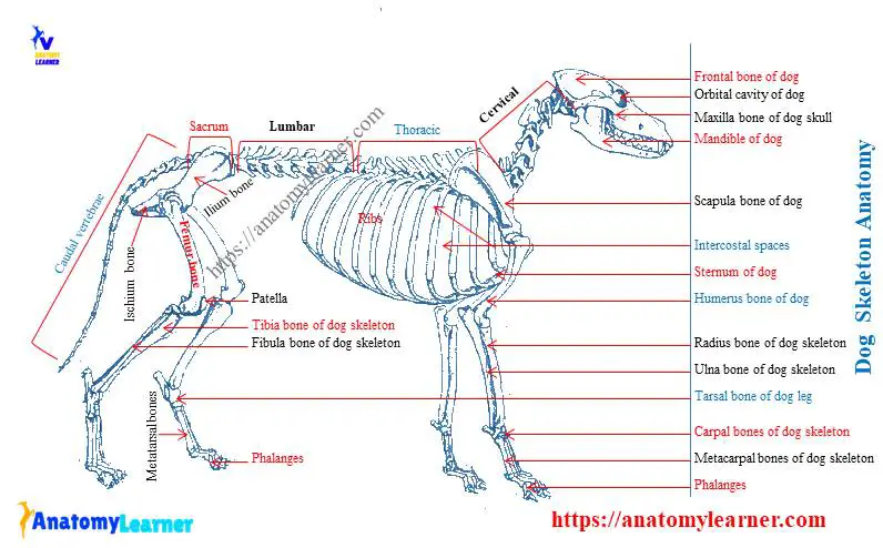
Dog skeleton anatomy labeled
You already got all the bones labeled diagram from the dog skeleton. Again, I would like to show you all the essential bones from the dog skeleton anatomy with the labeled diagram.
Here, in the dog skeleton labeled diagram, I tried to show you the different segments of the forelimb, hindlimb with their bones. Again, I tried to show you all the bones from the vertebrae column of a dog skeleton.
In addition, in the diagram, you will find a few identified skull bones. The sternum and the ribs are also identified in the dog skeleton labeled diagram. If you want to more updated dog skeleton labeled diagram, you may join anatomy learner on social media (get more images).
Frequently asked questions on dog skeleton bones.
So, again this part of the article, I will try to solve the common inquiries on the dog bone anatomy. Okay, let’s see the common questions on dog bone anatomy and their answers.
How many bones are present in the thoracic limb of the dog?
In the two thoracic limbs of a dog, you will find nearly ninety bones. Again, each thoracic limb of the dog consists of scapula, humerus, radius, ulna, carpal, metacarpals, phalanges, and sesamoid bones.
All the structures of the thoracic limb bones are described before. It will be better if you read the full article to get the basic idea of the thoracic limb of the dog.
What are thoracic limbs?
The forelimb of the animal is known as the thoracic limb.
What are the different parts of the segments of the canine thoracic limb?
The different parts of the segments of the canine thoracic limb are – shoulder girdle (thoracic girdle), arm or brachium, forearm or antebrichum, and forepaw or manus. You will find a detailed description of every single segment in the above section of this article.
Which part of the thoracic limb is also called the brachium?
The arm segment of the thoracic limb is also called the brachium. You will find the humerus bones in the brachium of the dog thoracic limb.
Conclusion
I hope you got the basic idea of every structure of the dog skeleton anatomy with the labeled diagram. But, it would be best if you learned the detailed anatomy of the bones from different segments of thoracic and pelvic limbs. Again, you might have a good piece of knowledge on the anatomy of the dog skull bones.
The vertebrae of the dog skeleton anatomy also possess some exceptional osteological features that a ruminant or horse. Now, you should try to identify all the bones from the dog skeleton from the actual samples of your anatomy laboratory. You may use the dog bones labeled diagram from the anatomy learner.