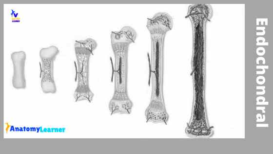
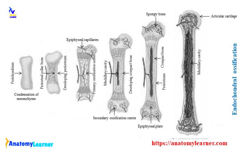
In endochondral ossification, bone is formed by replacing the calcified cartilage. In this article, I will discuss the detailed process of endochondral ossification with labeled diagrams.
Hi there, welcome back again, and many, many thanks for getting into this article. I hope this article is going to be one of the best and easiest articles on the internet to learn the whole endochondral ossification process with examples.
I tried to simplify every step of endochondral ossification so that you might understand easily. Here, you will know – when does endochondral ossification begins, how the primary center of ossification start, how the marrow cavity is formed, and much more.
So, if you are interested to learn the whole ossification process, then continue this article till the end.
Endochondral ossification definition
The process of bone formation in pre-existing cartilage models is known as endochondral ossification. In this ossification process, the calcified cartilage is replaced by the bone. This is a complex process to form a bone where you will find two ossification centers.
For your kind information, I have published the intramembranous ossification previously. If you don’t read that article, please go and know how intramembranous ossification occurs.
Endochondral ossification steps
First, I would like to summarize endochondral ossification – what happened in different steps or processes of this ossification. I will enlist the main changes in this type of ossification (for your better understand and memorization).
“Please try to understand and memorize the following points or important changes during intracartilagenious ossification process. These will help you to describe the whole process briefly.”
#1. Condensation of mesenchymal tissue and perichondrium formation.
#2. The proliferation of chondroblasts and maturation of this cell
#3. Formation of subperiosteal collar bone at the inner layer of perichondrium and conversion of perichondrium to the periosteum.
#4. Release of the organic matrix from chondroblasts and precipitate calcium salts at the matrix.
#5. Calcification causes the death of chondroblast and forms spaces (formation of primary areolae).
#6. Formation of periosteal bud and reaches the interior of the midsection of the cartilage model.
#7. Establishment of primary center of ossification.
#8. Osteoblast again synthesis and secrete osteoid and contributes to mineralization or calcification process (continue and form the bone trabeculae).
#9. Osteoclasts absorb the irregular mineralized structure and form large secondary areolae.
#10. These osteoblastic and osteoclastic activities help to develop the marrow cavity.
#11. Formation of Haversian system (also shaft or diaphysis formation)
#11. Formation of the secondary ossification center
#12. Epiphysis formation from the secondary ossification center
#13. Fusion of epiphysis and diaphysis.
I hope these points will help you to understand the whole process in a little. Let’s head to the entire process of Intra cartilaginous ossification.
Where endochondral ossification occurs?
Before going to detailed process of ossification, you might know the sites of endochondral ossification.
#1. The bone of the extremities (long bones of a skeleton)
#2. Bones from vertebrae column
#3. Pelvis bone of animals
#4. Bones that form the base of an animal’s skull
Above these bones, the hyaline cartilaginous model is developed initially and replaced by bone later.
Endochondral ossification process
In the endochondral ossification process, bone growth both in width and length. You will find the following steps of the process under this type of ossification.
#1. Condensation of mesenchyme and cartilage model formation
#2. Formation of primary center of ossification
#3. Secondary areolae and marrow cavity formation
#4. Formation of proper bone (compact bone – diaphysis or shaft of the bone)
#5. Secondary ossification center and epiphysis formation
#6. Fusion of epiphysis and diaphysis
“These processes are modified, I did so for your easy understand. You may follow any authorized book to learn this ossification process.”
Let’s start to know the details change on these mentioned processes.
Condensation of mesenchyme and cartilage model formation
This cartilaginous model (endochondral) of ossification begins with the condensation of mesenchyme tissue. The stellate-shaped mesenchymal cells become spherical and condense in the area where the long bone is to be formed.
Spherical mesenchymal cells become differentiate into chondroblasts and start laying cartilage matrix. Again, a perichondrium appears around the cartilage. Thus the mesenchymal model replaces by the hyaline cartilage model.
Now, let’s know how these changes occur? Fine, the chondroblasts or cartilage cells at the midsection of this model become proliferate by mitosis and arranges in rows towards the ends. They get mature and enlarge (hypertrophied), the intercellular matrix between the hypertrophied cells becomes thin.
At this time, the chondroblasts synthesize and secrets organic matrix, plasmalemma buds (known as matrix vesicles). Thus chondroblasts contribute to the calcification process within the cartilage matrix. The calcification prevents getting enough nutrition to these hypertrophied cells and results in degeneration and death of cells. This death of cartilage cells forms the spaces in the matrix that is known as primary areolae.
Again, the perichondrium is invaded by the numerous capillaries in this model. The chondrogenic layers changes to the osteogenic environment. The pro-osteoprogenitor cells of this layer differentiate into osteoblasts. These osteoblasts appear at the inner layer of the perichondrium and form a thin shell at the midsection of the cartilage model. This thin shell is known as the periosteal band or bony collar. Great, let’s go to the second step or process of endochondral ossification.
Formation of primary center of ossification
Now, you got the bony collar in the midsection of the cartilage model. Oxygen levels begin to rise due to invading numerous capillaries at the degenerating hypertrophied chondrocytes area. Thus the osteoprogenitor cells from the periosteum and undifferentiated mesenchymal cells.
The blood vessels and their associated cells form the periosteal buds, and it reaches the interior portion of the midsection of cartilage model. When it comes to the midsection of the cartilage model, the primary center of ossification is formed.
The osteoprogenitor cells differentiate into the osteoblasts under the influences of inductive bone-forming factors in the blood plasma. These cells remain in a cluster around the calcified cartilage and begin to synthesize osteoid.
Again, these cells also contribute to the calcification process. Their osteoblastic activities remain to continue until the bone trabeculae are formed.
Secondary areolae and marrow cavity formation
After forming the primary center of ossification, the cartilage model increases at each end by interstitial growth.
The capillaries form the primary center continue to invade this model towards both the epiphyses. Here, they initiate endochondral ossification. Chondroblast undergoes the repeated changes, and the bony collar increase in thickness.
The osteoclast starts to absorb the irregular calcified mass and forms the large secondary areolae. This secondary areola helps to develop the marrow cavity that becomes fills with the hemopoietic tissue.
Formation of true bone
In this step of endochondral ossification, the actual bone (compact) is formed. The osteoblasts lay down the lamellated bone in the matrix. Several longitudinal grooves appear on the outer surface of the circumferential bony lamellae, and the ridge of each groove proliferate.
The groove converts into the tunnels, and the osteoblast of the tunnel proliferates and differentiates into osteocytes. Thus the tunnel converts into the Haversian system, and compact bone is formed (diaphysis of bone).
Secondary ossification center and epiphysis formation
The secondary ossification center occurs at the ends of long bones in similar ways to forming the primary ossification center. But you will not find any perichondrium on articular cartilage and bony collar in here.
Secondary ossification center develops in the cartilaginous epiphysis of the larger long bone only. You know the connective tissue covers the epiphyseal cartilage. Again, this cartilage is well supplied by the arterioles, venules, and nerve fibers. These vessels do not enter the epiphyseal plate or the future articular cartilage.
The arterioles end in a capillary glomerulus. The initial ossification occurs as multiple foci adjacent to the glomeruli. Ossification start, and chondrocytes next to the glomerulus of the cartilage become hypertrophied and degenerated. In the meantime, the matrix becomes calcified.
From the secondary ossification center of this model, spongy epiphyseal bone is formed. But the ossification doest not replace all the epiphyseal cartilage. More cartilage remains to serve as a template for enlarging the end of the bone. Again, some remain to serve as the articular cartilage.
Fusion of epiphysis and diaphysis
You know, cartilage remains restricted into articular cartilage and epiphyseal plate. Mainly the articular cartilage remains cartilaginous throughout life and forming a smooth gliding surface for the synovial joint. The epiphyseal plate intervenes between diaphysis and epiphysis.
Continuous interstitial growth of the cartilage cells in the epiphysis, involving the chondrocyte hyperplasia, synthesis of organic material, and chondrocyte hypertrophy, lengthen the long bones. Thus bone growth in length by the development of epiphyseal cartilage.
The width of the bone increase by the deposition of subperiosteal membrane bone.
Some common inquiries on endochondral ossification
#1. What tissue forms the model for endochondral ossification?
Hyaline cartilage forms the model for Intra cartilaginous ossification.
#2. Where does endochondral ossification occur?
This is a very common question, and I have already mentioned it in the main part of the article. Again, I will answer – generally, this ossification occurs in the long bone of the extremities of animals’ bodies, the bone of vertebrae, and the base bone of the skull.
#3. What type of tissue are replaced in the embryo during intracartilagenoius ossification?
Calcified cartilage tissue is replaced by bone in the embryo during intracartilaginous ossification.
#4. What is primary spongiosa?
At the junction of epiphyseal and metaphyseal, the trabecular bone consists of the calcified matrix. These are collectively known as primary spongiosa.
#5. What is secondary spongiosa?
At the deep to the metaphysis, trabeculae of primary spongiosia decrease. But these trabeculae appear with trabeculae composed of lamellar bone. These trabeculae are known as secondary spongiosa.
#6. What is bone modeling?
During the growth process of bone, different changes in size and shape occurred; these changes are known as bone modeling.
FAQ on endochondral ossification
This part of this article will get the answers to frequently asked questions by students or anatomy learners.
What are the five (5) zones of endochondral ossification?
Well, you will find the following five zones or regions in a growing bone (from epiphysis to the metaphysis) –
#1. A narrow zone of resting cartilage
#2. Zone of cell proliferation
#3. Cell in the area of the matrix
#4. A small site of cell maturation and
#5. Zone of provisional calcification
Want to know more about these zone of a growing bone? Fine, let’s continue to understand the zone of growing bone.
The narrow zone of resting cartilage is adjacent to the bone and marrow cavity of the epiphysis. Here, you will find the small chondrocytes dispersed in an irregular pattern and are nourished by the vessels from the epiphysis.
In the zone of cell proliferation, you will find larger chondrocytes that form rows or columns at the right angle of the epiphysis. You will find different mitotic figures in this zone.
The cells in the zone of matrix synthesis possess all the organelles that need for the synthesis. All the cell separates from the adjacent cells by a layer of matrix.
In the maturation zone, cells arrange in column and increase in size, accumulate glycogen, and produce phosphate and initiate calcification.
You will find the hypertrophied cells surrounded by a wall of the calcified matrix in the zone of provisional calcification.
What is the endochondral ossification process?
The endochondral ossification process is a process of bone formation, where the bone is formed by replacing the calcified cartilage. In this process, you will find two ossification centers – a primary center of ossification and a secondary ossification center. The diaphysis of a long bone formed from the primary center of ossification. Again, the secondary center of ossification, the metaphysis of long bone forms.
What is the difference between intramembranous and endochondral ossification?
In intramembranous ossification, bone forms from direct condensation of mesenchyme tissue. In comparison, the bone in endochondral ossification replaces the calcified cartilage.
You will find one ossification center in intramembranous ossification. But in Intra cartilaginous ossification, you will find two centers of ossification.
What are the six (6) steps of endochondral ossification?
I have already described the six steps of endochondral ossification in the main part of this article. The 6 steps of this type of ossification are –
#1. Mesenchymal condensation and cartilage model
#2. Primary center of ossification formation
#3. Formation of areolae and marrow cavity
#4. Compact bone formation
#5. Formation of secondary ossification center and epiphysis of long bone
#6. Epiphysis and diaphysis fusion
What are the 2 types of ossification?
The 2 types of ossification are – intramembranous and intracartilaginous ossification. Intracartilagenious ossification is also known as endochondral ossification.
Which vitamins are required for the formation of bone?
For the normal development and maintenance of bone, adequate vitamin and mineral are required. Following are the vitamin that needs for normal bone formation –
#1. Vitamin A – for ossification
#2. Vitamin D – for absorbtion of calcium
#3. Vitamin C – for collagen synthesis
What is the hormone requires for bone formation?
You know, balanced endocrine activities are essential for the normal development and growth of bone. For bone formation, the following hormones require.
#1. Growth hormones – involves stimulation of the growth of the epiphyseal plate.
#2. Parathyroid hormone – it activates the osteoclasts to resorb the bone, and thus calcium increase in blood.
#3. Calcitonin – inhibits bone resorption by osteoclasts.
You might also read the following articles from anatomy learner (histology section) –
#1. Spongy bone identification under light microscope
#2. Histological features of spongy bone and their identification points under a light compound microscope.
#3. Characteristics of loose connective tissue under light microscope and slide identification.
Don’t forget to follow anatomy learner on social media to get more updated pictures on the ossification process.
Conclusion
Endochondral ossification is a complex process that occurs in the long bones of the extremities, vertebrae, and base of skull bones. I hope this guide helps you to understand the fundamental steps of endochondral ossification.
If you have any questions related to this ossification, please let me know. And if you think this article is helpful for you, then share it with your friends who also want to learn the ossification process.