As a veterinary student, you might know the horse anatomy as well as the ruminant. In this long article, I will discuss horse anatomy in a little. It is not possible to describe all the anatomical features of the horse body in one article. I will try to cover a horse’s most important anatomical features from different organ systems.
You will find the summary of anatomical features of horse bones, important internal organs, muscles, and few external features. I will try to cover the most important system from horse anatomy like osteology, myology, the horse’s digestive system, male genital, female genital system, and others.
If you want to know more about the specific organ’s system, you might find the related articles from the equine anatomy section of anatomy learner.
Fine, let’s enjoy the all-important and amazing anatomical features of horse anatomy with proper diagrams.
Horse anatomy
I know you already learn the general anatomy of animals. Now, it’s time to compare these anatomical features with a horse. You should cover the following organ’s systems from horse gross anatomy.
- #1. Osteology – horse bones anatomy
- #2. Myology of horse – special properties of horse muscles
- #3. Digestive system – exceptional anatomical features of the digestive tract of a horse
- #4. Syndesmology – special features in few joints
- #5. Respiratory organs of horse
- #6. Urogenital organs from a horse – this is most important, and you might know
- #7. Heart and aorta of horse
- #8. The nervous system of a horse
- #9. Endocrine glands of horse and
- #10. Exception in special senses organs
Let’s start to know all the exceptional features from these organ’s systems of horse.
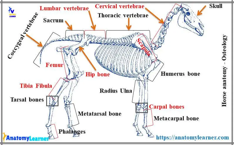
Osteology of horse
This is the basic and most important system of horse anatomy that you might not skip. You should learn all of the osteological features of horse bones. If you have good knowledge of general osteological features of animal bones (like – cow, goat, or sheep), you may easily compare the horse bones with them.
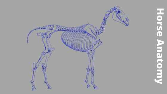
Please, try to identify all the bones from the horse skeleton below. You will find 200 bones in horse skeleton whereas in cow it is about 208 in number.
What are the differences between the horse and cow bone?
You may ask me the basic difference between the horse and cow’s bones. Great, I love to answer you. The main differences between horse and cow bones are –
#1. Horse bones are relatively larger than the cow’s bones
#2. They are rougher (surface) than the cow’s bones (for tight attachment of muscles).
#3. Important structures in bone-like trochlea, tuberosity, tubercle are more prominent than cow bones.
#4. Having more deep fossa, fovea or facet compare to cow bones (may vary, discussed in osteology section).
#5. All the bones have some special characteristics features (discussed in the osteology section)
These are the first ways to differentiate horse bones from cow bones. But if you want to identify any specific bone-like scapula or humerus of a horse, then you might know their characteristics features and compare them with other animal’s bones.
Let’s find some special osteological features from forelimb and leg bone of horse anatomy.
Bones of fore limbs from horse
You will find the following characteristics features in fore limb bones of a horse.
#1. There is no acromion process in horse scapula
#2. The coracoid process is well developed, and the glenoid notch is deeper in the scapula
#3. Tuber scapulae are larger in horse scapula in comparison to other animals.
#4. You will find very prominent deltoid tuberosity in the horse humerus bone
#5. The musculospiral groove is twisted and deeper in the horse humerus.
#6. An ill-developed ulna bone extends up to the distal third of the radius bone.
#7. There are seven bones of the carpus in the horse skeleton (four in proximal and three in distal row).
#8. There is one large metacarpal (III) and two small metacarpal bones (II and IV), small metacarpal located at both lateral aspects of the large metacarpal bone.
The small metacarpal bones of a horse are known as splint bones.
Horse anatomy leg bones
Following are the important osteological features from the horse anatomy leg bones.
#1. Gluteal lines are not prominent in horse hip bone
#2. The ventral tubercle is absent in horse hip
#3. The ischial tuberosity is not trifid as like the cow
#4. The femur of a horse is massive, and you will find an extra third trochanter on the lateral border.
#5. The patella is not as triangular bone as found in a cow.
#6. Tibia is the larger and longer bone of a horse.
#7. There is a grooved anterior tuberosity in the horse tibia bone
#8. There is six tarsal bone found in horse – tibial tarsal, fibular tarsal, central tarsal, first and second fused tarsal, third and fourth tarsal bones.
#9. Horse have one large metatarsal (III) and two small metatarsal bones (II and IV).
Horse anatomy head bone
You will also find some special osteological features in horse head bones compared to other animals. I will enlist the important osteological features from horse head bone anatomy.
#1. The skull is long and four-sided in a horse.
#2. There is no cornual process in horse head bone.
#3. There is an extensive foramen lacerum in the horse skull
#4. The interparietal bone is placed centrally and considered as a single bone.
#5. The fusion between the two halves of the mandible is complete (incomplete in cow).
If you want to know more about horse skull bones, you may read the article from anatomy learner. I hope you will know all the osteological features of horse skull bones.
Horse vertebrae and other bones
From a lot of osteological features, I will enlist the top features from horse vertebrae and others bones.
#1. The wing of the atlas vertebra is modified and curved in horse
#2. The spinous process of axis vertebrae is divided into two parts by two ridges and projected caudally.
#3. The mammillary processes of thoracic vertebrae are more developed in the horse
#4. There are five or six vertebrae found in lumbar vertebrae in a horse. The transverse processes of lumbar vertebrae are elongated plate and flattened dorso-ventrally.
#5. There are five, six, or seven bones fused to form a sacrum where the spinous processes are not fused as found in the cow.
#6. The sternum is boat-shaped and compresses laterally toward the cranial end and dorsoventrally at the caudal ends in a horse.
#7. There are 18 pairs of ribs in a horse (the first eight pairs are sternal, and the rest ten pairs are asternal ribs).
I know this little information is not enough to learn the osteological characteristics of bones from horse anatomy. But you might get a basic concept of bones with their special features from this article.
Syndesmology of horse
In an animal joint, you will find two or more articular ends of bones or cartilages, binding materials like ligaments or muscles. If you want to learn animal syndesmology in detail, then you may go syndesmology section.
The mandibular symphysis of the horse ossifies within few months and remains a single bone piece. In older horses, the pubic symphysis is also replaced by the bone. The ligaments and other binding materials of horse joints are stronger than a cow. In the hip joint of a horse, you will find the accessory ligament of the femur bone that is known as the check ligament.
This check ligament is a strong band detached from the symphysial tendon of the abdominal muscle. It is directed lateral, caudal, and dorsad, passes through the acetabular notch dorsal to the transverse acetabular ligament.
Muscles of horse
The muscles of a horse are well developed and tightly attached to bones. You will find similar types of muscles in horse body as you found in cow or goat. I have published an article on an animal muscle that will help you understand and identify the muscles from the animal body.
Here, I will show you some of the important muscles of the horse body. If possible, I will add some other horse muscle pictures in the future.
Horse anatomy digestive system
You will find many peculiar characteristics in the horse anatomy digestive system. I will provide some peculiar anatomical features from the horse’s digestive system.
The tongue of the horse is spatula-shaped, and the body is narrow. You will find the torus linguae in cattle tongue, but there are no torus linguae in horse tongue. A horse’s esophagus has three parts – cervical, thoracic and abdominal parts. The abdominal part of the esophagus is so small in a horse compared to a cow.
You will find a J-shaped simple secular stomach in a horse at the left side of the median plane of the body. There are both glandular and non-glandular parts in a horse stomach. The most important characteristic features of the horse stomach are margoplicatus. Margoplicatus is the structure that divides the glandular and non-glandular parts of a horse stomach.
There is a lot of variation found in the horse intestine. You will find a big comma-shaped cecum in the horse digestive tract. A horse’s colon is also a big tube and has several folds or parts. You will find the right ventral, sternal flexure, diaphragmatic flexure, right dorsal, left ventral, left dorsal, transverse colon, and pelvic flexure in the horse colon.
The parotid gland is the largest salivary gland in a horse. You will find the right lobe, left lobes, and middle lobes in the horse liver. There is no gall bladder in a horse, and hepatic ducts directly open into the duodenum.
The spleen and pancreas of a horse are both roughly triangular-shaped. I have described the digestive organs of horse separately here in anatomylearner.com
Respiratory organs of horse
There is also some peculiarity found in the respiratory organs of a horse. Please find out these special anatomical features from horse respiratory organs –
#1. The nasal cavity is larger and narrower compared to the cow.
#2. The trachea is comparatively larger than the cattle.
#3. Unlike those of the other domestic animals, a horse’s lungs are not subdivided into lobes.
#4. You will find three paired – arytenoid, corniculate, cuneiforms, and three unpaired – cricoid, thyroid, and epiglottis cartilages on the larynx of horse.
Urinary organs of horse
Some special anatomical features from horse urinary organs are found. The kidney of a horse has no lobulation, and the renal pyramid is not so prominent. You will easily identify the horse’s kidneys with their shape. The right kidney is heart-shaped and the left kidney is bean-shaped in a horse.
The renal pelvic is more dialated in the horse. Urinary bladder is comparatively small in horse compare to a cow.
Male genital organs from horse anatomy
You might know the anatomical features of male genital organs from horse anatomy. The testis is located in the prepubic region and ovoid, compressed side to side. The long axis of the testis is obliquely longitudinal.
The mediastinum testis is not well developed in the horse, but well developed in bull testis. Epididymis attached closely to the lateral surface of horse testis.
Pe-(enis) of a horse is a cylindrical structure, and there is no sigmoid flexure as found in the bull or goat pe-(enis. The glans is the enlarged free end and consists of corona glandis and fossa glandis. A bulbourethral gland is larger, but the prostate glands are indistinct in horse.
Organs of mare
Horse ovary is bean-shaped and located at the sub-lumbar region, ventral to fourth or fifth lumbar vertebrae contact with a lumbar wall of the abdomen. You will find characteristics features on the free border of the horse ovary. There is a depression on the free border of ovary and known as ovulation fossa in horse.
The horse of mare uterus is cylindrical and blunt at their cranial ends. Body of uterus located at abdominal cavity (partly in the pelvic cavity) and cylindrical, flattened dorsoventrally. The cervix is short, and the cervical canal is straight and simple. The endometrium is devoid of any cotyledon in the horse.
The va(g)zi-nya of a horse is straight, shorter, and less capacious. You will not find the sub-urethral diverticulum in horse. The ventral commissure is thick, and the mucous membrane is red. The urethra is shorter but thicker in comparison to a cow.
You will find two developed mammary glands located at the prepubic region in the horse. If you wish to read detailed anatomical features on mare organs, you may visit the equine anatomy section.
Anatomical features of horse heart
The horse heart occupies the great part of the middle mediastinal space. The shape of the horse heart is irregular, somewhat flatten cone. The base is attached with great vessels and located from the second intercostal sixth ribs or intercostal space. Apex is lying centrally dorsal to the last sternebra of a horse.
The base of the horse heart is wider, and the surface is more flatter than a cow’s heart. Cranial border is the more convex, and caudal border is nearly verticle.
There are 18 pairs of intercostal arteries in a horse. The celiac artery of horse mainly divides into three major branches – splenic artery, left gastric artery, and hepatic artery. There is no median sacral artery in horse.
Horse hoof anatomy and skin features
The hoof is the hard covering of the distal end of each digit. You will find only one hoof for each limb of the horse. There are three different parts of the horse hoof – wall, sole, and frog. These parts of horse hoof are well developed.
The wall is the part of the hoof visible when the foot is placed on the ground. Sole constitutes the greater part of the ground surface of the hoof. Again the frog is a wedge-shaped structure that occupies the angles bounded by the bars and sole.
The internal surface of the hoof wall is concave and consists of thin primary epidermal lamellae. I will show you the different structures from a horse hoof.
A horse’s skin is thinner than cattle and contains more well-developed sweat and sebaceous glands. You will find many in a horse (long hair in wither neck and forehead).
Again, some other peculiar characteristics are found in horse – eargots and chestnuts. Erogots is a small hard area located at the caudomedial surface of all the four fetlock joints in horse. Chestnuts are the structures located a little above on the medial surface of carpus and tarsus in all the limbs.
Endocrine glands of horse
There are also some variations in the endocrine glands of horse. Thyroid gland is located on the most cranial part of the trachea and loosely attached by deep fascia. They are dark brown in color, firm, and highly vascular glands. A narrow fibrous isthmus connects two lateral thyroid lobes.
The right adrenal gland is located medial to the caudal vena cave and between the vena cave and psoas muscles. The left adrenal gland of horse is shorter than the right one.
The pineal gland of horse is ovoid or fusiform in shape and red-brown in color. This pineal gland is located in the midline depression between the thalamus and rostral colliculi.
I will update this article regularly, and you will get notified at social media. If you did not follow anatomylearner on social media, let go and follow to get more updates.
You might read the other different articles related to anatomical features of different organs from the horse –
#1. Horse stomach anatomy – both external and internal anatomical features
#2. Anatomical features of horse pe-einis and uterus
#3. Horse leg anatomy (bone and muscle anatomy)
Conclusion
This is the summary of horse anatomy where I tried to provide valuable information. If you think you got a basic idea of the different organs system of a horse, then share this article with your friends who are also interested to learn horse anatomy.
I will update this article with other most interesting anatomical characteristics of different organs from horses. Please stay connected with anatomy learner and read the upcoming articles. In future, you will get more horse anatomy diagram, model pictures, worksheets and other from this article.