Connective tissue is the fundamental tissue that provides structural and mechanical support to other tissues and organs of an animal’s body. Based on the variation of quantity and arrangement of fibers within the matrix, the ideal connective tissue is classified into two main types – loose connective tissue and dense connective tissue.
Hey, do you love to know the histological features of loose connective tissue and identify them under the light microscope? Okay, in this article, I will discuss the histology of loose connective tissue with real microscope slide pictures.
After reading this article, you will know the types, examples and location of loose connective tissue with diagram and slide pictures. So, if you love to know then, you may continue this article till the end.
Loose connective tissue
Loose connective tissue is common and the most widely distributed connective tissue in animals. It is important to know for further studying of the different organ’s structures of animals’ bodies. You will find the loose connective tissue just beneath many epithelia, where it provides support and vascular supply.
Suppose you want to identify loose connective tissues slide under the light microscope. In that case, you might have a piece of good knowledge on different types of connective tissue cells, fibers and ground substances of connective tissue.
You might identify the following histological features from the loose connective tissue slide under the light microscope.
- #1. Collagen fibers bundles
- #2. Elastic fibers (branching)
- #3. Connective tissue cells (like fibroblasts, macrophages, mast cells, plasma cells, large and small lymphocytes; you will find more fibroblasts and macrophages in the sample tissue)
- #4. Capillary with blood cells
“In any connective tissue structure, you will find fixed and free cells (fibroblasts, reticular cells, adipocytes, mast cells, macrophages, pigment cells, plasma cells, free macrophages and leucocytes). Connective tissue fibers (collagen fibers, reticular fibers, elastic fibers and fibrous adhesive proteins) and ground substances (proteoglycans, glycoamioglycans and interistial fluid).”
Identification of loose connective tissue histology slide
If you are a beginner at histology learning, you might know the histological features of loose connective tissue and identify the microscope slide under the light microscope. I think the following identifying characteristics will help you to identify the loose connective tissues.
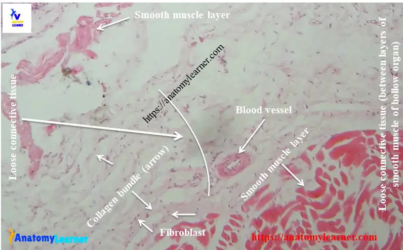
#1. The sample tissue section shows the loose arranged pink collagen fibers (collagen bundles) in different directions.
#2. Presence of branching and anastomosing networks of thin elastic fibers in different directions.
#3. There are a large number of connective tissue cells like fibroblasts, macrophages, mast cells, plasma cells and different types of blood cells.
#4. Presence of a large number of ground substances in the sample tissue section.
This is a slide of loose connective tissues slide. If you think you need other identifying characteristics, then you might add them.
Histology of loose connective tissue
Want to know the detailed histology of loose connective tissue? Here, you will know the different types of cells, fibers, and ground substances within loose connective tissue matrix.
Fibers and their arrangements
This type of connective tissue is characterized by a loose, irregular arrangement of connective tissue fibers within the abundant ground substances. You will find all kinds of fibers (collagen, reticular and elastic fibers) that loosely arranged. The amount and orientation of these fibers vary widely and depend on the particular tissue’s location and specific functions.
The pink collagen fibers (in routine staining) are the thickest, most extensive and most numerous fibers in the loose connective tissue structure that courses in different directions. You will also find the thin elastic fibers, fine single fibers and usually straight. They are light pink (in routine staining) or black (with special staining) in appearances and forms the branching and anastomosing networks within the this connective tissue matrix.
Cells and their histological features in connective tissues
Numerous connective tissue cells are found in the loose connective tissue histology where fibroblast and macrophages are more. Fibroblast is the most common flatten or fusiform in shaped with slender processes. These cells contain oval nucleus with prominent nuclei and are actively responsible for forming fibers and ground substances.
Fixed macrophages are irregular in shape, having eccentrically placed identical nucleus. You will also find the masy cells occur single or in group along the blood vessels of the tissue structure. There are also different blood cells; small and large lymphocytes are found in the matrix of loose connective tissues.
The faint background around the fibers and connective tissue cells is the ground substances that contains proteoglycans, glycosaminoglycans and interstitial fluid.
Location of loose connective tissues
Usually, you will find the loose connective tissue beneath many epithelial tissues (lining epithelium). It provides the support and vascular supply and allows easy movement, shifting different organs of animals’ bodies.
- #1. In between layers of smooth muscles of any hollow organs
- #2. In the papillary layer of dermis and hypodermis of thin and thick skin
- #3. The serous lining of peritoneal and pleural cavities
- #4. Around the nerves and skeletal muscles bundles
Diagram and pictures of connective tissue
I tried to show all the histological features of ideal connective tissue in the this connective tissues diagram. If you want, you may try to draw this same diagram.
Again, if you want more diagram and real slides pictures of connective tissues, you may follow anatomy learners on social media. You may find some mistakes in the labeled diagram of the provided sample tissue; please let me know.
You might like the other articles from anatomy learner (related to veterinary histology and anatomy of different organs system of animal’s body) –
#1. Histology of various epithelial cells with slide pictures.
#2. Special connective tissue – histology of bone, cartilage and blood cells with labeled images.
#3. Osteological features of a typical vertebra of animals – ox, sheep, goat, horse and others.
Dense connective tissue
You will find more dense connective tissue fibers (especially collagen fibers) and fewer cellular and ground substances in dense connective tissue. If you want to know more about dense connective tissue histology with slide pictures then, you may read the details guide from anatomy learner.
Reticular connective tissue
Reticular connective tissue is the unique or modified form of connective tissue proper that has reticular fibers (tiny and branching fibers) and cells. It provides the architectural frameworks (netlike supporting frameworks) of some organs of the animal’s body.
Reticular connective may be found in the muscles fibers, blood vessels, nerves, and different epithelial structures of the body.
Conclusion
This might be the best guide to know the basic of loose connective tissue histology with real slide pictures and labeled diagram. Now, you will identify the loose connective tissues slides under the light microscope.
If you think this guide is beneficial, share this with your friends who want to know the primary connective tissue structure.