As a human medical student or veterinary student, you might learn histology slides with proper identifying characteristics. This article will show you the essential histology slides from the different organ systems of an animal’s body.
Here, you will get the histology slides of epithelial tissue, digestive system organs, respiratory organs, endocrine glands, male and female genital organs, and others. I will try to cover the most important identifying characteristics with each slide. So, this article will be a great article on the internet to learn the histological features of different slides of an animal.
Histology slides guide
Histology is a visual and colorful science that is studied with the help of a light microscope. I hope you have a piece of good knowledge of permanent slide preparation, properties of different staining, and handling of the microscope. These are the prerequisites to study any histology slide. This article will show you histology slides from the following different organs system of an animal’s body with identifying features.
- #1. Histology slide of epithelial tissue
- #2. General connective tissue histology slide
- #3. Histology slides of special connective tissue (blood, bone, and cartilage)
- #4. Muscular tissue histology slide
- #5. Respiratory organ histology slides
- #6. Slides of the circulatory system (artery, vein, and heart)
- #7. Digestive system histology slides
- #8. Histology slides of exocrine and endocrine glands
- #9. Microscopic slides from lymphoid tissue
- #10. Microscopy slides from urogenital organs
- #11. Histology slide of skin and retina
You know, it is not possible to describe all the features from each histology slide in this single article. Please go to the specific article here if you want to learn the detailed histology of any slide. Okay, let’s start to learn and identify histology slides from different organ systems of animals.
Histology slides epithelial tissue
The epithelium is classified according to the number of cell layers and morphology of the surface cells. Basement membrane separates the epithelium from the connective tissue. This is the fundamental tissue, and you might learn them with great care.
From the epithelial tissue, I should learn and identify the following histology slides with their identification points.
- #1. Simple squamous histology slide
- #2. Simple cuboidal epithelium histology slide
- #3. Simple columnar epithelium histology slide
- #4. Stratified squamous epithelial slide (both keratinized and non-keratinized)
- #5. Stratified cuboidal eptiehlium slide
- #6. Pseudostratified ciliated columnar epithelium slide
- #7. Transitional epithelial tissue slide
I have a detailed guide on the identification of epithelium tissue here at anatomy learner. You will find more slides on that epithelial tissue identification guide.
“For more labeled pictures of epithelial tissue slides or others, please go to the specific articles.”
Identification of epithelium tissue slides
A single layer of flat or squamous cells rests on the basement membrane found in a simple squamous epithelial slide. Simple squamous epithelial lines the external surface of digestive organs, lungs, and heart. Again, it lines the inner surface of blood vessels, heart, and lymphatic organs.
You will find a single layer of round cells on the basement membrane in a simple cuboidal epithelium slide. It generally lines small ducts and kidney tubules.
A single layer of tall cells with cilia is found in the simple columnar epithelial slide. You will find the simple columnar epithelium in the digestive tract and gall bladder slides.
Multiple layers of cells are found in the stratified squamous epithelium (basal cells are columnar, and superficial cells are flattened). The nonkeratinized stratified squamous epithelium contains a live superficial cell layer, whereas keratinize stratified squamous contains a dead superficial cells layer. Nonkeratined stratified squamous epithelium lines on the esophagus, buccal cavity, and oropharynx slides.
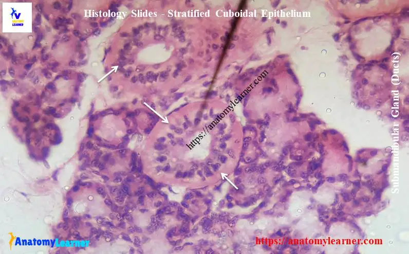
Multiple layers of cells are found in stratified cuboidal epithelial tissue slides. The superficial cells are round, and basal layer cells are low columnar type. You will find stratified cuboidal epithelium in the male urethra, conjunctiva, and different large excretory ducts.
In the pseudostratified ciliated columnar epithelial slide, you will find a single multinucleated layer of cells. All the cells reach to basement membrane but do not reach the surface. You will find this pseudostratified epithelium in respiratory passages, uterine tubes, and epididymis slides.
There are more cell layers in the transitional epithelium that changes shape in response to distension caused by fluid accumulation. These cells lines exclusively renal calyx, renal pelvis, ureter, and urinary bladder.
General connective tissue histology slide
Connective tissue originated from the mesenchymal cells that differentiate during development into cell types. It consists of cells, fibers, and ground substances. From the connective tissue, I will show you the following essential and basic slides with identifying features.
- #1. Loose connective tissue slide
- #2. Dense regular connective tissue slide and
- #3. Dense irregular connective tissue slide
You will also find the detailed guide on identifying different types of connective tissue microscope slides here.
Loose connective tissue is more prevalent in the animal body than dense connective tissue. It is characterized by a loose, irregular arrangement of connective tissue fibers and abundant ground substances.
You will find pink collagen fibers that are directed in a different direction. There are thin elastic fibers that branch and anastomoses to form the network.
In contrast to loose connective tissue, dense regular connective tissue contains densely packed collagen fibers that exhibit uniform, regular and parallel arrangement. You will find a parallel row of fibroblasts in between the collagen fiber bundles. Dense regular connective tissue uniformly found in the tendon and ligament histology slide.
Dense irregular connective tissue contains thicker and more densely packed collagen fibers, fewer cells, and less ground substances. You will also find numerous elastic fibers in dense irregular connective tissue. The collagen fiber in this type of tissue shows a random and irregular orientation. Dense irregular connective tissue present in the dermis of skin and capsule of different organs.
Please find all the labeled images of connective tissue histology slides.
Histology slides of special connective tissue
This part of this article will find the histology slides of special connective tissue like bone, blood, and cartilage. Bone is a special connective tissue consisting of cells, connective tissue fibers, and extracellular matrix. I will show you two different types of slides – compact bone and spongy bone from the bone histology.
- #1. Compact bone microscope slide and
- #2. Spongy bone microscope slide
You will find the Haversian system, Haversian canal, concentric lamellae, and interstitial lamellae in the compact bone histology slide. I have a detailed guide on compact bone histology with the identification points here at anatomy learner.
In spongy bone histology, there are numerous bony trabeculae separated by marrow cavities. Osteocytes are found in the matrix of the trabeculae of the spongy bone histology slide.
Cartilage is a special type of connective tissue that develops from the embryonic mesenchymal cells. Cartilage exhibits tensile strength provides firm structural support for the soft tissue of the animal body. You might learn three types of cartilage structure from the slides.
- #1. Hyaline cartilage slide
- #2. Elastic cartilage slide and
- #3. Fibroelastic cartilage slide
You know the hyaline cartilage is the most common type of cartilage in the body and serves as a skeletal model for the bones. The histology slide of hyaline cartilage shows the homogenous and glassy extracellular matrix. Within the matrix, you will find the isogenous chondrocytes group.
You will find the thin, branched, and anastomosing elastic fibers along with larger chondrocytes in the elastic cartilage slide. Elastic cartilage may found in the external ear, auditory tube, epiglottis, and part of the larynx.
The fibrocartilage slide shows the dense collagen bundles with a chain of chondrocytes. You will find fibrocartilage in intervertebral discs, pubic symphysis, and few joints.
Muscular tissue histology slide
Three types of muscle histology slides can be identified with their special morphology or structure under the light microscope. All muscle tissue contains the elongated cell (known as fibers). Here, you should learn and identify the three types of muscle tissue histology slides with identification points.
- #1. Smooth muscle histology slide
- #2. Skeletal muscle microscope slide and
- #3. Cardiac muscle histology slide
In the skeletal muscle histology slide, you will find long, cylindrical fibers, multinucleated cells with peripheral nuclei. Each muscle fiber consists of a smaller subunit (myofibrils) that extends the entire length of the fiber. The arrangement of actin and myosin filaments is very regular in skeletal muscle tissue. So, you will find a distinct cross-striation pattern under a light microscope. In the cross-section of skeletal muscle, you will find a muscle fascicle that contains myofibrils, connective tissue, and other structures.
“You might identify both the longitudinal and cross-sectional muscle tissue under a light microscope. For that, you may visit the muscle tissue histology section.”
In the smooth muscle tissue slide, you will find the fusiform-shaped fibers and a single central nucleus. There are actin and myosin, but no cross-striation pattern is found in smooth muscle. Smooth muscle is mainly found in the hollow organ structure and blood vessels.
The cardiac muscle fibers are also cylindrical but short and exhibit the branching pattern. As the skeletal muscle, cardiac muscle also shows distinct cross-striations because of a regular arrangement of actin and myosin filaments. The most characteristics feature of cardiac muscle tissue is the presence of intercalated discs. These are the dense junctional complexes in cardiac muscle tissue.
If you wish to learn more about muscular tissue histology, please find more information and more labeled images.
Respiratory organ histology slides
The respiratory system consists of two main parts – conducting portion and the respiratory portion. I will show you the essential histology slides from the respiratory system with their identifying features. Make sure you know the detailed histology of a hollow organ or any tubular organ. This structure will help you to understand the structure of some respiratory organ’s histology.
From the respiratory organ system, I will show you the following microscope slides with their identifying characteristics. Though I have a detailed guide on respiratory organ histology here in the anatomy learner blog.
- #1. Slide of lung and the associated structures within lung
- #2. Microscope slide of the trachea
- #3. Histology slide of the epiglottis and other laryngeal cartilage
Under the lung histology slide, you might identify the intrapulmonary bronchus, primary bronchiole, respiratory bronchiole, and terminal bronchiole. I have shown all these bronchus and bronchiole here on the lung histology slide.
The simple squamous epithelium lines the surface of the lung tissue. You will find numerous thin-walled alveoli that line with the simple squamous epithelium. There are also intrapulmonary bronchus, three different types of bronchiole present with the lung tissue histology slide.
You will find the mucosa, submucosa, hyaline cartilage layer, and adventitia in the trachea histology slide. The lining epithelium of the trachea is pseudostratified columnar epithelium with goblet cells. You will also find C-shaped cartilage and seromucous glands in the submucosa layer of trachea histology.
In the base of the epiglottis histology slide, you will find the pseudostratified ciliated columnar epithelium. On another surface, there is the stratified squamous epithelium. Central elastic cartilage forms the core of the epiglottis histology slide.
“Don’t forget to learn and identify the different types of bronchus and bronchiole from the lung tissue histology slide.”
Slides of the circulatory system
You know the cardiovascular system of an animal consists of heart and blood vessels. Before identifying the microscopic slides of the circulatory system, make sure you know the general structure of the artery. This might help you to understand the primary difference between muscular artery, elastic artery, and vein. You might identify the following histology slides from the circulatory system of an animal.
#1. Heart histology slide
#2. Larger artery or elastic artery slide
#3. Medium-sized artery or muscular artery slide and
#4. Vein histology slide
Microscope slide identification from circulatory systems
You will find details histology on these structures in the circulatory organs histology section. If you need more labeled images on circulatory organs, you may check these articles.
You will find tunica intima, tunica media, and tunica adventitia in the large artery histology slide. The tunica media of large arteries is very thin and contains many elastic laminae. Generally, there are two types of elastic laminae – internal and external laminae.
Internal elastic laminae are located between the tunica intima and tunica media. Again, the external elastic laminae are peripheral to the muscular tunica media. You will also find a thin layer of elastic fibers in the tunica adventitia of a large artery.
The tunica intima of the muscular artery is well developed, especially the internal elastic laminae which stands out prominently. You will find the circularly arranged smooth muscle layers in the tunica media of the muscular artery. The adventitia of the muscular artery consists of a thin layer of fibroelastic tissue.
The structure of the vein is similar to the basic structure of an artery. You will find a thin tunica media with few smooth muscle fibers and less elastic fibers in a medium-sized vein. But in a large vein structure, you will find thick tunica adventitia with longitudinally oriented bundles of smooth muscle fibers.
Digestive system histology slides
The animal digestive system consists of a long tube or tract that starts at the oral cavity and ends at the an-eus. From this system, I will show you a lot of histology slides with identifying features. But, I have published articles on digestive organs with descriptions and real slides pictures. If you love to know the histological features of any digestive organs, you may check these articles from anatomy learners. You might identify the following different microscope slides from the digestive system of animals.
#1. Microscope slide of tongue
#2. Esophagus histology slide
#3. Simple stomach histology slide
#4. Compound stomach (rumen, reticulum, omasum, and abomasum)
#5. Histology slide of duodenum
#6. Jejunum histology slide
#7. Ileum histology slide
#8. Caecum histology slide
#9. Colon microscopic slide
#10. Re-actum microscope slide
#11. Histology slide of liver
#12. Slide of gall bladder and
#13. Microscope slide of the pancreas
“I would like to suggest you read these slides from the specific articles published on anatomy learner previously. You will find lots of histology slide images with identifying characteristics.”
The stratified squamous epithelium covers the upper surface of the tongue. You will find the filiform and fungiform papillae on the tongue histology slide. Again, there are skeletal muscles that run in a different direction in the tongue histology structure.
Before identifying the hollow organs (digestive tract) from the digestive system, I would like to remind you again to read the general structure of a tubular organ.
You will find all four layers (mucosa, submucosa, muscularis, and adventitia) in the esophagus structure. You might describe the following points in each tubular organs from the digestive system –
#1. The lining epithelium of the tract (parts)
#2. Layers and their structure (tract)
#3. Special features in any layer
Liver, pancreas, and gallbladder microscope slides
From the liver histology slide, you might identify the polygonal hepatic lobules, hepatic cords, portal tried central vein and other different vital structures. You will find the detailed guide on liver histology here in anatomy learner.
In the pancreas histology slide, you will find the darkly stained serous acini and lightly stained Langerhans. You will also find the interlobular ducts, interlobular septum, artery, vein, and nerves in the pancreas microscope slide.
The simple columnar epithelium lines the fold of the gallbladder. You will find a fibromuscular layer in the gallbladder histology slide.
Histology slides of exocrine and endocrine glands
This part of this article will show you the exocrine and endocrine histology slides with their identification points. Make sure you know the structures and function of different types of acinus like – mucous acini, serous acini, and seromucous acini.
Please, try to identify the following histology slides of exocrine and endocrine glands –
- #1. Parotid salivary gland histology slide
- #2. Submandibular salivary gland slide
- #3. Sublingual gland histology slide
- #4. Mammary gland histology slide
- #5. Pituitary histology slide
- #6. Thyroid gland histology slide
- #7. Adrenal gland microscope slide
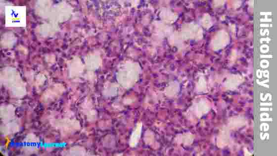
You will find serous acini and many striated ducts in the parotid salivary gland histology slide. In the submandibular salivary gland histology slide, there are many serous acini and few mucous acini. There are also many striated ducts, and serous demilunes are found in the submandibular salivary gland slide.
In the sublingual salivary gland slide, there are many mucous acini and tubules. Please check the specific articles from anatomy learner if you want to know more about the histology of these three types of the salivary gland.
The pituitary gland histology slide contains the pars distalis, pars nervosa, pars intermedia, and hypophyseal cleft. In the pars distalis, you will find the chromophobes and chromophil cells. The pars intermedia separated from the pars distalis by hypophyseal cleft.
In the thyroid histology slide, different thyroid follicles contain thyroglobulin. There are parafollicular cells present in between the thyroid follicles.
You will find the three distinct zones in the cortex of the adrenal gland histology slide. In the medulla of the adrenal gland, there are chromaffin cells and sympathetic ganglionic cells. If you want to know more about the histology of the endocrine gland with natural slides picture, please check these articles from anatomy learner.
Microscopic slides from lymphoid tissue
The lymphoid system consists of tissue and organs made with lymphocytes. From the lymphoid system of an animal, I will show you the following microscope slides.
- #1. Thymus histology slide
- #2. Lymph node histology slide
- #3. Microscope slide of spleen and
- #4. Tonsil histology slide
In the thymus histology slide, many lymphoid tissue lobules contain darkly stained cortex and lightly stained medulla. You will find Hassall’s corpuscles in the medulla of the thymus histology structure.
There are lymphatic nodules and subscapular sinus found in the cortex of the lymph node histology slide. Again, in the medulla of the lymph node, there are medullary cords and sinus.
You will find the thick capsule, thick trabeculae, white pulp, and red pulp in the spleen histology slide. The white pulp of the spleen contains lymphatic nodules and central arterioles. Again, in the red pulp, you will find the splenic cords and sinusoids.
In the tonsil histology slide, there are subepithelial lymphatic nodules and mucous glands. The stratified squamous epithelium lines the crypts of the tonsil.
Microscopy slides from urogenital organs
You might learn and identify the following histology slides with their valid identification points from the urinary system of an animal. I want to suggest you check all these articles separately from the all guide pages or learning resources page.
- #1. Kidney histology slides, and it’s associate structures
- #2. Ureter microscope slide
- #3. Urinary bladder histology slide
- #4. Urethra histology slide (both male and female urethra histology)
There are renal corpuscles, proximal convoluted tubules, and distal convoluted tubules in the cortex of the kidney histology slide. Don’t forget to learn the detailed histological features of kidney corpuscles with description. In the medulla of the kidney slide, you will find the collecting ducts, thick and thin loop of Henle.
In the urethra histology slide, you will find a star-shaped lumen and the mucosa lined with transitional epithelium. A thick muscular layer made with discrete bundles of smooth muscle is found in the histology of the urethra.
Male organs histology slides
Learn the detailed histological characteristics of the following organs of male animals. For that, I like to suggest you go to this page and learn details of histology with anatomy learner.
- #1. Testis histology slide
- #2. Epididymis histology slide
- #3. Vas deference microscope slide
- #4. Histology of pe-neais
- #5. Prostate gland histology slide
- #6. Seminal vesicle histology slide and
- #7. Bulbourethral gland histology slide
You will find the seminiferous tubule in the testis histology slide. Please try to find out the spermatogenic cells at various stages of maturation from the testis histology cells. You might also find out the interstitial cells of Lydig and Sertoli cells from testis histology slides.
The pseudostratified stratified columnar epithelium lines the ductus epididymis. You will find the smooth muscle fibers surrounding the ducts and sperms in the lumen of the duct.
In the vas deference histology slide, you will find a stellate-shaped lumen and thick muscular coats. The muscles are arranged in three different layers in the vas deference structure. You will also find sperms in the lumen of the vas deference histology slide.
The mucosa of the seminal vesicle forms branching and anastomosing folds that lines with simple columnar epithelium. You will find a well-developed muscular coat in the histology structure of the seminal vesicle.
You will find a unique feature in the prostate gland histology slide – prostatic concretions or corpora amylacea.
Female organs microscope slides
I will show you the following histology slides with their labeled diagrams from the female organ system of an animal. Make sure you learn the detailed histology of the different organs from female animals.
- #1. Ovary histology slide and different follicles structures
- #2. Fallopian tube histology slide
- #3. Histology of va(g)zi-nya
The surface of the ovary covers with the germinal epithelium (simple cuboidal). There are different growing follicles at various stages of maturation on the ovary histology slide. I showed the different structures in a mature follicle from the actual histology slide.
You will find the highly folded mucosa in a uterine tube or fallopian tube histology structure. There is a thick muscular layer found in the uterine tube histology.
In the uterus histology slide, there are perimetrium, endometrium, and myometrium. There are numerous cells and uterine glands found in the endometrium of the uterus. The Myometrium of the uterus has thick middle circular smooth muscle layers.
You might differentiate the proliferative phage and secretory phages of the uterus from the histology slide under a light microscope.
Histology slide of skin
Skin is composed of two different layers – the superficial epidermis and the deep dermis. You will also find the hypodermis in the histology slide of the skin. I will show you the different structures from the epidermis and dermis of the skin histology slide.
The epidermis of the skin is very thin and lines with the keratinized stratified squamous epithelium. You will find the papillary layer and reticular layer in the dermis of the skin. You might find out the dermal papillae, sebaceous glands, sweat glands, arrector pili muscles from the skin histology slide.
If you want to know more about the skin histology slide, please check the article from anatomy learner (skin histology slide).
Most Frequently asked questions on histology slides
How do you identify histology slides?
To identify the histology slide, you may follow the following steps. I hope these simple eight steps will help you to identify the histology slides.
#1. Identify the coverslip by touching with your fingertip.
#2. Fix the slide with the microscope’s clip (coverslip facing up)
#3. Switch on the light of the microscope.
#4. Adjust the condenser of the microscope.
#5. Keeping an eye on the eyepiece.
#6. Fix the low-power object to bring it with the line of the object.
#7. Focus and locate the targeted tissue by using the coarse adjustment.
#8. Moving the stage and find the target histological features of your specimen.
How do you make a histology slide?
To visualize the histological features of any specimen, you might go through the permanent slide preparation. There is a long process of permanent slide preparation containing – tissue collection, fixation, processing, sectioning, and staining. You might perform the following steps to make a histology slide.
#1. Tissue collection
#2. Fixation of the collected tissue
#3. Dehydration of the tissue
#4. Clearing and infiltration of the tissue
#5. Embedding f the infiltrated tissue
#6. Sectioning and attachment of the sectioned tissue on a glass slide
#7. Staining processes
What are tissue slides?
Tissue slides are the sectioned tissue that cut into five to seven micrometer thick and picked up on the glass slides under a water bath.
What is the best way to study animal histology slides?
There are no specific ways of studying histology slides. But you may follow these points while studying histology slides.
#1. Find out the all structures from a histology slide (get help from books or labeled images)
#2. Read the details histological features from books or the internet
#3. Make short notes on the specific slide that you read
#4. Join the class lectures of your tutors
#5. Read articles on the internet
#6. Go to anatomy learner (histology section) and read all histology slides
#7. Do it regularly (minimum 30 days) and practice
If you want to get any updates on any labeled images or articles from anatomy learner, make sure you join me on social media.
Conclusion
I hope this article might help you to get all the histology slides from different organs system of animals. I want to suggest you again check all the articles on specific histology slides to learn more.
All the histology slides from the epithelial tissue, digestive system, respiratory system, and endocrine glands are essential for beginners. Make sure you learn these slides well, and let’s practice with anatomy learner.