Skin is the heaviest organ of an animal’s body that covers the surface of the body. It comes into direct contact with the external environment. In the skin histology slide, you will find two main layers – epidermis and dermis. If you want to identify and understand the histological features from both thick and thin skin histology slides, this article is for you.
This article will show you the different histological structures from the epidermis and dermis of skin histology slides. You will get a full guide on these layers of skin with a labeled diagram.
Skin histology
First, I would like to introduce you to the most important structures from the skin microscope slide. From both thick and thin skin histology slides, you might identify the following features. I hope these will help you to understand the skin structure primarily.
Let’s enlist the histological features from the skin microscope slide –
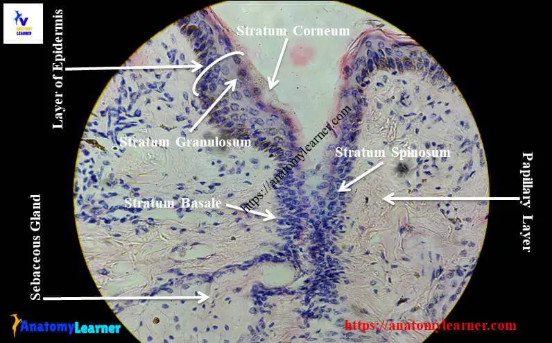
- #1. Stratum corneum of epidermis
- #2. Stratun lucidum of epidermis layer
- #3. Stratum granulosum cells layer of the epidermis
- #4. Stratum spinosum cells layer of the epidermis
- #5. Stratum basale cells layer of the epidermis
- #6. A papillary layer of skin
- #7. Reticular layer of skin
- #8. Dermal papillae of the skin
- #9. Duct and sweat glands on papillary layer
- #10. Arrector pilli muscles
- #11. Sebaceous glands on the reticular layer
- #12. Hair follicles
- #13. Hypodermis of skin (if present in section)
You will find most of these histological features in the provided skin slide labeled picture. If you don’t find any structures on the skin labeled diagram, please let me know.
Skin histology slide identification
You will find two different types of skin histology slides – thick and thin skin. Generally, skin is thickest over the dorsal surface of the body and on the lateral surface of an animal’s body. You will also find the thickest skin on the palmar and plantar surface, where the stratum corneum is thickest.
Again, thin skin is found on the ventral surface of the body and the medial surface of the limb. Thin skin has a protective hair coat. In the histology slide, you will find the thinner epidermis in thin skin.
Let’s identify the thick and thin skin histology slides under a light microscope. First, talk about the thin skin microscope slide identification.
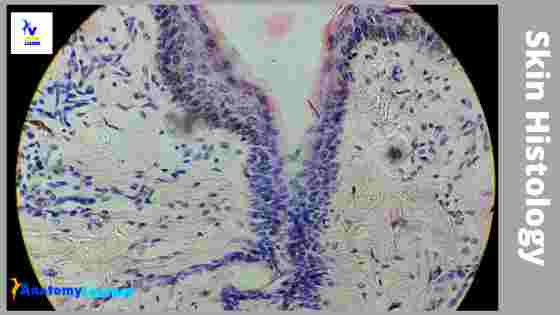
- #1. The provided tissue section shows two distinct layers – the epidermis and dermis.
- #2. Presence of thin epidermis that lines with keratinized stratified squamous epithelium.
- #3. There are dermal papillae found on the dermis layer.
- #4. Presence of sweat glands, sebaceous glands, and hair follicle on the provided tissue section.
This is the microscopic slide of thin skin. Great, now, let’s try to identify the thick skin microscope slide with identifying points.
- #1. Presence of very thick epidermis that lines with keratinized stratified squamous epithelium.
- #2. The stratum corneum of epidermis is thicker.
- #3. The dermis layer shows dermal papillae and contains only sweat glands.
- #4. Absence of hair follicles and sebaceous glands on the sectioned tissue.
So, this is the thick skin microscope slide. Fine, if you want to know the detailed histological features of the skin, please continue this article.
Structure of thin skin of an animal
In the structure of the thin skin of animals, you will find the two distinct layers – the epidermis and dermis. I will show you the different histological characteristics from the epidermis and dermis of a skin microscope slide.
#1. The epidermis of skin (you might learn the general features, layers of the epidermis, and cells of the epidermis)
#2. The dermis of the skin (you might learn the features of the papillary layer and reticular layers of dermis)
Histology of epidermis
The epidermis of the skin is made of keratinized stratified squamous epithelium. It projects into the dermis as an epidermal ridge. The epidermis is the superficial layer and avascular that are nourished by diffusion. There are free nerve ending found in the basal layer of the epidermis.
In the thin skin histology slide, you will find the following five (5) layers and four (4) different types of cells. I will go through the detailed description of these layers and cells of the epidermis.
- #1. Stratum basale layer
- #2. Stratum spinosum layer
- #3. Stratum granulosum layer
- #4. Stratum lucidum layer and
- #5. Stratum corneum layer
You might know the detailed features from these layers of the epidermis. Let’s know the histological features of epidermal layers of skin.
Stratum basale layer of epidermis
This basale layer of the epidermis consists of a single layer of columnar or cuboidal cells that rest on the basal lamina. They attached laterally to each other and overlaying stratum spinosum cells by desmosome. The nucleus is larger and oval that occupies most of the cell.
Different mitotic figures may found in the cells of this stratum basale layer of the epidermis. The newly formed cells move towards the superficial layer of the dermis.
Stratum spinosum layer
This stratum spinosum layer of the epidermis is also known as the prickle cells layer.
It consists of several layers of irregular polyhedral cells. The cytoplasm of the cells contains more prominent tonofilaments (bundle of keratine filaments).
The tonofiliaments of the cells are more distinct than the basal cells layers.
You will find a centrally located nucleus in these spinosum cells layer. The two layers of the epidermis are stratum basale, and stratum spinosum together called stratum Malpighi or Malpighian layers. All the mitotic activities of the epidermis occur in this stratum Malpighi.
Stratum granulosum layer of epidermis histology
In this granulosum layer of skin epidermis, you will find several layers of flattened or polygonal or diamond-shaped cells. The cytoplasm of these granulosum cells contains irregular, membrane-bound, electron-dense keratohyalin granules. They play a role in keratinization and barrier functions.
Again, you will find some lamellated bodies or membrane coating granules known as lamellar granules. These lamellar granules occur near the Golgi bodies and smooth endoplasmic reticulum. The number and size of these granules are increased and move towards the cell membrane. These lamellar granules release lipid content by exocytosis and thus make a waterproof barrier.
Stratum lucidum layer of epidermis
This stratum lucidum layer of the epidermis is found only in the thick skin histology and in the hairless region of the animal’s body. This is the thin, translucent, homogenous line between the stratum granulosum and stratum corneum layers of the epidermis.
Stratum lucidum consists of keratinized, closely compacted, dense cells that devoid of nuclei and cytoplasmic organelles. The cytoplasm of these lucidum cells contains protein-bound phospholipid and eleidin. Do you know what eleidin is? Well, in short, eleidin is a derivative of keratohyalin.
Stratum corneum layer from skin epidermis histology
This is the most superficial layer from the skin epidermis histology slide. You will find several layers of completely keratinized dead cells (flattened) on this layer. The plasma membrane of these cells layer is thicker, and the cytoplasm is filled with keratin.
Cells of this layer continuously shed from the superficial surface. The thickness of this stratum corneum layer varies in different areas of the body and animals. Keratinized cells are surrounded by a plasma membrane and a thick submembranous layer. It contains protein (involucrin).
This involucrin protein provides structural support to the cells. It helps cells to resist invasion by microorganisms and destruction by environmental agents.
Great, you have completed the five layers of the epidermis. Now, you might know the different cells of the epidermis layer of a skin microscope slide.
Cells of the epidermis layer of skin histology
You will find the following different types of cells in the epidermis layer of the skin histology slide. I will provide a short description of these epidermal cells of the skin.
- #1. Keratinocytes
- #2. Melanocytes cells
- #3. Langerhan cells and
- #4. Merkels cells in the epidermis layer of skin
Let’s go to the detailed description of these different types of cells of the epidermis.
Keratinocytes of epidermis
Keratinocytes are the most numerous cell types that undergo the keratinization process.
It forms the epidermal layer of skin. Keratinocytes produce tough, complex scleroprotein known as keratin. You know, keratin is composed of amorphous protein and fibrilar protein.
Keratinocytes migrate from the stratum basale towards the surface of the epidermis.
They begin to undergo the keratinization process. Keratinocytes lose the mitotic potential and synthesize keratin.
The plasma membrane of these cells becomes thick. The disintegration of nuclei and organelles occurs in this process of keratinization.
Again, the cornification and desquamation of these cells occurred within this process. Dead, cornified keratinocytes are shed periodically from the surface.
Melanocytes of skin epidermis histology
The melanocytes of the epidermis are derived from the neural crest. You will find melanocytes in the basal layer of the epidermis. You will also find melanocytes in the external root sheath, hair matrix of hair follicles, sweat, and sebaceous gland duct.
Melanocytes have several dendritic processes. These processes extend between adjacent keratinocytes or run parallel to the dermal surface.
Melanocytes have a spherical nucleus and contain typical organelles. The cytoplasm of melanocytes is so clear except in melanosomes. Melanocytes lack monofilaments and desmosomes. They require the tyrosinase enzyme to produce melanin. You know, albino animals lack tyrosine.
Langerhans cells of the epidermis layer of skin
In the stratum spinosum layer of the skin histology slide, you will find the Langerhans cells. They also find the stratified squamous epithelium of the upper digestive tract, female genital tract, and sheep rumen.
Langerhans cells are derived from the bone marrow. These cells are functionally and immunologically related to the mononuclear phagocytic system of the body.
These are the star-shaped cells that have an identical nucleus. The cytoplasm of Langerhans cells contains typical organelles. The Langerhans cells lack of tonofilaments and desmosome.
These cells contain Langerhans or Birbeck cell granules. Birbeck is the distinctive rod or rocket-shaped granules in the Langerhans cytoplasm. These cells also have a long dendritic process that traverses intercellular space up to the granular cell layer.
Merkel cells of the epidermis
You will find Merkel cells in both hairy and hairless skin of animals. The Merkel cell is located in the basal layer of the epidermis layer of skin. You will find a lobulated and irregular-shaped nucleus in the Merkel cell.
The cytoplasm of Merkel cells is clear and lacks tonofilaments. Vacculation is found in the cytoplasm of Merkel cells and contains electron-dense spherical granules.
You will find a Merkel cell nutrient complex in the special area of electron-dense granules. These are known as tactile hair discs.
Merkel cells are believed to function as slow adapting mechanoreceptors for touch.
Dermal and epidermal junction of a skin histology slide
The junction between the epidermis and dermis of skin histology is known as the dermal ridge or papillae. You will also find the epidermal ridge in the skin microscope slide.
Histology of dermis
Now, let’s known about the histology of the dermis of animals. This dermis is made of vascular connective tissue. You will find fibroblast, mast cells, macrophages, plasma cells, fat cells, and chromatophores in the dermis layer of skin histology.
The dermis of thick or thin skin is divided into two different layers –
- #1. A superficial papillary layer of the dermis and
- #2. Deep reticular layer of the dermis
Want to know the histological features from these two different layers of dermis? Let’s find the important histological characteristics of the papillary and reticular layers from the dermis.
A papillary layer of dermis histology
The papillary layer of skin histology (dermis) is thinnest and consists of loose connective tissue in contact with the epidermis layer. This papillary layer can protrude into the epidermis and giving rise to dermal papillae. The dermal papillae contain either blood capillaries or meissner’s corpuscles. They also contain anchoring fibrils.
The epidermis eva(g)zi-nates into the dermis and formed the epidermal ridge or pegs.
Reticular layer of dermis histology
This is the thickest and consists of dense irregular collagenous connective tissue with fewer cells. You will also find the elastic fibers in the reticular layer of the dermis.
Smooth muscle fibers are located near the hair follicles that are known as arrector pilli muscle. There are numerous sweat and sebaceous glands. You know, sebaceous glands are the holocrine glands that secrets sebum. Sebum has antibacterial and antifungal properties.
You will also find numerous blood vessels and lymph vessels in the dermis layer of the skin histology slide. There are two types of plexus found in the dermis layer of skin –
#1. Papillary plexus (between the papillary and reticular layer of the dermis) and
#2. Cutaneous plexus (between the dermis layer and hypodermis of skin).
You will also find various cutaneous receptors in the deep part of the reticular layer –
#1. Pacinian corpuscles (pressure receptors) and
#2. Krause end bulbs (cold receptor).
“You might identify the histological structures from the skin microscope slide labeled diagram.”
Hypodermis or subcutaneous tissue
In the hypodermis of skin, you will find the loose arrangement of collagen fibers and elastic fibers. These structures allow skin flexibilities and free movement over the underlying structure.
There is fatty tissue in the hypodermis that may form a small cluster of cells or large masses. Adipose tissue creates a cushion or pad of fat in the hypodermis layer of skin.
If you want to know more about the hypodermis layer of skin, please let me know. I will try to provide more information on the hypodermis layer of skin histology. Or you may check the article at anatomy learner (histology section or skin histology section).
Thick and thin skin histology labeled diagram
I would like to show you the different histological features from both thick and thin skin histology slides with a labeled diagram. I hope these skin microscope slide labeled diagrams might help you to identify and learn all the structures.
If you need more skin microscope slide labeled diagram, please follow anatomy learner on social media. I will update or upload a new skin slide labeled diagram on social media (if any correction).
Functions of skin
I would like to provide the main functions of skin in a little. You might know the skin’s function in detail.
- #1. Skin protects against mechanical trauma, invasion of microorganism evaporation, and ultraviolet ray.
- #2. The sensory receptor of skin contains many receptors for pain, touch, temperature, and pressure.
- #3. The skin helps in thermoregulation performed by the glands and by the blood vessels and adipose tissue.
- #4. The epidermis of the skin involves in synthesis of vitamin D.
- #5. Skin helps to excrete catabolic waste products and water from animal’s body.
- #6. It helps to regulate blood pressure.
- #7. The skin helps to store glycogen, cholesterol and absorb certain lipid-soluble substances.
If you want, you may read other articles from anatomy learner like –
#1. Loose connective tissue histology slide with their identifying features
#2. Identification of dense regular connective tissue microscope slide with labeled diagram
Frequently asked questions on Skin microscope slide
What is skin histology?
There are two different main layers in skin histology – the superficial epidermis and the deep dermis layer. The superficial layer of skin is stratified keratinized squamous epithelium, where you will again find five different layers. In the deep dermis, there are two parts – the papillary layer and the reticular layer.
What is the histology of the epidermis?
In the histology of the epidermis, there are five different cell layers.
- #1. Stratum basale cells layer
- #2. Stratum spinosum cells layer
- #3. Stratum granulosum cells layer
- #4. Stratum lucidum and
- #5. Stratum corneum cells layer
What are the 7 layers of skin?
There are five different layers found in the skin’s epidermis (given in the previous question). Again there are two other layers – dermis and hypodermis. So there are seven different layers of skin.
- #1. The basal layer of the epidermis
- #2. Spinosum layer
- #3. Granulosa layer
- #4. Lucidum layer
- #5. Corneum layer
- #6. Dermis layer (papillary and reticular)
- #7. Hypodermis layer
What are the 4 layers of skin?
In any thick and thin skin, you will find two different main layers. The 4 layers of skin are –
- #1. Epidermis layer
- #2. The papillary layer of the dermis
- #3. Reticular layer of dermis and
- #4. Hypodermis layer of skin
Conclusion
This simple guide might help you to learn skin histology. I hope the skin histology slide labeled diagram was helpful for you. Please make a difference between the histology of thin and thin skin with diagrams.
If you want, you may share this article with your friends who want to learn and identify the different structures from skin histology slides. Don’t forget to join social media to get more updates on skin microscope slides labeled diagrams.