The hock joint in cattle is the composite joint among tibia, tarsals, and metatarsals. Here, I will describe the anatomy of a cattle’s hock joint with the labeled diagram.
Quick overview: The hock joint in cattle is a compound joint comprising tibiotarsal, intertarsal, and tarsometatarsal articulations. It is a synovial type of joint responsible for extension and flexion movements of the cattle’s hind limb.
I will describe the bony involvement and articulations of the cow’s hock with their binding materials (ligaments). You will also find a short description of the muscles from this guide that are forming cattle’s hock.
So, let’s get started to learn the detailed anatomy of the cow hock with a diagram.
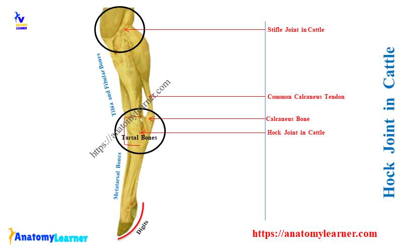
Hock joint in cattle
First, I would like to provide an overview of the cattle’s hock. The hock of a cattle consists of several articulations –
- A tibiotarsal articulation,
- The intertarsal articualtion, and
- The tarso-metatarsal articualtion,
Type of joint: here, the tibiotarsal articulation is ginglymus (synovial), and other articulations are gliding types.
Bones involvement in the cattle hock: the distal end of the tibia, tarsal bones, lateral malleolus, and a proximal end of metatarsals. Let’s see the specific bone involvement of 3 articulations of the cattle’s hock from Table 1 –
| Hock joint in cattle | Bones involvement |
| Tibiotarsal joint | Trochlea of talus, and The corresponding surface of the distal end of the tibia |
| Proximal intertarsal joint | Talus and calcaneus, and Central and 4th tarsal bone |
| Distal intertarsal joint | Central tarsal, and Distal tarsal on either side |
| Tarsometatarsal joint | Distal tarsal and The proximal end of the metatarsal bone |
Here, the intertarsal joint of the hock divides into proximal and distal intertarsal articulations.
Movement of the cattle hock: the tibiotarsal articulation is responsible for extension and flexion. In contrast, the other articulations of the cattle hock are responsible for gliding movement.
Binding materials (ligaments) of the cattle hock: you will find the below-mentioned common ligaments in the cattle hock structure –
- Joint capsule or capsular ligament,
- Lateral collateral ligament – divides into long lateral and short lateral ligaments,
- Medial collateral ligament – consists of long medial and short medial ligaments,
- Triangular dorsal ligament,
- Flat and strong plantar ligaments, and
- A few special ligaments of the cattle hock – these are the short bands that connect adjacent bones of the tarsus and metatarsus.
You will find the details of the attachment of these ligaments from the cattle hock in the description section.
What is the hock of a cow?
The hock of a cow is the hindlimb’s joint located between the lower end of the tibia and the upper end of the metatarsals. It consists of multiple joints among the tibia, tarsal, and metatarsal bones of the cow.
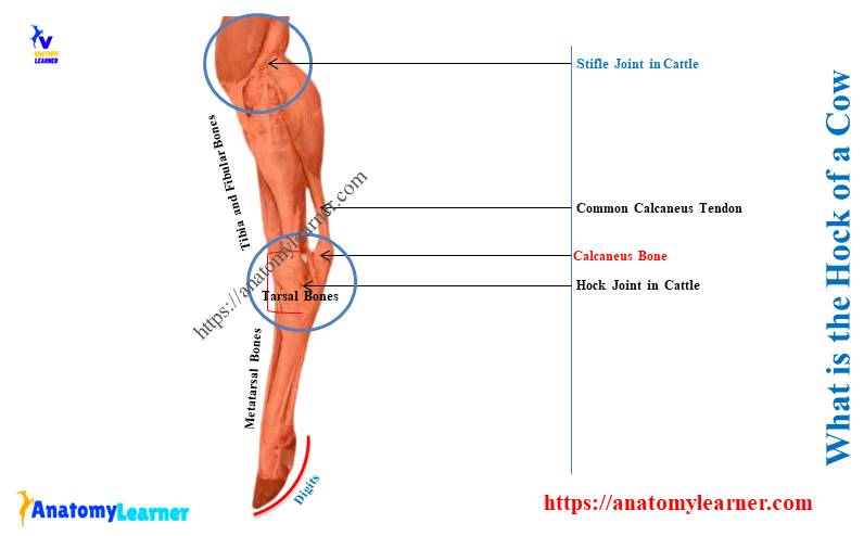
This region (from the distal end of the tibia to the proximal end of the metatarsals) is known as the hock region of a cow. It is a true joint of the hindlimb of a cow that follows all the features of a typical joint.
Let’s see the typical features of the animal joint from the below-mentioned article –
“The cattle hock is also known as the tarsal or pedal articulation.”
What type of joint is the cattle hock?
The cattle’s hock is the ginglymus (synovial) and gliding type of hindlimb joint. You will find the typical features of the synovial joint in the tibio-tarsal articulation of the cattle’s hock.
Again, the intertarsal and tarsometatarsal joints of the hock are gliding type.
As a synovial joint, the tibiotarsal articulation shows the extension and flexion movement. I have already described the features of the typical synovial joint in animals.
Let’s see the below-mentioned article for more details about the animal’s synovial joint –
What are the joints of the fore and hind legs in cattle?
Here, the diagram shows the joints from the cattle’s fore and hind legs. You will find the below-mentioned joints in the forelimb of the cattle –
- Shoulder joint (humeral articulation),
- Elbow joint – consists of humero-radial, humero-ulnar, and radio-ulnar joints,
- Carpal or knee joint – consists of ante-brachiocarpal, intercarpal, and carpometacarpal,
- Intermetacarpal joint,
- Metacarpophalangeal joint (fetlock joint),
- Proximal interphalangeal joint (pastern articulation) and
- Distal interphalangeal joint (coffin articulation),
Again, the hind leg of a cattle shows the below-mentioned joints –
- Sacro-iliac articulation of the hindlimb,
- Pelvic symphysis – consists of pubic and ischial symphysis,
- Coxal articulation (hip joint of the cattle),
- Stifle joint or genual articulation of the cattle’s leg – consists of femoropatellar and femorotibial joints,
- Tibiofibular articualtion – proximal and distal articualtions,
- Tarsal or hock or pedal articulation (describe here in detail) and
- Fetlock, pastern, and coffin joints of hindlimb like forelimb,
You may know more about the different joints of the animal’s body with their bone involvement –
Description of hock joint in cattle anatomy
The tibiotarsal joint of the hock of a cattle is a typical ginglymus. It is formed by the trochlea of the tibial tarsal and the corresponding surface of the distal end of the tibia bone.
The ridges and grooves of these articular surfaces are directed obliquely forward. Here, the articular surface of the trochlea is about twice as extensive as that of the tibia bone.
Suggested articles from anatomylearner related to this hock joint –
- Tibia and fibula of ox – osteological features of the leg bone of animals, and
- Cow metatarsal bone – how many metatarsals does a cow have?
The other two joints – intertarsal and tarsometatarsal, are arthrodia. They also have articular surfaces and ligaments that allow them a minimal amount of gliding movement.
The intertarsal joint of the cow’s hock consists of –
- Proximal intertarsal joint – is the articulation between talus and calcaneus, and central and 4th tarsal bone, and
- Distal intertarsal joint – is the articulation between central tarsal and distal tarsal on either side,
Finally, the tarsometatarsal articulations of the cattle hock are formed between the distal tarsal and proximal end of the metatarsal bone.
Cow tarsal bones and joints
To understand the hock joint, you might also have a clear idea of the tarsal bones of a cow. There are five pieces of tarsal bones in the cow tarsus –
- Tibial tarsal (also known as talus),
- Fibular tarsal (also known as calcaneus),
- Central and fourth tarsal of the cow and
- The first tarsal and fused second and third tarsals,
These tarsal bones of the cow are arranged in three rows. Here, the diagram shows the arrangement of the cow tarsal bones from the lateral to the medial aspect.
The first row of the tarsus consists of tibial tarsal and fibular tarsal. Here, the tibial tarsal faces medially, whereas the fibular tarsal faces laterally.
The middle row of the cow tarsus consists of the central and fourth tarsal bones. Again, the distal of the third row consists of the medial first tarsal and fused lateral second and third tarsal.
The tibial tarsal is long, narrow, and flattened dorsolaterally. It bears trochlea on either end.
The calcaneus of the cow tarsus is longer and more slender. Distally, this bone possesses a projection that articulates with the lateral malleolus bone.
The central and fourth tarsal bones are in the form of quadrilateral plates. They extend across the entire width of the cow’s tarsus.
The first tarsal of the cow tarsus is also quadrilateral but smaller than the central and fourth tarsal. Finally, the second and third tarsal of a cow tarsus are rhomboid in shape and face medially.
Ligaments of the hock joint in cow
For description purposes, the whole ligaments of the cattle hock joint divide into –
- Common ligaments – consist of joint capsule, lateral, medial, dorsal, and plantar ligaments, and
- Special ligaments – you will find several short bands or ligaments in the cow hock,
The special ligaments of the cow hock include –
- 4 ligaments between the tibia and fibular tarsals,
- Short ligament among central, third, and fourth tarsals,
- Ligament between tarsal and tibia bone, and
- Ligaments between distal ends of tarsals and metatarsal bones,
First, let’s describe the common ligaments of the cow’s hock joint with the labeled diagram.
The joint capsule or capsular ligament of the cow hock
This ligament of the cow hock lines with the synovial membrane. The fibrous part of the joint capsule attaches around the margin of the tibial and metatarsal articular surfaces.
The part of this ligament is also attached to the collateral ligament of the hock. This capsular ligament of the cow hock divides into –
- The dorsal part – known as the anterior ligament and
- The plantar part – is also known as the posterior and tarsometatarsal ligament,
The dorsal part of the joint capsule or capsular ligament is thin. Whereas the plantar part is thick and immediately attaches to the tarsal bones.
There are four synovial sacs in the joint capsule of a cattle hock– –
- Tibio-tarso sac – it is larger and lubricates the proximal part of the hock,
- Proximal intertarsal sac – it communicates with the tibiotarsal sac cranially,
- Distal intertarsal sac – it lubricates the joint between central and other tarsals and
- Tarso-metatarsal sac – it lubricates the joint formed between the tarsal and metatarsal bones,
The lateral collateral ligament of the cow hock
The cattle hock shows two distinct bands in the lateral collateral ligaments –
- Long lateral ligament – superficially, and
- Short lateral ligament – deeply,
The long lateral ligament arises on the posterior part of the lateral malleolus. It is almost straight downward and attaches to the fibular and fourth tarsal.
This long ligament also attaches to the large and lateral small metatarsal bones of the cattle. Finally, this ligament forms a canal for the lateral extensor tendon.
The short lateral ligament arises on the anterior part of the lateral malleolus. It directs backward and ends on the lateral surface of the tibial tarsal and adjacent fibular tarsal.
Medial ligament of the cattle hock joint
The medial ligament of the cattle hock also consists of two parts that cross each other –
- Long medial ligament – superficially, and
- Short medial ligament – deeply,
The long medial ligament of the cattle hock arises on the posterior part of the lateral malleolus. It becomes wider and attaches to the distal tuberosity of the tibial tarsal. This long medial ligament also attaches to the large and medial small metatarsal.
The short medial ligament of the cattle hock lies under the long medial ligament. It extends from the anterior part of the medial malleolus to the proximal tuberosity on the medial surface of the talus.
This short medial ligament divides into two parts. One part of this ligament ends on the talus, and the other ends on the sustantaculum tail.
The dorsal oblique ligament of the cow hock
This is a triangular ligament of the cow hock that attaches to the distal tuberosity of the tibial tarsal. It spread out below the central and third tarsal bones.
Finally, the dorsal ligament attaches to the proximal end of the large and outer small metatarsal bones.
Plantar ligament of the cattle hock
This is the strong and flat ligament of the cattle hock anatomy. It covers the outer part of the plantar surface of the tarsus.
This ligament attaches to the plantar surface of the fibular and fourth tarsals of the cow. It also attaches to the proximal part of the lateral metatarsal bone.
Special ligaments of the cow hock joint
You will find the below-mentioned special ligaments in the cow hock joint –
In tibial and fibular tarsal bones:
Four short bands unite the tibial and fibular tarsal bones –
- Medial ligament – extends from the sustentacular tail to the adjacent part of the tibial tarsal,
- Lateral ligament – extends from the trochlear process of the fibular tarsal to the adjacent part of the trochlea,
- Proximal ligament – extends from the caudal margin of trochlea to the fibular tarsal and
- Interosseous ligament – located between the two adjacent tarsal bones,
Among central, third, and fourth tarsal bones:
- Interosseous and oblique ligaments – located between central and third tarsal bones,
- Lateral transverse and interosseous ligaments – located between the central and fourth tarsal bones,
- Plantar transverse liagement – present between the third and fourth tarsal bones,
Ligaments in the proximal row of tarsus:
- Plantar and interosseous ligament – remains between the central tarsal and tibia,
- Short oblique ligament – locates between the fibular and central tarsals,
- There are other different interosseous and plantar ligaments in the cattle hock structure.
Ligaments in distal tarsal and metatarsals:
- Tarso-metatarsal ligament – remains between distal tarsal and metatarsal bones and
- Interosseous ligament – present between the third tarsal and metatarsal bones,
Special features of ox hock joint
Let’s see the special features of the ox hock joint –
- The joint capsule of the ox hock joint is roomy,
- There is very considerable mobility at the proximal intertarsal joint,
- A short lateral ligament attaches distally on the tibial tarsal only,
- There is a strong transverse ligament attaches the lateral malleolus to the tibial tarsal bone,
- The dorsal oblique ligament of the ox hock is narrow and thin,
Conclusion
The hock joint in cattle is a complex structure made up of tibiotarsal, intertarsal, and tarsometatarsal joints. It is the combination of ginglymus and gliding types of joint.
The tibiotarsal joint of the hock exhibits extension and flexion movement. In contrast, the other two joints have a gliding movement.