The ovaries of a cow and a mare are a little different anatomically. Let’s know the answer to the question – what is the difference between the ovaries of a cow and a mare?
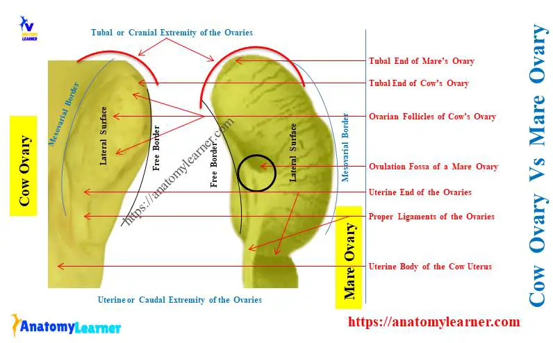
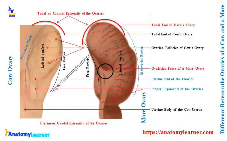
Quick answer: the ovaries of a cow are oval and smaller than the bean-shaped ovaries of a mare. An ovulation fossa is present on the free border of a mare’s ovary, whereas it is absent from the cow’s ovary.
You will also find differences in the location and internal structures of cow and mare ovaries. Here, I will provide the unique features of bovine and equine ovaries with their anatomical differences.
Let’s know the differences between the ovaries of these two species in detail.
What is the difference between the ovaries of a cow and a mare?
Cow’s ovaries are two oval bodies on each side, located a little above the middle of the pelvic inlet. The caudal extremity is connected to the horn of the animal’s uterus by the proper ligament of the ovary.
The mare’s ovaries are larger and bean-shaped bodies located at the sublumbar region. Both cow and mare’s ovaries present two surfaces, two borders, and two extremities.
But, you will find important differentiating features between the cow and mare’s ovaries. Let’s see the difference between the ovaries of a cow and a mare from Table 1 –
| Features of ovary | Cow ovaries | Mare ovaries |
| Location | Little above the middle of the lateral margin of pelvic inlet | In the sublumbar region, Usually, ventral to fourth or fifth lumbar vertebrae, |
| Shape of ovary | Oval-shaped in cow, Pointed in uterine end, | Bean-shaped in mare |
| Size of ovary | Length – 1 – 1.5 inches, Thickness – 1 inch, | Varies much in different subjects – Length – upto 3 inches Thickness – upto 1 inch |
| Weight | 19 – 25 grams | 75 – 90 grams |
| Free border of ovaries (Ovulation fossa) | Free border of cow ovary is convex, Have no ovulation fossa, | Free border of mare ovaries are marked by notch, These notches lead into a narrow depression, They are known as ovulation fossa, |
| Germinal epithelium | Greater parts of cow’s ovaries are covered by germinal epithelium | Only ovulation fossa is covered by germinal epithelium |
| Cortex and medulla | Cortex – outer zone of a cow ovary, Medualla – inner zone of a cow ovary, | The position of cortex and medulla are reverse in mare’s ovary Cortex – inner zone, and Medulla – outer zone of ovary, |
| Location of follicles | Follicles locates on the outer zone of ovary, Follicles of various sizes are often projecting from the surfaces, | Inner zone of ovaries (cortex) |
| Corpus luteum | Pronounced yellow color | Yellow mass |
Gross anatomy of cow and mare’s ovary
Grossly, the ovary of a cow shows 2 surfaces, 2 borders, and 2 extremities. Let’s identify the below-mentioned gross features from the cow’s ovary –
- Right and left ovaries of the cow,
- Medial and lateral surfaces of the ovaries,
- Attached and free borders of the cow ovaries,
- Tubal and uterine extremities of the ovaries,
- Ovarian follicles on the surfaces of the cow’s ovaries,
- Corpus luteum on the ovaries,
- Proper ligament of the cow ovaries (ovarian ligament) and
- Meovarium (part of the broad ligament) of a cow ovary,
All these structures mentioned above are identified in the cow ovary labeled diagram. You may find more labeled diagrams on bovine ovaries here.
Overview of gross anatomy of the ovary of a cow
For description purpose, the cow ovary present 2 surfaces, 2 borders, and 2 extremities –
- Surfaces – medial and lateral,
- Borders – attached or mesovarial and free borders, and
- Extremities – tubal or cranial and uterine or caudal extremities,
Both the lateral and medial surfaces of the cow ovaries are smooth and rounded. You will find follicles of various sizes on the smooth lateral surface.
These follicles remain in different stages of the development. Mostly, you will see rounded, fluid-filled graafian follicles on the lateral surface.
These follicles are scattered at the periphery of the cow ovary. Rupture of these follicles occurs, and you will see the remnant of these ruptured follicles.
The remnant of the ruptured follicles is known as the corpus luteum. They are yellowish bodies and well-developed in the cow ovary.
Sometimes, the follicles disappear from the surface and form the scar tissue.
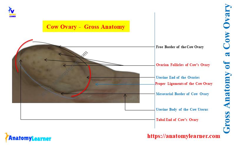
The attached or mesovarial border of the cow ovary is convex. The cranial part of the broad ligament encloses this border. It is the mesovarium of the cow’s ovary.
The free border of the cow ovary is also convex and doesn’t present any identifiable or unique features.
The tubal or cranial extremity of the cow ovary is rounded. It is connected to the fimbriated end (infundibulum) of the uterine tube.
Again, the uterine or caudal end of the cow ovary is also rounded. It connects to the horn of the cow uterus by the proper ovarian ligament.
You may know the details of anatomical features of the cow uterus from the below-mentioned article –
What is the structure of the ovaries in a cow?
The germinal epithelium externally covers the structure of the normal ovaries in a cow. This germinal epithelium is continuous with the peritoneum.
You will find the tunica albugenia, which is a fibrous layer just below the germinal epithelium. The whole structure of the cow’s ovaries may divide into –
- Cortex – outer area of the cow ovary and
- Medulla – inner or central area of the cow ovary,
The cortex of the cow ovary consists of follicles in various stages of development. You will also find the corpus luteum and connective tissue stroma in the cortex of a cow ovary.
The medulla of the cow ovary consists of vessels, nerves, lymphatics, smooth muscle, and connective tissue.
The ovaries of a cow undergo maximum histological changes in different stages of life. Thus, you may also know the histological structures of the cow ovaries.
What are Graafian follicles in a cow’s ovary?
Answer: Graffian follicles are the rounded bodies scattered mostly at the peripheral part of the cow ovary. They are found in the ovarian surfaces in different stages of development.
These follicles of different stages are called primary, secondary, tertiary or graffian follicles. You may feel the graafian follicles during the palpation of a cow’s ovary.
Let’s find the details of histological features of the Graafian follicles from the ovary –
What is the corpus luteum in a cow ovary?
Answer: The remnant of the ruptured follicles of a cow ovary reorganize to form the corpus luteum. It is well-developed in a cow ovary and reddish yellow color.
They are the outgrowths from the surface of the cow ovary for a certain period. Thus, you may easily identify this corpus luteum in a cow ovary grossly.
How many ovaries does a horse have?
Answer: a horse has two ovaries (right and left ovary) that attach to the corresponding uterine tubes. They are bean-shaped, and their size varies much in various subjects.
Typically, you will find larger ovaries in the young horse than in the older horses. One ovary is larger than the other in the horses.
The position of the ovaries in a horse is very inconstant within the abdominal cavity. It depends on the mode of attachment with the sublumbar region of a horse.
What is unique about the equine ovary?
Answer: the unique features of the equine ovaries are –
- The ovaries are larger and bean-shaped in equine,
- The free border (margo liber) of an equine ovary is marked by a notch that leads into a narrow depression,
- This depression is present in both the right and left ovaries of an equine and is known as the ovulation fossa,
- Both right and left ovaries of equine are situated in the sublumbar region (ventral to 4th or 5th lumbar vertebrae),
- They (ovaries of the equine) are usually in contact with the lumbar wall of the abdomen,
- Each ovary of an equine is attached to the sublumbar region by the cranial part of the broad ligament,
- The uterine end of the equine ovaries connects with the uterine horns by ovarian ligament,
- The greater part of the surfaces of the equine ovaries have a peritoneum covering,
- You will not see the peritoneal investment at the attached border of the equine ovary,
- The ovulation fossa of the equine ovary is covered by a layer of short polygonal cells (germinal epithelium),
- The position of the cortex and medulla of the equine ovary is reversed compared to the cow’s ovary, and
- In the new-born foal, the ovaries are larger and oval in form,
Key differentiating points of ovaries of a cow and a mare
According to the gross features of the ovaries of a cow and a mare, following are the key differentiating points –
- Shape of the cow and mare ovaries (oval and bean),
- Condition of the free border of the ovaries (presence or absence of ovulation fossa) and
- The internal structure of the ovaries (position of cortex and medulla),
By these features, you may easily differentiate the cow ovary from the mare ovary grossly. Now, let’s see the video where I tried to explain the difference between the ovaries of a cow and a horse.
Conclusion
The answer to the question – ‘what is the difference between the ovaries of a cow and a mare?’ was explained. Thus, the main difference between the ovaries of a cow and a horse is in their shape and border.
The shape of the cow’s ovaries is oval and pointed, whereas the horse shows bean-shaped ovaries. Again, the ovulation fossa is unique in equine ovaries, which are absent from the cow ovaries.