The stomach of a horse is the large dilation of the alimentary canal just caudal to the diaphragm. In this short article, I will discuss the horse stomach anatomy with a labeled diagram. So, here you will get all the external and internal features from equine stomach anatomy. Again, I will try to cover all the inquiries on horse stomach anatomy and other organs of the equine digestive system.
If you are interested to know equine stomach anatomy, then continue this article. I hope this article will help you to know everything about horse stomach.
Peculiar features from horse stomach anatomy
First, I would like to provide you the most peculiar features of horse stomach anatomy with a diagram. Then I will show you all the anatomical features you might identify grossly at the veterinary anatomy laboratory.
The horse’s stomach is placed at the left slide of the median plane on the dorsal aspect of the abdomen behind the liver and diaphragm. This stomach intervenes between the esophagus and the small intestine.
Now, let’s find these peculiar anatomical features from the horse stomach –
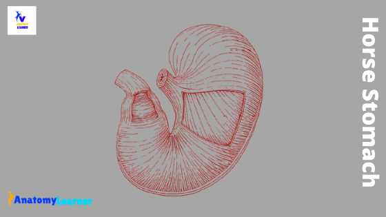
- The stomach horse is in the form of a simple saccular structure (curved J-shaped sac).
- The capacity is about twelve liters (may vary)
- Presence of short lesser, and greater curvatures in horse stomach.
- At the left extremity of the horse’s stomach, close to the entrance of the esophagus, there is a rounded cul-de-sac (saccus cecus).
- The glandular and non-glandular parts of the horse stomach are separated by a rough line known as margo plicatus.
- Again the glandular part divides into cardiac, fundic, and pyloric gland regions according to the presence of different types of gland in the mucous membrane.
- There is a well-developed pyloric sphincter in the horse stomach.
These are the very short features of equine stomach anatomy. Now, let’s know which structures you might identify grossly from the horse’s stomach.
- Saccus cecus of horse stomach
- Short lesser curvature
- The greater curvature of horse stomach
- Non-glandular part of the stomach (esophageal region)
- Margoplicatus of horse stomach
- Cardiac gland region of the stomach
- Fundus gland region of equine stomach
- Pyloric gland region of horse stomach
- Pyloric sphincter at the pyloric orifice
These are the basic anatomical features from the equine stomach that you might identify grossly. For more anatomical features of the horse stomach, you may continue this article.
Horse stomach anatomy diagram
Fine, now I will go over the detailed features of horse stomach anatomy with labeled diagrams. The horse stomach is a sharply curved, J-shaped sac-like structure. The right part of the stomach is very much short than the left part.
The convexity of the horse stomach is directed ventrally. There is a small constriction that divides the horse’s stomach into right and left sacs.
Do you want to know the exact location of the horse’s stomach? The horse stomach is located in the dorsal part of the abdominal cavity, caudal to the diaphragm and liver, and mainly left to the median plane of the body.
You will find two surfaces, two curvatures, and two extremities in the horse’s stomach. Now. I will discuss the external features of equine stomach anatomy.
Surfaces of horse stomach
Let’s discuss the surfaces of the horse stomach. There are two different surfaces in a horse’s stomach – the parietal surface and the visceral surface. The parietal surface of the horse’s stomach is convex and directed cranially, dorsally, and towards the left. This surface lies against the diagram and the liver.
The visceral surface of the equine stomach is also convex and faces in the opposites direction. This visceral surface relates to the terminal part of the colon, the small intestine, and the greater omentum.
Curvatures of horse stomach
There are two curvatures (lesser and greater) in the horse’s stomach. Do you know how these curvatures form? The borders between the visceral and parietal surfaces create these curvatures.
The lesser curvatures are very short and extend from the esophagus’s terminal part to the junction with the small intestine. On the other hand, the greater curvature of the horse stomach is very extensive.
First, the greater curvature is directed dorsally and curves over the left extremity of the stomach. Then it descends, passes to the right, crosses the median plane, and curves dorsally. Finally, the greater curvature ends at the pylorus.
The left part of the great curvature relates to the spleen. Again the ventral aspect of the greater curvature rests on the left parts of the great colon.
Extremities of horse stomach anatomy
There are two extremities in horse stomach anatomy – the left extremity and pyloric extremity. You will find a special feature on the left extremity of the horse stomach.
There is a round form structure in the left extremity of the horse stomach. This rounded form structure is known as cul-de-sac or saccus cecus.
This saccus cecus lies ventral to the left crus of the diaphragm and so beneath the dorsal part of the sixteenth and seventeenth ribs.
Caudally it is related to the pancreas and the terminal part of the great colon. Again, it connects with the spleen laterally.
You will find a smaller pyloric extremity in the horse stomach. This pyloric extremity of the horse stomach continues with the duodenum. There is a marked constriction between the junction of the pyloric extremity and the duodenum.
The pyloric extremity of the stomach lies just to the right of the median plane. It is in contact with the visceral surface of the liver. You will find a constriction that marks off the pyloric antrum from the rest of the right sac.
The pylorus is the opening into the intestine, and its position indicates externally by a distinct constriction. Internally, there presence a circular ridge (ring of muscular tissue) in the pylorus. You know, this structure is called the pyloric sphincter.
The esophageal orifice or ostium cardiac locates at the left end of the lesser curvature. But the position of the cardia varies with the excursion of the diaphragm. It usually lies to the left of the median plane and ventral to the vertebral end of the fourteenth rib.
The esophagus joins the horse’s stomach very obliquely. Again, the opening is closed by the sphincter cardiae and numerous folds of the mucous membrane.
How the stomach held in position in a horse?
While studying horse stomach anatomy, it is also important to know how the stomach held in its position? The stomach of a horse is held mainly by the pressure of the surrounding viscera and the esophagus. You will find the following peritoneal folds that connect the stomach with the adjacent parts.
- The gastrophrenic ligament of the horse stomach
- Lesser and greater omentum of horse stomach
- Gastrosplenic ligament and gastropancreatic folds
The gastrophrenic ligament connects the greater curvature of the stomach from the cardia to the left extremity. It also connects the crura of the diaphragm. But you will find a narrow area that is uncovered by the peritoneum. In this part, the stomach is attached to the diaphragm by the areolar tissue.
The lesser omentum connects the lesser curvature of the horse stomach and the first part of the duodenum with the liver.
The gastrosplenic ligament passes from the left part of the greater curvature of the horse stomach to the hilus of the spleen. This ligament is continuous ventrally with the greater omentum.
The greater omentum connects the ventral part of the greater curvature and the first curve of the duodenum. Again, it connects with the terminal part of the great colon and the initial part of the small colon.
The gastropancreatic fold extends from the left sac dorsal to the cardia to the duodenum. This ligament attaches to the liver and dorsally, pancreas ventrally.
Structure of horse stomach
Now, I will show you the structure of the horse’s stomach. The wall of the horse stomach consists of four different layers or coats – mucosa, submucosa, muscularis mucosa, and serosa. I will describe each coat with important information.
Serous coat of horse stomach
The serous coat covers the great part of the horse stomach. It closely attaches with eh muscular coat, except in the curvatures. It partially bridges over the lesser curvature and covers the elastic tissue that assists in retaining the next from the stomach.
Muscular coat of horse stomach anatomy
The muscular coat of horse stomach anatomy consists of three incomplete layers. You will find the following incomplete layers of muscle in the horse stomach –
- An external longitudinal muscle layer
- A middle of the circular muscle layer and
- An internal of the oblique muscle layer
The external longitudinal muscle layer is very thin and present only along the curvatures and at the antrum. You will not find this longitudinal muscle layer in the saccus cecus and middle of the greater curvature.
You will find the elastic fibers in the middle of the greater curvature instead of longitudinal muscle fiber. But in lesser curvature, you may find the longitudinal muscle fibers. In the pyloric antrum, there presents a well-developed longitudinal muscle fiber in a horse.
You will find the middle circular muscle layer only on the glandular part of the horse stomach. The oblique muscle fibers are arranged in coarse bundles into two layers.
The external oblique muscle fibers are found in the left sac that is the continuation of the longitudinal fibers of the esophagus. Again, the internal oblique fibers also find in the left sac and continuous with the circular fibers of the esophagus. Here the stomach and esophagus exchange their fibers with the external oblique muscle fiber. They help to form a remarkable loop around the orifice of the cardiac (cardiac sphincter).
Submucosa and mucosa coat of horse stomach
The submucosa coat of the horse stomach is a layer of loose connective tissue that connects the muscular and mucosa coats. You will find a lot of blood vessels and nerves in this submucosa layer.
The mucosa coat is divided into two parts – the proventricular part and the glandular part. The proventricular part lies the greater part of the left sac and resembles the esophageal mucous.
It is white and covers with a thick, stratified squamous epithelium. There is no glands in the proventricular part of the horse stomach.
At the cardia orifice, you will find numerous folds. These folds terminate abruptly, forming an irregular, sinuous, raised edge known as margo plicatus.
Below this margo plicatus ridge, you will find the glandular part of the horse’s stomach. There are different gastric glands in the glandular part of the horse’s stomach.
The glandular part of the horse stomach is divided into three different zones according to its glands.
- Cardiac gland region,
- Fundic gland region and
- The pyloric gland region
Regions of glandular part of horse stomach
The cardiac gland region of horse stomach anatomy contains the short tubular cardiac glands. It is the narrow zone along the margo plicatus but does not extend to the greater curvature. In fresh condition, you will find the yellowish-gray color in the cardiac gland region of the horse stomach.
The cardiac gland region of horse stomach is extremely narrow at the greater curvature but becomes wide towards the pyloric part.
The fundic gland region of the horse stomach contains the fundus glands. This is a large area of the glandular part that has a mottled, reddish-brown color. You will find two different types of fundus glands in the fundic gland region of the horse stomach.
There is a very thick mucous membrane and vascular system present in the fundus region. This fundus region resembles the fundus of the stomach in a dog.
The pyloric gland region has a very thin mucous membrane, reddish-gray or yellowish-gray color.
The pyloric sphincter s the thick ring at the pyloric orifice. You will also find another ring at the left end of the pyloric antrum. You will find circular folds that cover the pyloric sphincter (pyloric valve)
The folding of the stomach wall at the lesser curvature produces a prominent ridge that projects into the stomach cavity.
Blood and nerves supply to the equine stomach
The stomach of the horse receives blood supply from all the branches of the celiac artery. The nerves are derived from the vagus and sympathetic nerves in the horse stomach.
Horse stomach diagram
Now, I will show you again all the anatomical features of the horse stomach with a diagram. You might practice with the real sample of horse stomach. For more digrams on horse stomach, you may follow anatomy learner on social media.
Horse digestive system diagram
Here I will show you every single organ from the horse digestive system diagram. But you may know the detailed anatomy of all organs from the horse digestive system from the horse anatomy section.
Horse stomach capacity
The stomach of a horse is relatively small. The capacity of horse stomach varying from eight to fifteen liters (average twelve liters).
The size, form, and position of the horse’s stomach are subject to considerable variation. In the empty condition, the saccus cecus of the horse stomach contains gas and strongly contracted. The middle part of the saccus cecus contains ingesta and preserves its rounded features.
In the empty condition, the colic of the small intestine usually lies ventral to the stomach and may separate from the colon.
When the stomach is moderately full, its most ventral part lies opposite the ninth intercostal and tenth ribs.
Frequently asked question on equine stomach
So, in this part, you will get the frequently asked question on horse stomach anatomy. If you have any inquiries on horse stomach, please let me know.
Do horses have 4 stomach?
You have known, there are four different parts in the stomach of ruminant (rumen, reticulum, omasum, and abomasum). But do horses have these 4 stomachs? The answer is no; horses have a simple stomach like dogs and cats. You will find glandular and non-glandular parts in a horse’s stomach (already discussed). Again, there are three different parts (cardiac, fundic, and pyloric) in the glandular part of the horse’s stomach.
How many stomachs does a horse have?
The horse has only one simple secular-type stomach. You will find glandular and non-glandular parts in that simple saccular stomach of a horse. Again, there are three different regions in the glandular part of the horse stomach.
Where is the stomach located in a horse?
The horse stomach is located mainly to the left of the median plane of the horse body, in the dorsal part of the abdominal cavity. Cranial to the horse’s stomach, you will find the diaphragm and liver.
What type of stomach does a horse have?
The horse has a simple saccular structure stomach. It is J-shaped and located at the left side of the median line on the dorsal aspect of the abdomen.
Conclusion
There is a great variation in the anatomical features of horse stomach compare to ruminant. You will find the four-chambered stomach in ruminant, whereas you will find a simple saccular stomach in a horse. I hope this simple guide helps you a lot to learn horse stomach anatomy. Please use the horse stomach anatomy labeled diagram and real sample for practices.