The horse skeleton is the rigid framework of the body that consists of bones, cartilages, and ligaments. There are two hundred and five bones found in horse skeleton. In this long article, I will discuss the osteological features of all bones from the horse skeleton anatomy labeled diagram.
I will also try to cover all possible inquiries on the horse skeletal system at the end of this article. If you are interested to learn anatomical features of bones from the axial and appendicular skeleton of a horse, you may continue this article.
Special features of horse skeleton anatomy
I will enlist few special features of few bones from horse skeleton anatomy. These special features might help you to get a basic idea of horse bones. But, you might learn the detailed anatomical facts of every single bone from the horse skeletal system.
Few special osteological features from the axial and appendicular skeleton of a horse –
- The skull of a horse is long and four-sided. You will find an extensive foramen lacerum in the horse skull.
- There is no cornual process in horse skull. The fusion between the two haves of the mandible is complete.
- There is no acromion process in the scapula of a horse. The glenoid notch is distinct and deep.
- The musculospiral groove is more deep and twisted in a horse.
- The ulna bone is ill-developed in a horse, and you will find an extensive semilunar notch at the proximal end.
- There is one large metacarpal and two small metacarpal bones in a horse.
- The gluteal line on the horse ilium bone is not prominent. Likewise, you will not find any ventral tubercle in horse hip bones.
- You will find the third trochanter at the proximal end of the lateral border of the femur. The fovea capitis is deep and notched in a horse.
- The anterior tuberosity of the horse tibia is grooved.
- You will find seven carpal and six tarsal bones in the skeletal horse system.
- The wings of the atlas are pierced posteriorly by the foramen transversarium.
- The sternum of a horse is boat-shaped, and compressed laterally.
- There are eighteen pairs of ribs in the horse skeleton, out of which the first eight pairs are sternal, and the rest ten pairs are asternal ribs.
Now, you might learn the detailed anatomy of every single bone from a horse.
Horse skeletal system anatomy
Thanks for continuing this article; now, I will discuss the osteological features of every single bone from horse skeletal anatomy. For description purpose, any skeleton is divided into two parts – axial and appendicular skeleton.
- The axial skeleton of the equine and
- An appendicular skeleton of equine
The appendicular skeleton of a horse
The appendicular skeleton of a horse consists of forelimb and hindlimb bones. I hope you know the name of all bones from the forelimb and hindlimb of a horse.
Forelimb bones of horse
In forelimb of the horse consists of the following bones –
- Scapula of horse
- Humerus of horse
- Radius and ulna bones of horse
- Carpal, metacarpal, and phalanges of horse
Again, in the hindlimb of the horse consists of the following bones –
- Bones of the pelvic girdle (ilium, ischium, and pubis bones of a horse)
- Femur of horse
- Tibia and fibula bones of horse
- Tarsal, metatarsal, and phalanges of horse
So, what are the peculiar features of these bones? Okay, let’s start to know the peculiar features of these bones.
Scapula of horse anatomy
The pectoral girdle of the horse consists of the scapula and fused coracoid. There is no clavicle in a horse’s pectoral girdle. Instead, the scapula of a horse is a flat, well-developed bone that locates on the cranial part of the lateral wall of the thorax.
This scapula is also triangular in outline and posses two surfaces, two borders, and three angles like ox scapula. The lateral surface divides into two unequal fossae by the spine of the scapula – supraspinous fossa and infraspinous fossa.
The spine of the horse scapula is directed backwards and not reach upto the distal end. Therefore, you will not find any acromion process like ox scapula. But other osteological features (like surfaces, borders, and angles) are similar to ox scapula.
There is a hollow subscapular fossa at the costal surface of the horse scapula. In a ventral or glenoid angle, you will find an oval glenoid cavity for articulation with the head of the femur. The glenoid notch is more deep and round in horse.
The supraglenoid tubercle is the large, rough prominence in horse scapula. You will find a small but well-developed coracoid process at the medial aspect of the supraglenoid tubercle.
So, the main characteristics features of horse scapula are –
- The spine of the scapula does not contain the acromion process
- A subscapular fossa is more concave
- Supraglenoid tubercle is larger
- Presence of more deep and distinct glenoid notch
- Presence of well-developed coracoid process
I hope you will identify the horse scapula with the help of the above mentioned special osteological features.
Humerus from horse skeleton
The humerus is a long bone of horse skeleton anatomy that possess some peculiar osteological features. It consists of a body and two different extremities. The body or shaft is irregularly cylindrical and has a twisted appearance.
There presence four different surfaces in the shaft of the humerus of a horse. The lateral surface is smooth and is spirally curved, forming the musculospiral groove. This musculospiral groove is deeper in horse humerus compare to an ox.
The nutrient foramen is in the distal third of the medial surface of the humerus. The crest of the humerus is more distinct and bears a large deltoid tuberosity on its middle.
The proximal extremity of the horse humerus consists of the head, neck, two tuberosities, and the intertuberal groove. In addition, you will find an almost circular articular surface on the head of a horse humerus.
The intertuberal groove or bicipital groove locates cranially in between the greater and lesser tubercle. But this bicipital groove is further divided by an intermediate tubercle or ridge.
There are trochlea and capitulum at the distal extremity of the horse humerus. You will also find the olecranon fossa, radial fossa, lateral epicondyle, and medial epicondyle at the distal extremity of the humerus.
So, the most characteristics features of horse humerus are –
- Presence of twisted and more deep musculosprial groove
- There is a very prominent deltoid tuberosity in horse humerus
- The intermediate tubercle subdivides the intertuberal groove.
I hope the above mentioned special features might help you to differentiate the horse humerus from an ox.
Radius and ulna bones of horse
The radius bone is larger than the two bones of the forearm in the horse. The body of the radius bone is curved in its length, somewhat flattened craniocaudally and wide at its ends.
You will find the two surfaces and two borders in the shaft of the horse radius bone. The proximal extremity is flattened craniocaudally and wide transversely.
You will find the humeral articular circumferences, which corresponds to that of the distal end of the humerus. The medial surface of the olecranon process is more concave compare to an ox.
There is a well-marked radial tuberosity at the medial slide of the dorsal surface of the radius bone. The lateral tuberosity is more salient in horse radius bone.
The distal extremity posses trochlea and compressed craniocaudally. You will find three carpal articular surfaces at this distal extremity of radius bone – medial facet, lateral facet, and middle facet.
Okay, now I will enlist the salient features of horse radius bone –
- Presence of well-marked radial tuberosity at the proximal end.
- The olecranon process is more concave on its medial surface
- Presence of more extensive semilunar notch in horse radius
Let’s discuss the most reduced long bone from horse skeleton anatomy. The ulna is the most reduced long bone in a horse that locates caudal to the radius bone. You will find a three-sided shaft and two extremities in horse ulna bone.
The cranial surface applies the caudal surface of the radius bone that forms the interosseous space of the two bones.
The proximal extremity is the major part of the ulna bone and bears the olecranon process. Again the distal extremity posses the styloid process.
Carpal, metacarpal and phalanges of horse
The carpus consists of seven or eight carpal bones arranged into two rows – proximal and distal rows. The radial carpal is the largest bone of the proximal row in a horse. It is compressed transversely and six-sided bone.
The intermediate carpal is somewhat wedge-shaped, wider dorsally than primarily. Again, the ulnar carpal bone is the smallest and more irregular bone in the proximal row in the horse skeleton.
The accessory carpal bone locates palmar to the ulnar carpal and the lateral part of the trochlea of the horse radius bone.
You will find three metacarpal bones in horse skeletal anatomy. The third metacarpal is well-developed and termed as the large metacarpal of a horse. The other two metacarpals (second and fourth) are reduced bones and called small metacarpal or splint bones.
The body of the horse’s large metacarpal bone is semicylindrical and possesses two surfaces and two borders. The proximal extremity bears articular surfaces for the distal row of carpal bones. Again, the distal extremity posses an articular surface for the proximal phalanges and proximal sesamoid bone.
Each of the small metacarpal bone locates either side of the palmar aspect of the large metacarpal bone. Thus, the body of small metacarpal bones are three-sided and tapers to the distal end.
The proximal extremity of these small metacarpal bones is large, whereas; the distal extremity is usually small.
You will find three phalanges and sesamoid bones in the digit of the horse forelimb. The body of the proximal phalange is wider and much thicker.
The middle phalange is dorsopalmarly flattened, and its width is greater than its height. In the distal phalange of a horse, you will find three surfaces, three borders, and two angles.
Hindlimb bone of a horse
The hindlimb of the horse consists of the following bones –
- Hip bones (ilium, ischium, and pubis bones of horse)
- Femur of horse
- Tibia and fibula bones of horse
- Tarsal, metatarsal, and phalanges of horse
Now, I will discuss on details anatomy of hindlimb bones from horse skeleton. But if you want to know more anatomical features of any specific bones, please go to the general anatomy section and learn details about them.
The pelvic girdle of the horse consists of the ossa coxarum (os coxae of both sides), the sacrum, and the first three or more caudal vertebrae.
Os coxae or hip bones from horse skeleton
The os coxae or hip bones are the largest flat bone in horse skeleton anatomy. It consists primarily of three bones – ilium, ischium, and pubis that meet to form a large acetabulum.
The ilium of a horse is the largest of the three bones and posses three surfaces and three borders. The wide part of the ilium bone is a wing. Its gluteal surface face dorsolaterally and caudally. You will find a curved indistinct gluteal line in the gluteal surface of horse ilium bone.
The psoas tubercles are not so well developed in horse ilium bone. Tuber sacral curves dorsally and a little caudally.
A hemispherical and wide articular cavity (acetabulum) forms by the junction of three bones. You will find a deep notch and secondary notch in the acetabular cavity of the horse.
The ischium forms the caudal part of the ventral wall of the bony pelvis. The pelvic surface is smooth and slightly concave. The caudal border of the ischium is thick, rough and form the ischiatic arch.
The ventral surface of ischium bone is nearly flat. Therefore, you will not find any ventral tubercle on the ischium bone.
The pubis is the smallest bone of three parts of os coxae in a horse. It forms the pelvic floor’s cranial part and possesses a head, two surfaces, three borders, and two branches.
So, the summary of the os coxae bones of a horse is –
- Tuber sacral inclines backwards in horse
- Presence of ill-developed psoas tubercle in horse ilium
- The gluteal line is not prominent in the ilium bone.
- The acetabular cavity is very wide.
- There presence deep acetabular notch and secondary acetabular notch.
- Absence of ventral tubercle in horse
I hope these above characteristics features might help you to identify the hip bones of a horse.
The femur of horse anatomy
The femur is the largest and more massive bone in a horse skeleton. It extends obliquely distally and cranially. The femur articulates with the acetabulum proximally and the tibia and patella distally.
You will find a cylindrical body and two large extremities in the femur of a horse. The body is cylindrical but flattened caudally and larger distally in the horse femur. The body is convex side to side and posses four different surfaces.
The medial border bears a thick lesser trochanter on its proximal part. Again the lateral border contains a prominent eminence on its proximal part (trochanter tertius or third trochanter). Finally, at the distal part of the lateral border of the horse femur, you will find a deep supracondyloid fossa.
The proximal extremity of the horse femur is large and consists of the head, neck, and greater trochanter. In addition, you will find a deep notch in the head of the horse femur (known as fovea capitis femoris).
The trochanteric ridge is not oblique in the femur of a horse. Instead, it directs along the length of the femur.
The trochanteric fossa is deeper in horse compare to ox or goat.
The distal extremity of the horse femur is also large in both directions. This extremity consists of trochlea cranially and two condyles caudally.
There is a deep intercondyloid fossa in between the lateral and medial condyles of the horse femur. The medial epicondyle is the rounded prominence on the medial surface, whereas the lateral epicondyle is less distinct.
Horse femur identification points
So, what are the special osteological features of the horse femur? Fine, I will enlist the important osteological features of horse femur for you –
- It is more massive bone and contains a third trochanter
- Fovea capitis is deep and notched
- Trochanteric ridge directed along the length of bone
- The supracondyloid fossa is deeper than an ox.
Patella of horse
The patella of a horse is a large sesamoid bone that articulates with the trochlea of the femur. It is not as triangular as in an ox. Instead, you will find two surfaces, two borders, base, and apex in the horse patella.
The cranial surface of the patella is free and quadrilateral in shape. It is also convex and rough for muscular and ligamentous attachment.
The articular surface is also quadrilateral but less extensive. You will find the vertical ridge and two concave areas at the articular surface of the patella.
The medial and lateral borders converge to the apex distally, and each forms an angle at the base. The base patella is convex transversely and concaves craniocaudally. You will find a blunt apex in the patella of a horse.
Tibia from horse skeleton
The tibia is the longer and larger bone in horse skeleton anatomy that extends from the stifle joint to the hock. This bone articulates proximally with the femur, distally with tarsus, and laterally with the fibula bone.
The shaft of the tibia is large and three-sided proximally. It became smaller and flattened at the distal extremity. Thus, you will find the three surfaces and three borders in the tibia bone of the horse.
The lateral surface of the tibia is smooth and somewhat spiral in a horse. You will find a broad surface in the medial surface proximally. The caudal surface of the tibia bone is flattened and divided into different parts by the popliteal lines. You will find nutrient foramen on or near the popliteal line.
The cranial border of the horse tibia bone is very prominent proximally and forms the tibial crest. The medial border is rounded, and the lateral border is concave in a horse.
You will find a large and three-sided proximal extremity in the horse tibia bone. In addition, there are lateral and medial saddle-shaped articular eminence at the proximal part of the tibia.
You will also find the intercondyloid eminence or spine in between lateral and medial eminence.
There is a large cranial eminence (known as tuberosity of tibia) in the tibia. This tuberosity of the tibia is marked cranially by a groove in a horse.
Identifying features of horse tibia
The distal extremity of the tibia is much smaller and quadrangular in a horse. It presents an articular surface for the talus that consists of two grooves. A distinct ridge separates these two grooves.
Here in the groove and ridge, you will find peculiar osteological features that might help you identify the horse tibia bone. The ridge and the groove of the distal end of the tibia are directed obliquely, cranially and laterally.
Fibula of horse
The fibula of a horse is a much reduced long bone that finds along the lateral border of the tibia bone. In addition, you will find a slender rod-shaped shaft in a fibula that forms the wide interosseous space of the leg.
The proximal extremity of the horse fibula is relatively large and flattened transversely. Its medial surface presents a narrow area along the proximal border for articulation with the lateral condyle of the tibia bone.
The distal extremity of the fibula is fused with the tibia bone. Now, I would like to summarize the special features of tibia and fibula bones of horse –
- The tibia of a horse is a larger and longer bone in the skeleton.
- A ridge grooves the anterior tuberosity of the tibia bone.
- You will find a wide interosseous space between the tibia and fibula bones of a horse.
Pes of horse skeleton anatomy
The pes of horse skeleton anatomy consists of tarsal, metatarsal, and phalanges bones. In addition, you will find six or exceptionally seven short tarsal bone in the tarsus of a horse.
The fibular tarsal or calcaneus bone is the largest bone in the tarsus of a horse. It is elongated, flattened from side to side and forms a lever for the muscles.
You will find an enlarged portion at the proximal end of the fibular tarsal. This enlarges part is known as the calcaneal tuber. The third tarsal bone is small and triangular in outline.
There are three metatarsal bones in a horse that have some general features as the metacarpal bones. But the large metatarsal bone is about one-sixth longer than the metacarpal bone.
The body of the metatarsal bone is more cylindrical and almost circular in cross-section.
The proximal extremity of the metatarsal bone is much wider dorsoplanterly than that of the metacarpal bone. The second and fourth metatarsal bones (small metatarsal bones) are slightly longer than those of small metacarpals.
You will find proximal, middle and distal phalanges in the digit of the hindlimb of a horse. The main difference between the hindlimb and forelimb phalanges is their forms and size.
The proximal phalange is shorter, wide proximally, and narrow distally. A narrow and slightly longer middle phalanges present in the digit.
Axial skeleton anatomy of a horse
Now, I will discuss the bones from the axial skeleton of a horse. You already know that the axial skeleton consists of bones from the skull, vertebrae, ribs and sternum. So, in this part of the article, you will know –
- Peculiar features of skull bones of a horse
- Characteristics features of vertebrae of a horse
- Special osteological features of ribs and sternum of a horse.
Okay, first, start with the special osteological features of the skull bones of a horse.
Skull of horse
The skull of a horse is a long and four-sided structure. Here, I will only discuss the important features of the horse skull anatomy.
The frontal surface of the horse skull forms with the squamous part of occipital, interparietal, parietal, frontal, nasal, and incisive bones. You will not find any cornual process in the skull of a horse.
The zygomatic process meets malar as well as the supraorbital process in a horse. In a horse, the zygomatic process is very large and strong compare to an ox. The nasal region is convex side to side, wide caudally, and narrow rostrally.
You will find an osseous nasal aperture and interincisive canal at the incisive region of the horse skull. The interparietal bone is centrally placed and considered as a single bone in a horse.
You will not find any frontal crest or temporal line in the horse skull. In cattle or other ruminants, you will find a facial tuber, but it is known as the fascial crest in a horse.
There is an extensive foramen in the horse skull, and that is the foramen lacerum. The premaxilla bone presents six alveolar sockets for upper incisors. You will find a complete orbital cavity or rim in horse skull anatomy.
The fusion between the two haves of the mandible is complete. At the junction between the axial and premaxilla, there is an alveolus for a canine tooth.
So, what are the special osteological features of a horse skull that might help you identify it easily?
- The skull is long and four-sided.
- There is no cornual process in the skull.
- A single interparietal bone present in the central the skull
- Presence of an extensive foramen lacerum in the skull
- The two-part of the mandible fused completely.
So, this is a skull of a horse.
Vertebrae from horse skeleton
In the horse skeleton anatomy, you will find seven cervical, eighteen thoracics, six lumbar, five sacral, and fifteen to twenty-one caudal vertebrae. Now I will discuss the important osteological features of these vertebrae of a horse.
Special osteological features of cervical vertebrae of horse
Osteological features of thoracic vertebrae of horse
Characteristics features of lumbar vertebrae of horse
A unique characteristic of the sacrum of horse and
Caudal vertebrae of horse
Okay, first, I will discuss the cervical vertebrae of a horse with labelled diagrams.
Cervical vertebrae of horse
The cervical vertebrae of a horse is seven in number. The first and second cervical vertebrae of a horse are highly modified in conformity. These cervical vertebrae have some special functions of support and movement of the head of a horse.
Again, the sixth and seventh cervical vertebrae have some special characteristics. All the cervical vertebrae (except first) are quadrangular, massive, and longer than other vertebrae of a horse skeleton.
The body of these cervical vertebrae is long as compared with those other vertebrae of a horse. The dorsal surface has a flat central area narrow in the middle of the vertebrae and wider at either end.
You will find a median ventral crest at the ventral surface of the cervical vertebrae of a horse. The cranial extremity of the horse cervical vertebrae has an oval articular surface. Again, the caudal extremity is larger and has a nearly circular cotyloid cavity.
The arch of the cervical vertebrae is larger and strong in a horse. There are extensive articular surfaces on the articular process of cervical vertebrae. The transverse process is a larger and plate-like structure in the cervical vertebrae of a horse.
The transverse process of six cervical vertebrae of a horse has three branches. You will find a large transverse foramen in the six cervical vertebrae of a horse. The ventral crest is less prominent and caudally.
The body of the seventh cervical vertebrae of a horse is flattened dorsoventrally and wide, especially caudally. There is no division in the transverse process of the seventh cervical vertebrae. Again, there is no transverse foramen in the seventh cervical vertebrae of a horse. A pair of tubercles replace the ventral crest.
Thoracic vertebrae from horse skeleton
The thoracic vertebrae is usually eighteen in number, but sometimes you may find nineteen in some horse skeleton. In addition, you will find the following special features in the thoracic vertebrae of a horse.
The body of the thoracic vertebrae is short and constricted in the middle. The ends are expanded and have articular surfaces which are not strongly curved. The body gradually diminishes in length and width to the middle of the region and then increase slightly.
You will find the small arch in the thoracic vertebrae of a horse. But the caudal notch of these thoracic vertebrae are relatively large and often converts into the foramen.
There are small articular processes in the thoracic vertebrae of a horse. The transverse process is short, thick and tuberous at the free ends.
There is a large and narrow spinous process in the thoracic vertebrae that directs dorsally and caudally.
The transverse process diminishes in size and is placed more ventrally as they are traced caudally. Again the spinous processes increase in length to the third and fourth and gradually diminish to the fifteen.
The last thoracic vertebrae is distinguished by the absence of caudal pair of costal facets. Instead, in the last four or five thoracic vertebrae, you will find a distinct mammillary process.
Lumbar vertebrae of horse
There are six lumbar vertebrae in a horse, but sometimes you may find five. These lumbar vertebrae are characterized by the form and size of their transverse processes.
You will find a semi-elliptical body on the cross-section of the first three lumbar vertebrae of a horse. From the fourth caudally, they become wider and flatter, and the ventral crest diminish.
The first three lumbar vertebrae arches are equal in size and similar to that of the last thoracic vertebrae of a horse. You will find a deep notch at the caudal part of the pedicle of the lumbar vertebrae of a horse.
The cranial articular process fuses with the mammillary processes and presents dorsally concave surfaces. You will find the elongated and dorsoventrally flattened transverse process in the lumbar vertebrae of a horse.
The spinous process resembles those of the last two thoracic vertebrae. They are usually about equal in height, and width diminish at the last three.
Sacrum from horse skeleton
The sacrum is usually formed by the fusion of five sacral vertebrae in a horse skeleton. It is triangular, and the long axis gently curved and slightly oblique.
There are two surfaces, two borders, a base and an apex in the sacrum of a horse. The dorsal surface contains five sacral spines that direct dorsally and caudally. The ends of these spines are tuberous and sometimes bifid.
You will find four dorsal sacral foramina in the sacrum bone of the horse. In addition, there is two lateral sacral crests in the horse sacrum. You will not find any median sacral crest in horse scarum except in older horse.
The pelvic surface is concave on its length and wide cranially. The lateral borders are rough, thick cranially, and then thin caudally. The wing of the sacrum is a strong prismatic mass with pointed ends.
Caudal vertebrae of horse
The caudal vertebrae of a horse may vary very considerably in number. For example, you may find fifteen to twenty-one caudal vertebrae in a horse. These caudal vertebrae are very ill-developed in a horse. From first to last, they gradually become reduced in size and consists of the body only.
Now, I would like to enlist the exceptional osteological features of vertebrae of horse –
The bodies of the cervical vertebrae are longer.
The foramen transversarium pierces the wings of the atlas.
The mammillary process is well-developed in the last three or four thoracic vertebrae of a horse.
The number of lumbar and caudal vertebrae may vary in a horse.
The spinous process of the sacrum is not fuse like an ox.
Ribs and sternum of horse
Ribs are the elongated and curved bone that forms the lateral thoracic wall. Usually, you will find eighteen pairs of ribs in the horse skeleton.
The body of a typical rib is bandlike and varies in length, breadth and curvature in a horse. The ventral part of the rib is twisted and inclined medially.
The lateral surface is convex, and the medial surface is smooth and concave. You will find the costal groove caudally at the medial surface.
The vertebral extremity of the horse rib consists of the head, neck and tubercle. The head is the actual end of the rib and rounded and somewhat enlarged.
You may easily identify the first rib of a horse. The first rib of a horse is short and posses a wide shaft. But the last rib is slender and have a regular curve.
The length of these rib increase from the first to tenth and then diminish. The cranial border is thin and sharp from the second to eight ribs, and the caudal to this become thick and rounded.
You will find a canoe-shaped sternum in a horse. It is compressed laterally except in its caudal part. The dorsal surface of the sternum is concave longitudinally and flattened transversely.
Again the lateral surfaces of the sternum are convex dorsally, slightly concave ventrally and diminish in extent caudally. The xiphoid cartilage of the caudal extremity is a thin plate-like structure.
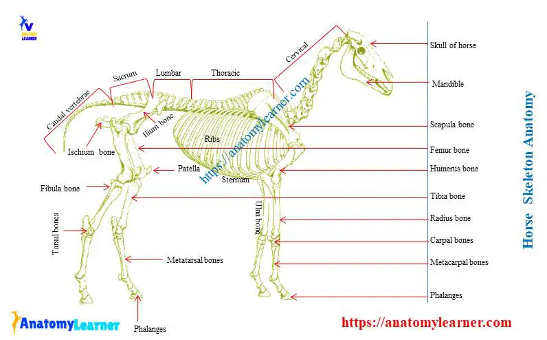
Horse skeletal system labelled diagram
In this part of this article, I would like to provide a complete labeled diagram of the skeletal system of a horse. For more updates on the horse skeletal anatomy, you may follow anatomy learner on social media.
If you need any labelled diagram on specific bones, please let me know.
Frequently asked question on horse skeleton anatomy
In this part of the article, you may find your common inquiries on the skeletal horse system. But if you don’t find your desire inquires, please let me know.
How many bones are in the horse skeleton?
You will find a total of two hundred and five (205) bones in the skeleton of a horse. But these number of bones may vary with other horse.
- Bones in the skull of horse – 34
- Bones in the vertebrae of horse – 54
- Ribs of horse – 36
- Sternum of horse – 1
- Bones in the thoracic limb of horse – 40 and
- Bones in the pelvic limb of horse – 40
What are the three largest bones in a horse?
The pelvic bone, femur and tibia are the three largest bones in a horse. The pelvic bone is the largest flat bone in the skeletal horse system. The femur of the horse is the largest long bone, and the tibia is the massive and longest bone in a horse.
How big is a horse skeleton?
The size of the skeleton depends on the external appearance of any animals. The larger animals have a larger skeleton, and the smaller animal has a small skeleton.
How many bones do horses have in their skull?
There are thirty-four (34) bones in the horse skull. If you want to learn these skull bones’ anatomical facts, you may read the full article from anatomy learner.
What kind of skeleton does a horse have?
You will find both the axial and appendicular skeleton in a horse. You already know the axial skeleton consists of the bones of vertebrae, skull, ribs and sternum. Again the appendicular skeleton of any animal consists of the bones from the forelimbs and hindlimbs.
How many bones are in a horses neck?
There you will find seven bones in the horse neck. I have already provided some information about the seven neck bones (cervical vertebrae) of a horse.
How many bones does a horses skeleton have?
There are two hundred and five (205) bones in the skeletal horse system. Please find the specific numbers of bones from the different region in the first question.
Conclusion
I hope this simple guide might help you to get a basic idea of horse skeleton anatomy. Then, you might follow the horse skeleton model and labelled diagrams properly. I will update this article with more resources in future.
If you get the basic idea of every single bone from the skeletal horse system, I would like to suggest you learn the detailed anatomy of these bones separately. For this, you may get help from the general anatomy section or horse anatomy section.