Ileum Histology Slide with Labeled Diagram and Identification Points

Ileum histology slide diagram

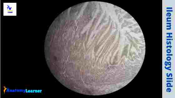
The ileum histology slide consists of the four layers like tunica mucosa, submucosa, muscular, and serosa. Here, I will show you the detailed histological features of the wall of the ileum slide with a labeled diagram. I will also provide you with the important identification points for the ileum histology slide so that you may identify it so quickly.  … Read more

Duodenum Histology Slide with Labeled Diagram

Duodenum histology diagram

There are three different parts –duodenum, jejunum, and ileum in the small intestine of an animal. You will find some salient microscopic features of each region of the small intestine. In this short article, I will discuss duodenum histology in detail, along with its slide identification points.  I will also show you the fundamental difference in the … Read more