The inguinal canal contents in male and female animals like bull cows differ slightly. However, the boundaries of the inguinal canal in bulls and cows are almost similar.
Here, I will show you the boundary and contents of the inguinal canal from various animals. I will describe all the canal structures from a male bull and then compare them with a female cow.
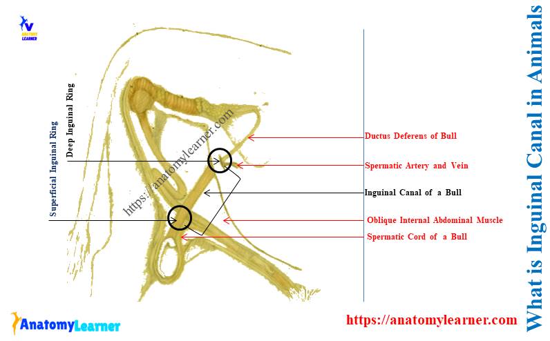
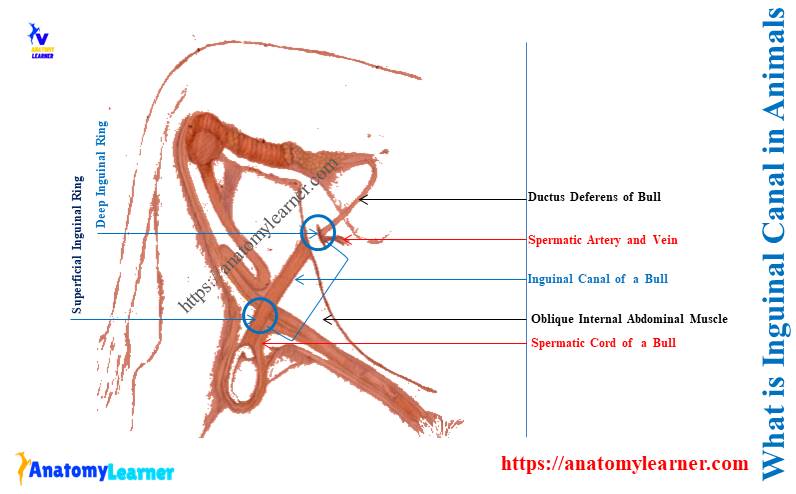
The inguinal canal in animals is an oblique passage through the lateral abdominal wall bounded by muscles and ligaments. It contains the spermatic cord in the male bull and the round ligament, ilioinguinal nerve, and vessels in a female cow.
So, with a diagram, let’s see the anatomy and contents of the inguinal canal in animals.
What is the inguinal canal in animals?
The inguinal canal in animals is the slit-like oblique fissure through the lower abdominal wall. These are the two passages placed in front of the corresponding pubis bones of animals.
Here, the diagram shows the inguinal canal of the bull. There are 2 inguinal canals in a bull, just cranial to the right and left part of the pubis bone.
The below-mentioned guide helps you to know more about the features of a bull’s pubis bone –
So, the key features of the inguinal canal in animals like bulls, cows, dogs, and goats are –
- It is a slit-like oblique passage,
- Located just cranial to the pubis bone, and
- Two in number in an animal,
Animal inguinal canal anatomy
Anatomically, the animal inguinal canal consists of two rings –
- Superficial inguinal ring – external opening of inguinal canal, and
- Deep inguinal ring – internal opening of the inguinal canal,
Here, the superficial inguinal ring faces externally towards the lateral abdomen. It is located 2 centimetres lateral to the linea alba of animals.
Let’s know details of an animal’s linea alba from the below-mentioned article –
The superficial ring of the animal inguinal canal contains the following features –
- It is merely a slit in the aponeurosis of the external oblique abdominal muscle,
- This ring represents where the animal’s abdominal wall formed around the gubernaculum in the fetus,
- The external visual part of the bull’s spermatic cord exists through this superficial inguinal ring,
- The opening of the superficial inguinal ring of an animal is larger than the deep ring,
Again, the deep inguinal ring of the animal is the abdominal opening of the inguinal canal. It is also known as the opening of the cavity of tunics.
Let’s see the unique features of the deep inguinal ring from the bull –
- It is the small and slit-like internal abdominal opening of the inguinal canal,
- The fascia of transverse abdominal muscles forms this deep inguinal ring,
- It is also bounded by the internal inguinal ligament,
- Different essential structures, like contents of the spermatic cord (male) and round ligament (female), exist through it,
What are the boundaries of the inguinal canal in cattle?
The boundaries of the inguinal canal in cattle are formed by the following –
- The anterior wall of the cattle’s inguinal canal: is formed by the transverse abdominis and internal abdominal oblique muscle. Again, the aponeurotic part of the external oblique abdominal muscle also forms the cranial wall of the animals inguinal canal.
- Caudal wall of inguinal canal: the external oblique abdominal muscle form the caudal wall of inguinal. You will also find the aponeurotic parts of the other abdominal muscles of the animals.
- The medial boundary of the cattle inguinal canal: the medial boundary of the cattle inguinal canal is formed by the straight abdominal muscle.
- Lateral boundary of the cattle inguinal canal: both the right and lateral boundary of the inguinal canal of cattle is formed by the external oblique abdominal muscle.
The cattle’s deep and superficial inguinal ring is attached to the inguinal ligaments. As abdominal muscles bound the inguinal canal, you might know their details.
The below-mentioned guide might help you to understand the anatomy of animal abdominal muscles –
- Dog abdomen anatomy – abdominal muscles and organs with diagram and
- Cow muscles anatomy – (the abdominal muscles part),
What are the contents of the inguinal canal in animals?
The contents of the inguinal canal in male animals are –
- Spermatic cord and its associated structures,
- Ilioinguinal nerve (second lumbar spinal nerve),
- Branch of genitofemoral nerve (third lumbar spinal nerve),
- Spermatic artery and other vessels,
- Peritoneum of the tunics,
- Internal and external cremaster muscles and
- Internal and external spermatic fascia,
A spermatic cord of a male animal in the inguinal canal
The cattle’s spermatic cord extends from the deep inguinal ring to the bottom of each testis. There are 2 spermatic cords in cattle, but you will see only one spermatic bundle externally.
Let’s know the details of bull spermatic cord from this article –
Here, the diagram shows the extension of the spermatic cord of an animal through the inguinal canal. I also try to show the contents of the spermatic cord that passage through this oblique canal.
Branches of ilioinguinal and genitofemoral nerves in inguinal canal
These are two lumbar spinal nerves in the animals that also give off branches to the inguinal region. The formation of the ilioinguinal and genitofemoral nerves is almost similar in animals.
Let’s see the diagram where I tried to show you the formation and distribution of the ilioinguinal nerve. This diagram also shows the distribution of genitofemoral nerves in the animal’s abdomen.
Small branches of these 2 spinal nerves pass through the inguinal canal and femoral area. But, they mainly innervate the muscles and skin of the areas of the lumbar and abdomen.
Let’s see the formation of the ilioinguinal nerve from the animal. This might provide a clear concept of the perfect distribution in the animal’s abdomen.
The dorsal and ventral roots come from the respective horns of the spinal cord. Here, the dorsal root forms the spinal ganglion and joins with the ventral root.
They form the spinal nerve together, which further divides into dorsal and ventral ramus. Both the dorsal and ventral ramus consist of lateral and medial branches.
The lateral branches of both ramus innervate the skin, whereas the ventral ramus innervate the abdominal muscles.
Let’s get the full guide on the formation and distribution of lumbar spinal nerve –
Spermatic artery and other vessels
Different arteries like the spermatic, artery of the cord, and artery of the vas will pass through the inguinal canal. Here, the spermatic artery of animals directly comes from the main aorta.
The external artery gives off the branches that pass through the inguinal canal. Finally, the branches of the external artery reach the animal’s spermatic cord.
Again, the umbilical artery of animals gives off the small branches for the vas deferens. This is the artery for the vas and runs within the inguinal canal.
You will also see the veins around the spermatic artery that form a convoluted plexus. These veins also travel through the inguinal canal of the animal.
The plexus of the veins around the animal spermatic cord is known as the pampiniform plexus. They remain very close to the deep ring of the cattle inguinal canal and drain caudal vena cava.
Cremaster muscles in the animal inguinal canal
The creamster muscle of the animal consists of two parts – internal and external. This cremaster muscle is the caudal fasciculus of the internal oblique abdominal muscle.
This cremaster muscle of the animal lies adjacent to the peritoneum tunic between the spermatic fascia. The major part of the internal cremaster muscles is found in the cranial bundle of the animal spermatic cord.
Spermatic fascia in the inguinal canal
The animal’s spermatic cord is surrounded by the modified layer of peritoneum passed through the inguinal canal. A transversalis fascia is reflected in them, known as the internal spermatic fascia.
The modified peritoneum tunics emerge from the inguinal canal. Now, the superficial and deep abdominal fascia are reflected in this structure.
Finally, this fascia lies superficial to the internal spermatic fascia. Then, the structure is known as the external spermatic fascia.
The deep and superficial fascia come from the external surface of the external oblique abdominal muscle.
Do female animals have an inguinal canal?
Answer: yes, the female animals have the inguinal canal. But, it has no external opening like the male animal, and the contents differ.
They are also two in number in female animals. You will find the inguinal canal in the female cows in the lower abdomen and just the cranial to the pubis.
Let’s see the content of the inguinal canal in a female animal –
- Round ligament of the female animal’s uterus,
- Ilioinguinal and genitofemoral nerves of female animals and
- Different vessels in of the female animals,
You may know the details of the round ligament of the female animal’s uterus from this article –
Where is the inguinal region of an animal?
Answer: the inguinal region of an animal is the part of the lower and lateral abdomen. It is just a few centimeters caudal to the umbilicus of the animals.
So, the inguinal region of an animal extends from the umbilicus to the front of the pubis bone. In most domestic mammals, you will find the inguinal canal within this region.
Where is the inguinal region of a dog or a goat?
Answer: the inguinal region of a dog or a goat is just the front part of the pubis. You may easily identify this inguinal region of a dog or a goat.
Here, the diagram shows the different regions of the body of a goat. The inguinal region of the goat body is identified in the labeled diagram.
What is the inguinal canal of a dog?
Answer: like other animals, it is the oblique passage through the abdominal wall. The dog’s inguinal canal extends between the deep and superficial inguinal rings.
The dog’s abdominal muscles form the boundaries of the inguinal canal. Anatomically, you will find similar features in the dog inguinal canal to other animals.
What goes through the inguinal canal dog?
Answer: The spermatic cord, ilioinguinal nerve, genitofemoral nerve, and various vessels go through the inguinal canal of a male dog. The female dog’s inguinal canal contains the round ligament, ilioinguinal nerves, and various vessels.
So, the branches of the lumbar spinal nerves and various vessels are common for both male and female animals.
Conclusion
So, you got the idea of the question – what is the inguinal canal in animals with its contents? The animal’s inguinal canal is the oblique passage in the inguinal region between two rings.
Both male and female animals have the inguinal canal with various contents. The spermatic cord of male dogs and cattle runs through this inguinal canal.