Do you want to know about the liver histology?
If yes, then you are in the right place now where you will get the ultimate guide to learn the normal liver histology in details.
In this article, I will discuss about the liver from start to end alone with lots of liver histology diagram. Hope, that will help you to understand all structure clearly.
I have also a gift for you at the end of this article where you will able to grab the liver histology pdf guide and also histology of liver ppt. You will able to practice and evaluate your learning with these resources.
So, if you are really interest to learn liver histology then I would like to request you to continue this long article so carefully. I am sure, if you follow all and read this article once attentively, you will get the all basic of liver.
Liver histology
First, I would like to inform you what actually learn in this article. I will help you to learn the followings –
- #1. What is liver?
- #2. What are the functions of liver?
- #3. Component and structures of liver
- #4. Details about the hepatocyte
- #5. Vessels and bile duct
If you want to know any of the specific part, then you may jump and continue to read.
I would like to provide a summary of the liver histology. Youmight get an idea and take your decision whether you want to learn more or not or learn specific part.
Hope, this summary will help you a lot to understand what actually you should know about the normal liver histology.
Okay; let’s notice the image or video and try to find out the following structures from the liver. In practice, you should identify the following structures under the light microscope.
- #1. Capsule, stroma and parenchyma
- #2. Liver lobule (for now the classical lobule) along with interlobular connective tissue
- #3. Hepatic lamina along with hepatocyte
- #4. Central vein
- #5. Portal canal or central canal
- #6. Hepatic artery, portal vein, bile duct and lymph capillary (portal tired)
- #7. Sinusoids
- #8. Perisinosidal space or space of disse
- #9. Macrophage cell or kupffer cell
You should identify and understand these structures first. I think you have completed 25% of liver histology. But, you should know the details about them to understand clearly. If you learn details it will remain every green in your mind.
So, will you interest to learn liver histology with diagram? Okay; let’s continue to read for more information and images.
Normal liver histology
You know liver is the largest gland in the body and characterized by multiple and complex functions. I am not interest to discuss details on its functions as my article is about its histology. If you want to know the functions of liver then you may go through the following points –
- #1. Excrete the waste products
- #2. Secrete bile and storage lipid, vitamin and glycogen
- #3. Synthesize fibirinogen, globuilin, albumin
- #4. Detoxified lipid soluble drugs
- #5. Conjugate steroid hormones and toxic substances
Hope, these are enough for now. Let’s discuss on the histological structure of liver. If you want to know about the liver anatomy then you may read this article here in anatomy section.
Component and structure of liver histology
For description purpose, there are three lobules present in liver
- #1. Hepatic lobule or classical lobule
- #2. Portal lobule and
- #3. Liver acinus
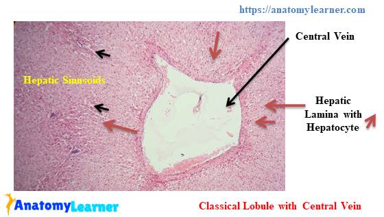
Notice the schematic diagram of the liver lobules. The hepatic lobule or classic liver lobule is the anatomical unit of liver.
The features of classic lobule in liver –
#1. Hexagonal shaped lobule
#2. Presence of hepatic lamina – consist of hepatocytes (branching pattern)
#3. Presence of central vein (cell arranged from central vein to periphery in branching pattern)
#4. Portal canal present three of the six angles of the lobule
#5. Presence of sinusoid (free surface of hepatocyte cell faced sinusoids)
The features of portal lobule in liver –
#1. This is the functional unit of liver (exocrine function)
#2. Triangular area of the parenchyma of three adjoining hepatic lobules
#3. Presence of bile ducts in the center of the lobule
Hepatic sinus
The main features of the hepatic sinus are –
#1. Diamond shaped region with the two central veins at each end and two portal canal at the other two end (see the schematic diagram, hope, you could understand)
#2. Diamond is divided into two triangles by a line of connecting the portal canal
#3. Flow blood from distrusting vessels in a single portal canal
Hope, you could understand these lobules. As there is no actual lobule, they are presented for easy studies.
Capsule and stroma of liver histology
The outer layer of the liver is covered by thin layer of connective tissue capsule. Connective tissue from capsule extends into the liver lobule and form the interlobular connective tissue which surrounds the individual lobules of the liver.
It is so hard to see the interlobular connective tissue septa under light microscope. This is because of presence of scanty connective tissue. But, in pig liver, you will find distinct interlobular connective tissue septa among the lobules.
Fine reticular fiber surrounds the hepatocytes cell and sinusoids. There are also presence of smooth muscle cell in the capsule and interlobular connective tissue.
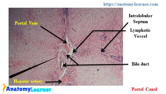
An expand area in the interlobular connective tissue which is actually know as portal canal or portal area. The contents of portal area or canal are –
#1. Branch of hepatic artery
#2. Branch of portal vein
#3. Bile duct and
#4. Lymph vessels
Let’s see the image and find these structures from the liver histology. Hope, you know the basic histology of artery, vein and duct and could able to identify them easily. If you want to know the basic difference of the artery, vein and duct under light microscope then you may get help from this article here.
Okay, fine; let’s move to the next part – the parenchyma of liver.
Parenchyma of normal liver
Before to start, if you want to get details guide about stroma and parenchyma then I would like to request you to read the article.
The ultimate guide of stroma and parenchyma at here in the histology of connective tissue section.
In parenchyma, you will find the row of hepatocytes. Actually, hepatocytes are arranged in a radiating manner on the hepatic lamina.
Hepatocytes of liver
Here, I will discuss on the following parts and structures –
#1. Heaptocytes and its structure
#2. Surface of hepatocytes
#3. Hepatic sinusoids or sinusoidal space
#4. Perisinusoidal space or space of disse
#5. Macrophage or kupffer cells
Okay, let’s start to know these structures in details.
Hepatocytes are the main cell of the liver histology lobule. The main features of the hepatocyte cells are –
#1. Hexagonal in shaped
#2. Presence of centrally placed spherical nucleus with one or more prominent nucleolus and scattered clump of heterochromatin
#3. Occasionally, binucleated hepatocytes are seen in the liver lobule
#4. The cytoplasm of the hepatocyte varies with wide limit
#5. Presence of abundant mitochondria, golgi complex and rough endoplasamic reticulum in the cytoplasm of the hepatocyte
#6. Presence of glycogen in the cytoplasm of hepatocyte – appears irregular shaped empty space in histological preparation
#7. There are more than six surfaces and they are three of different types –
Microvillus surface that faced the perisinusoidal space of the parenchyma. Don’t know what is perisinusoidal space? Okay, I am going to discuss on it later; let’s continue this.
Canalicular surface that border the bile canaliculi of the liver
Contact surface of the adjacent
Hepatic sinusoids and perisinusoidal space
The hepatic sinusoids are the blood capillaries which is located between the hepatic lamina. They carry blood from the terminal branch of interlobular hepatic artery and interlobular hepatic venule to the central vein of liver.
Sinusoids are lined by two types of cells –
#1. Endothelium cells and
#2. Larger macrophage or kupffer cells
You know, endothelium cells are actually the simple squomas epithelium cells lining. If you want to more about epithelium tissue then read this article at here.
Among the sinusoidal cells some cells are larger and scatter and often sending pseudopodia through the endothelium pore or between the cells – they are called the macrophage. The macrophage of liver is also known as kupffer cells of liver.
The functions of the kupffers cells of liver are –
#1. Metabolized the older erythrocytes
Help to digest haemaglobulin
#3, secrete protein related to immunoglobuilin process ‘
#4. Helps to destroy bacteria that eventually enter the portal blood through large intestine
The porous sinusoidal epithelium rest on the discontinuous basal lamina. The endothelium is separated from the hepatocytes by a space. This space is called the perisinusoidal space or space of disse.
The basolateral side of the hepatocyte lined the space of disse contains the microvillus.
Perisinusoidal space contain small amount of reticular fibers and also perisinusidal adipocytes which is actually known as ito’s cells
The main function of perisinusoidal space is the permit an easy exchange of macromolucles from the sinusoidal lumen to the hepatocytes and vice versa
Congratulations, you have completed the basic of the normal liver histology. Now, you may also know about the vasculatures of liver if you want.
Others information about liver histology
Blood supply of liver
Liver has dual blood supply –
#1. Portal vein system and
#2. Arterial system
Portal vein brings blood from intestine and associated organs, hepatic arteries supply the liver cells with oxygenated blood.
The portal veins branches repeatedly and send small portal venules (interlobular portal vein and interlobular hepatic arteries) to the portal spaces.
Interlobular portal venules give raises to the small branches. This small branches is called distributing venules. Short terminal venules arise from the distributing venules which end directly at the sinusoids.
Hepatic sinusoid carries blood from terminal branches of interlobular hepatic arteriole and interlobular portal venule to central vein.
Blood in the sinusoids leave the lobule through central vein. Again the central vein connects with the sublobular vein at the periphery of the lobule. Sublobular vein join and fuse, forming two or more larger hepatic vein. Hepatic vein directly empty the blood into the caudal vena cava.
Lymph and lymph vessels
Lymph in the liver is formed in the perisinusodial space and flows towards to the periphery of the lobule. Then it enters the intercellular space of the portal canal and interlobular connective tissue
Biliary tract
In liver, bile is produced by the hepatocyte cells and flows is occurred through the bile canaliculi then bile ductules and finally into the bile ducts.
Bile canaliculi are minute canals between the adjacent hepatocytes. These are actually the expanded intercellular spaces between hepatocytes.
They form a complex anastomosing network progressing along the hepatic lamina of the hepatocytes of the liver lobule and terminate at the region of portal canal
It carries bile form the central of the lobule to the periphery
At the periphery, bile enter to the bile ductules (which is actually known as the hering’s canal)
The ductules cross the limiting hepatocytes of the lobule and end in the bile ducts in the portal canal
Under light microscope, the bile ducts are easily visible. You may also identify them.
Identification of bile ducts
#1. They are lined by cuboidal or columnar epithelium
#2. They have distinct connective tissue sheath
#3. Located at the portal canal area and receive bile from the bile ductules
#4. They enlarge and fused to form right and left hepatic ducts and leave the liver at porta hepatis
You may also learn –
The ultimate guide of gallbladder histology
Ultimate guide of parotid gland histology
Full guide of smooth muscle histology
Conclusion
Hope, you got better idea and ultimate guide to learn the liver histology along with best liver histology diagram and liver histology pdf guide.
Now, you may practices and evaluate your learning with the help of liver histology ppt. Here, you will find the liver histology labeled images and other slides for your practices.
If you think this article was informative and valuable for you then I would like to request you to share this article with your friends who are interest to learn normal liver histology or with others members from anatomy society.
I used to publish this kind of article related to anatomy and histology in a regular basis. If you want to know more about the anatomy and histology of different organs or structures then you may follow this blog. For quick and fast update about the articles, images and videos you may follow at social media.
If you think this information is not enough or if you found any mistake or if you have any suggestion about liver histology; then I would like to request you to inform me at comment box or send me direct message. I will really happy and always appreciate your suggestion.