
The dog nose anatomy comprises external features and the structures of the nasal cavity. From the external part of a dog’s nose, I will show you the features of cartilages, vomeronasal organs, and ligaments. Again, you will find the details anatomy of the nasal conchae, meatus, paranasal sinuses, and glands from the dog’s nasal cavity.
Throughout this article, you will see many labeled diagrams on dog nose anatomy. These might teach you the dog nose structure perfectly.
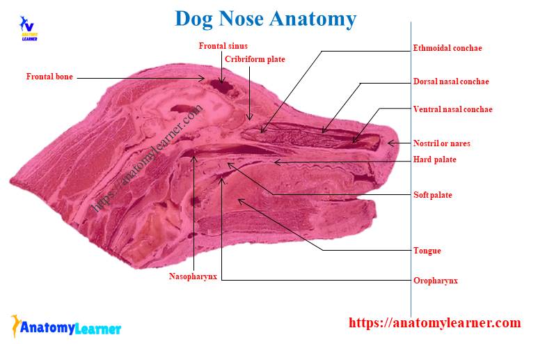
Dog nose anatomy
In a broad sense, dog nose anatomy refers to the external nose and its associated nasal cartilages. Again, it also refers to the internal nose or nasal cavity with its conchae and meatus.
A muzzle is the common structure of the facial part of the respiratory system and the rostral part of the digestive system. You will find a long muzzle in the dolichocephalic dogs and a shorter in the brachycephalic breeds.
Now, I will provide the main anatomical features from the external part of the nose and the nasal cavity in short. But, make sure you will learn the details anatomical facts of the dog nose from the specific part of this article.
Okay, first, let’s know the features from the external nose of the dogs.
The external part of nose
The skin around a dog’s nostril is moist and divides by a median philtrum that continues ventrally to the groove of the upper lip. A thick keratinized epidermis covers a nasal plate. You will find the irregular plaques and sulci in the nasal plate that make it unique for identification.
The nasal plate of the dog is kept moist by an overflow of the secretion of the nasal glands. But, you will not find any nasal gland in the nasal plate of a dog.
There is curved alar cartilage that supports the roof and wing of the dog’s nose. Again, on the nose floor, you will find accessory cartilage.
A dog’s nostril is comma-shaped, which is more thickened in its dorsolateral aspect. This dorsolateral thicken part is a wing of the nose. You will find an alar fold (an extension of the ventral nasal conchae) that terminates within the nasal vestibule.
You may find the congenital malformation of the nasal plane in some brachycephalic dogs. In this condition, the cartilage supporting the nose is so weak that it leads to the collapse of the wing. And thus, this condition leads to narrowing the nostril during inspiration.
So, as a veterinarian, you might have a piece of good knowledge on the external part of the dog nose with its details structures.
Nasal cavity structure
The nasal cavity of a dog extends from the nostril to the eye level. You will find a tubular nasal vestibule at the rostral part of the nose, which is caudal to the level of infraorbital foramen.
The nasal cavity divides into two halves by the nasal septum, where the dorsal and caudal parts become ossified. Again, the rostral part of the nasal cavity remains cartilaginous and accounts for the passive mobility of the tip of the nose. You will find a membranous middle part in the dog’s nasal cavity.
In the dog nasal cavity anatomy, you will find four nasal conchae (dorsal, ventral, middle, and ethmoidal). These nasal conchae are tightly filled the nasal cavity compared to that of the other animals.
You will find the dorsal and ventral nasal conchae at the rostral part of the cavity. The ventral nasal conchae of dog nose anatomy is thick but short and arises from the maxilla. Again, the caudal half of the nasal cavity almost fills with the ethmoidal conchae.
A nasolacrimal duct opens on the floor of the nasal vestibule and meets the alar fold. If the nostril spreads, you may easily see the opening of the duct.
You will find much smaller nasal glands on the rostral part of the septum. These glands open at the caudal limit of the vestibule and contribute marginally to wetting the nose.
Again, the nasal plate becomes wet by the secretion of lacrimal and nasal glands. In a dog’s nasal cavity, you will find the branches of the internal carotid artery and maxillary artery.
Special features of paranasal sinuses
The paranasal sinus is not so well-developed in a dog. You will find the largest sinus in a dog’s nose is the frontal one. But, the total paranasal sinuses of a dog include the frontal, sphenoid, and maxillary. The maxillary sinus communicates with the dog’s nasal cavity freely.
You will find a small sphenoid sinus in a dog nasal cavity. But, this cavity of the dog’s nose fills with the ethmoturbinates.
Please read the other important anatomical features of the different parts of the dog nose from the specific part of this article.
So, these are very short information on the dog nose structure. Now, you may learn the details anatomical facts of the dog nose with the labeled diagrams.
Dog nose external anatomy
The dog nose external anatomy consists of the fixed bony case and a cartilaginous framework. You will find the maximally and incisive bones in the lateral wall of the bony part of the dog’s nose. The paired nasal bones form the roof of the dog’s nose.
Again, the concave rostral end of the nasal bone and incisive bone form the bony nasal apparatus. This aperture of the dog’s nose is wider ventrally and lies in an oblique plane.
The cartilaginous part of the dog nose is the moveable part due to the presence of skeletal muscle along with the cartilages. There are four different cartilages in the structure of a dog nose. Again, there is a vomeronasal organ located at the rostral base of the nasal septum.
From the external part of the dog’s nose structure, you will learn the following parts in detail –
- Nasal plane or planum
- Bones of the dog nose
- Cartilages of the dog’s nose
- Structure of a vomeronasal organ, and
- Ligaments of the nose
Nice, let’s start to learn the anatomical features of the structures mentioned above from a nose of a dog.
Nasal planum of a dog
You will find short hair on the skin of the dog’s nose that directs caudally on the middorsal surface. Gradually, these hairs slope in a caudoventral direction laterally and continue with the lip. A flattened part in the apical portion of a dog’s nose lacks hair.
This flattened hairless portion of the nose is the nasal plane or planum of a dog. You will find two nostrils and a groove (philtrum) that separates these nostrils from each other.
The nasal planum also lacks glands and is covered with the epithelium.
Bones of the dog nose
The bony case of a dog nose consists of the maxilla, nasal, and incisive bones. I have previously described all the anatomical features of these nasal bones in anatomy learners. You may memorize the features of these dog’s nasal bones from that article.
The nasal bone of a dog is long, slender, and narrow caudally. Again, the dog’s nasal bone is wider rostrally. You will find a variation in the size and shape of the dorsal or external surface of the nasal bone in a different breed of a dog.
A mucous membrane covers the ventral or internal surface of the dog’s nasal bone. You will find the dorsal nasal conchae and dorsal nasal meatus in the internal surface of the nasal bone. There is an ethmoidal crest at the caudal half that serves to attach the dorsal nasal concha throughout its rostral half.
The incisive is another important bone that forms the nasal case. This bone has a small body with three processes (alveolar process, nasal process, and palatine process).
You will find three alveoli for the three superior incisor teeth on the alveolar process. There is a curved, tapering nasal process called the caudolateral part of the incisive bone. The palatine process of the nasal bone of a dog is laterally compressed and pointed structure.
The maxilla is the largest facial bone in a dog skull that possesses a body and four processes (frontal, zygomatic, palatine, and alveolar).
You may read more at – nasal, maxilla, and incisive bone features from a dog skull anatomy.
Cartilages of the dog nose
In the dog’s external nose anatomy, you will find different types of cartilages from the mobile part. These comprise the unpaired septal cartilage, paired dorsolateral and ventrolateral cartilages, and paired lateral accessory cartilages. In addition, you will also find the vomeronasal cartilage close to the ventral part of the septal cartilage.
So, let’s discuss these four types of cartilages from the dog nose structure –
- Unpaired septal cartilage
- Paired dorsolateral cartilages
- The ventrolateral cartilages (paired), and
- Paired lateral accessory cartilages
Septal cartilage of the dog nose
The unpaired septal cartilage of the dog nose separates the nasal cavity into right and left halves. It is the perpendicular plate-like structure that continues the ethmoid bone. A small portion of the rostral area, the septa, lacks the cartilage and forms the membranous nasal septum.
This membranous nasal septum is the mobile part that connects the cartilaginous immovable caudal part. You know the caudal part of the cartilaginous nasal septum is thicker ventrally and lies in the septal groove of the vomer bone.
You will find the prominent caudal process at the median plane of the cartilaginous nasal septum. Again, the rostral part of the cartilaginous septum continues a median course from the membranous part of the nasal septum.
You will find right and left laminae at the rostral border of the cartilaginous nasal septum of a dog. From the dorsal portion of each lamina give rises the dorsolateral nasal cartilage. Again, the small ventrolateral nasal cartilage arises from the ventral part of the rostral septal cartilage.
The accessory nasal cartilage of the dog nose connects the ventrolateral nasal cartilage by collagenous tissue.
Dorsolateral nasal cartilage
The dorsolateral nasal cartilage is the most expanded structure in a dog nose. This cartilage is the continuation of the dorsal part of the septal cartilage. It forms a tube that covers ventrally, laterally, and medially.
The caudal part of the dorsolateral nasal cartilage connects with the dorsal part of the bony aperture. It joins with the concave border of the nasal bone with the help of fibrous tissue.
You will find a thicker part in the dorsolateral nasal cartilage on its rostral end. Here, you will also find the blood vessels that form a meshwork in the collagen tissue directly caudal to the nostrils. In addition, the dorsolateral nasal cartilage of the dog nose becomes thinner at the caudal end.
The medial border of this cartilage is free and curves ventrally that joins to the ventrolateral cartilage of the nose.
Ventrolateral nasal cartilage of the dog
The ventrolateral cartilage of a dog nose is shorter and approximately one-fourth as wide as dorsolateral cartilage. It is the continuation of the rostral part of the lateral half of the septal cartilage. Again, its origin moves obliquely dorsal on the lateral surface of the ventral part of septal cartilage.
Rostrally, this cartilage runs into the apex of the nose. Again, it ends caudally to the lateral leaf of the septal cartilage adjacent to the articulation with accessory cartilage.
The caudal free border joins with the free border of the dorsolateral cartilage and forms the cartilaginous basis of the alar fold. You know this alar fold continues the ventral nasal concha to the vestibule.
Accessory nasal cartilage of a dog
This is a laterally convex leaf-like structure that articulates with the ventrolateral angle of the ventrolateral cartilage. It extends dorsocaudally to the lateral surface of the expanded portion of the dorsolateral nasal cartilage.
This cartilage also directly lies deep to the ventral surface of the midlateral slit of the nostrils. You may find second small accessory cartilage in the dog nose structure. This second accessory cartilage directly locates dorsal to the septal cartilage in the groove.
Vomeronasal organ of the dog nose
The other name of the vomeronasal organ of the nose is Jacobson’s organ. You will find two vomeronasal organs in the dog nose structure. They are located in the rostral base of the nasal septum as a tubular pocket of the olfactory epithelium.
You will find vomeronasal cartilage beside the other cartilages of the dog’s nose. This vomeronasal cartilage partially covers the vomeronasal organ.
Each of the vomeronasal organs opens rostrally into an incisive duct. The incisive duct, called the nasopalatine or Stenson duct, passes through the palatine fissure and connects the nasal and oral cavity.
Lateral to the incisive papilla, you will find the oral orifices of each incisive duct. Again, the paired incisive ducts pass dorsocaudally to open each nasal cavity.
You will find the extensive venous plexus at the lateral wall of the vomeronasal organ of a dog.
Ligaments of the dog nose
You will find only three major ligaments in the dog nose structure – one paired and one unpaired. These ligaments bind the dorsal portion of the bony nose to the mobile part of the nose. Let’s find the following ligaments from the dog nose –
- Dorsal nasal ligament (single) and
- Paired lateral nasal ligament of dog nose
The dorsal nasal ligament is a band of collagenous tissue that runs from the caudodorsal end of the dorsolateral nasal cartilage to the dorsum of the nasal bones. Again, the lateral nasal ligament also is the band of collagenous tissue on either side of the nose.
The lateral ligament runs from the midlateral surface of the caudal aspect of the dorsolateral nasal cartilage to the border of the bony nasal aperture.
Muscles of dog nose anatomy
In the dog nose muscle anatomy, you will find a few muscles like levator labii superioris, caninus, and levator nasolabialis. The levator labii superioris is the flat muscle that lies deep to the apical end of the levator nasolabialis. It arises from the maxillary bone caudoventral to the infraorbital foramen.
The insertion fibers of the levator labii superioris enter the dog’s nasal ala and the superior lip.
Again, the caninus is a small muscle that lies immediately ventral to the levator labii superioris muscle. This caninus muscle of a dog extends rostrally deep to the labial end of the levator nasolabialis muscle. You will find the terminal part of the caninus muscle at the superior lip of a dog.
This caninus muscle increases the diameter of the external naris. It also lifts the apical part of the superior lip of a dog.
The levator nasolabialis is the thin, flat, and broad muscle of the dog nose structure. It lies immediately deep to the skin on the lateral surface of the nasal and maxillary bones.
Again, it arises from the frontal region between the orbits from the nasofrontal facia and maxillary bone. This muscle’s most dorsal and rostral fibers insert into the external naris.
This muscle also increases the diameter of the external naris and lifts the apical part of the superior lip of the dog.
The nasal cavity of the dog
The nasal cavity is the facial part of the respiratory passages that consists of the vestibule and nasal cavity proper. It extends from the external nostril to the inner nasal concha and meatus. You know, a nasal septum divides the nasal cavity into two parts. This nasal system also consists of the bony case and the membranous part.
So, both two nasal cavities begin at the nostril and end at the inner nasal conchae and meatus. You will find four main air channels and some small channels in each nasal cavity of the dog nose. The lateral and dorsal nasal wall of the nasal cavity forms the lamina that forms the conchae.
Okay, let’s discuss the two main parts of the dog nasal cavity –
- Vestibule of the nasal cavity and
- Nasal cavity proper of the dog
Vestibule of the dog’s nasal cavity
The nasal vestibule of the dog is a large chamber obliterated by the large bulbous end of the alar fold. At the entering part of the nasal vestibule, you will find the nostrils where the air diverter medially and ventrally into the largest meatus of the nasal cavity.
You know nostrils are the curved opening into the nasal vestibule of the dog’s nose. These opening are wider dorsomedially than it is ventrolaterally.
You will also find the alar fold in the dog’s nose vestibule structure. This alar fold of the dog nose is the extension of the ventral nasal conchae. It terminates within the vestibule by a bulbous enlargement that fuses to the wing of the nostrils.
Do you know what the wing of the nostril is? Well, this is the thick dorsolateral part of the nostril. You will find the dorsolateral and accessory nasal cartilage in the wing of the nostril part of a dog nose.
Again, this part of the nasal vestibule receives the fibers of levator labii superioris and levator nasolabialis muscles. A nasolacrimal duct conducts the lacrimal secretion from the eye-opening into the vestibule. You will also find the small duct of the lateral nasal gland that opens on the oblique fold into the dorsal vestibule of the nose.
The nasal cavity proper of the dog’s nose consists of the nasal conchae, nasal meatus, paranasal sinuses, nasal mucosa, and nasal glands. Let’s discuss these structures that form the nasal cavity proper.
Nasal turbinates of dog nose anatomy (conchae)
The nasal conchae of the dog nose anatomy are the cartilaginous structure covering the nasal mucosa. These nasal conchae occupy most of the portion of the nasal cavity proper. You will find dorsal, ventral, middle, and ethmoidal conchae in the structure of the dog nasal cavity proper.
- Dorsal nasal conchae or dorsal nasoturbinates
- Ventral nasal concha or ventral maxilloturbinate
- Middle nasal concha or middle endoturbinate, and
- Ethmoidal conchae or ethmoturbinate
Dorsal nasal turbinates
The dorsal nasal conchae or turbinate is a long, slightly curled scroll of the endoturbinate. This dorsal nasal conchae of the dog nose cavity proper attaches to the ethmoidal crest of the ethmoid and nasal bone. You will find a mucosal fold that continues the conchae into the vestibule of the dog’s nose.
So, you may say the dorsal turbinates extend from the ethmoidal bone and attach to the nasal bone’s ethmoidal crest. This is the largest turbinates in the dog nose structure and possesses a pointed anterior end.
“You may learn more about the turbinates bones of the dog skull from anatomy learner’s guide.”
Ventral nasal conchae or turbinate
The ventral nasal conchae are broad and occupy the ventral part of the nasal cavity proper. It is a tightly folded series of scrolls that attaches to the conchal crest on the medial surface of the maxilla bone.
This ventral nasal conchae of the dog nose extends from the level of the first to third premolar teeth. You will find an extension of the ventral nasal conchae. This extension of the ventral nasal conchae is the alar fold that directs into the vestibular part of the nasal cavity.
Middle nasal turbinate
The middle nasal conchae is the small structure in the dog nasal cavity proper. You know the second endoturbinate of the ethmoidal labyrinth provides the rostral extension. This rostral extension of the endoturbinates is the middle nasal conchae in the dog nose.
It fulfills the short space between the scrolls of the ventral nasal conchae and ethmoid labyrinth.
Ethmoidal conchae of the dog nose
The ethmoidal conchae locates between the dorsal and ventral ethmoid bones at their caudal portion. It forms the ethmoidal labyrinth that properly fills the caudal part of the dog’s nasal cavity.
They are the outgrowth of the ethmoidal bone covered by the nasal mucosa. You will find numerous delicate, bony scrolls known as the ethmoturbinates. These ethmoturbinates are again divided into ectoturbinates and endoturbinates portions.
All these ectoturbinate and endoturbinate will attach with the ethmoid bone’s orbital lamina and cribriform plate. You will find six ectoturbinates and four endoturbinates in the structure of the ectoturbinate of a dog.
The six ectoturbinates are small and lie on the dorsal aspect of the bony labyrinth. Again, the four endoturbinates lie ventrally and fill the caudal portion of the nasal cavity.
The first ectoturbinate extends rostrally and from the bony scroll for the dorsal nasal conchae. Again, the second ectoturbinate forms the middle nasal conchae in the dog’s nose. The other ectoturbinates usually extend into the frontal sinus of the dog.
Nasal meatus of the dog nose
The space above the dorsal turbinate, between the dorsal and ventral turbinates, and below the ventral turbinate are known as the dorsal, middle, and ventral meatus, respectively in the dog nose structure. The inhaled air leaves the vestibule and travels the longitudinal nasal meatus to reach the nasal part of the pharynx.
So, you will find the below-mentioned meatus in the dog nose structure –
- Dorsal nasal meatus of dog nose
- Ventral nasal meatus of the dog nose
- Middle nasal meatus of the dog nose
- The common nasal meatus in the dog nose, and
- Nasopharyngeal meatus in the dog nose structure
Fine, let’s discuss the nasal meatus from the dog nose structure.
Dorsal nasal meatus
The dorsal nasal meatus of the dog nose is a passage through the dorsal part of each nasal cavity. You will find this dorsal nasal meatus between the dorsal nasal conchae and the ventral surface of the nasal bone.
Again, you will find a close association with the common nasal meatus on the medial aspect of the dorsal nasal meatus of the dog. The ethmoidal crest forms the lateral aspect of the dorsal nasal meatus. You know from this ethmoidal crest, the dorsal nasal conchae arises.
Ventral nasal meatus of the dog’s nose
The ventral nasal meatus of the dog nose anatomy locates between the ventral nasal conchae and the dorsal surface of the hard plate. You will find a narrow part in the rostral end that leaves the nasal vestibule. It becomes widened caudally and continues ventrally to the basal lamina of the ethmoidal bone as the nasopharyngeal meatus.
Middle nasal meatus structure
The middle nasal meatus of the dog lies between the dorsal nasal conchae and ventral nasal conchae. You will get a small wide in the middle nasal meatus of a dog compared to others. There is a dilated portion at the rostral end of the middle nasal meatus.
Again, the lateral part of the middle nasal meatus of a dog divides into several parts. An ellipsoidal dilation is found in the middle nasal meatus that connects the nasal vestibule. This ellipsoidal dialated structure is the atrium of the middle nasal meatus of the dog.
You will find a narrow handle of the club-shaped mucosal alar fold at the ventral part of the nasal’s atrium. Again, the dorsal part of the atrium has a connection with the rostral portion of the dorsal nasal concha.
The caudal part of the middle nasal meatus divides by the scrolls of the middle nasal conchae.
Common nasal meatus of the dog’s nose
The common nasal meatus of the dog’s nose is a narrow longitudinal space on either side of the nasal septum. At the lateral part of the common nasal meatus, you will find the boundary of the dorsal nasal conchae and ventral nasal conchae.
Again, at the dorsal aspect of this meatus, you will find a connection with the dorsal nasal meatus. There is also a connection between the ventral nasal meatus and the middle nasal meatus with the common nasal meatus of the dog’s nose.
The nasopharyngeal meatus of the dog
It is a short passage with a much longer lateral than a medial wall. This nasopharyngeal meatus of the dog extends on either side of the caudal dilated part of the ventral nasal meatus to the conchae.
Lateral to the nasopharyngeal meatus of the dog, you will find the maxillary and palatine bones. Again, the dorsal aspect of this meatus bounds by the basal lamina of the ethmoid bone.
The ventral part of the meatus surrounds by the palatine bone, whereas the medial part surrounds by the vomer bone. All the four nasal meatuses (dorsal, ventral, middle, and common) continue as the nasopharyngeal meatus to the pharynx (nasal part of the pharynx).
Paranasal sinuses of dog nose anatomy
The paranasal sinuses are also important structures of the dog nose anatomy. In this structure, you will find the maxillary sinus, frontal sinus, and sphenopalatine sinus. These paranasal sinuses have a direct connection with the respiratory passages.
The maxillary sinus is the cavity that extends inside the maxilla, lacrimal, and malar bone. It connects with the palatine sinus by a large oval opening. Again, it communicates with the middle nasal meatus of the nasal cavity at the dorsal aspect. The ventral surface of the maxillary sinus is rough in the dog.
The frontal sinus divides into a dog’s rostral, medial, and lateral components. Again, the sphenoid sinus of the dog is only the potential cavity that fills by an ethmoturbinate scroll. This is a very small sinus and has a connection with the ethmoidal meatus of the dog’s nose.
“The sinuses are the cavities in the bone of the skull that lines with the mucous membrane and directly or indirectly communicates with the nasal cavity. “
You may also learn more about these paranasal sinuses of the dog’s nose from another article of anatomy learner.
Maxillary and frontal sinuses of the dog
The maxillary sinus is the large and lateral diverticulum of the nasal cavity. This cavity is bounded by the maxilla, ethmoid, palatine, and lacrimal bones.
You will find a rounded fundus at the caudal part of this maxillary sinus of a dog.
The orbital lamina of the ethmoid bone forms the medial wall of the maxillary sinus in a dog. Again, the maxillary, palatine and lacrimal bones form the lateral wall of the maxillary sinus in a dog.
You will find some lateral glands at the medial wall of the maxillary sinus of a dog nose.
In addition, the frontal sinus of a dog locates between the outer and inner tables of the frontal bone. It varies more in size and divides into lateral, medial, and rostral compartments. The size and shape of the frontal sinus depend on the skull of different dog breeds.
The lateral compartment of the frontal sinus occupies the whole truncated enlargement of the frontal bone that forms the zygomatic process. You will find a nasofrontal opening at the lateral compartment of the frontal sinus of a dog.
The medial part of the frontal sinus of a dog is more irregular and has a great variation in size than the lateral one. In addition, the rostral compartment of the frontal sinus is very small in a dog.
Nasal mucosa of dog
The nasal mucosa of a dog nose consists of various types of epithelium that line the nasal cavity. In the respiratory organ histology section, you will get the details to guide on the nasal mucosa (various types of epithelium lining). Please, read the details guide on nasal mucosa histology from that article of anatomy learner.
You will find the receptor for the sense of smell on the ethmoturbinates of the nasal cavity. There is a highly vascularized mucosa present in the nasal cavity. This vascularized mucosa, vomeronasal organs, and a small nasal septum area from the olfactory organs in the dog nose.
Glands of the dog nose
Different serous, mucous, and mixed tubuloalveolar glands in the dog’s nose. The lateral nasal gland is a serous gland that lies in the mucous membrane of the maxillary sinus. You will find the thickest part of the lateral nasal gland at the fourth superior premolar teeth level.
The gland’s ducts unite and pass rostrally to from major duct that opens on the lateral wall of the vestibule. Again, the opening duct of the lateral nasal gland into the dorsal vestibule remains hidden due to the presence of the alar fold.
You will also find the serous, mucous, and mixed tubuloalveolar glands in the mucosa of the respiratory part of the dog nose. These glands are also present in the mucosa of the nasal vestibule of the dog. Again, you will find the goblet cells and olfactory glands throughout the respiratory region of a dog nose.
The nasolacrimal duct carries the serous secretion from the conjunctival sac to the nasal vestibule. So, you will see a moist nose in a healthy dog.
A nasal portion of the pharynx
You know there is a nasal portion of the pharynx in each animal. The nasal portion of the dog pharynx is the nasal pharynx or nasopharynx. This nasopharynx of a dog extends from the nasal conchae to the intrapharyngeal ostium.
The digestive passageway of the pharynx and respiratory passageway of the pharynx cross each other and form the intrapharyngeal ostium at the rostral end. Again, you will find the hard palate ventrally, vomer dorsally, and the palatine bones bilaterally around the nasal pharynx.
The middle and caudal part of the nasal pharynx surrounds the base of the skull and muscles. You will find the pharyngeal opening of the auditory tube on each lateral wall of the nasal pharynx just dorsal to the middle of the soft palate.
An auditory tube extends between the middle ear cavity and nasal pharynx cavity. This tube serves the equalize the atmospheric pressure on the two sides of the tympanic membrane of the dog.
Dog nose shape
You will find different shapes of the nose in the different breeds. Even you may find the difference in the shape of a dog nose within the same breed. So, what are the factors that might change the shape of a dog’s nose?
Well, several factors might change the shape of a dog’s nose. Here, I will provide the factor of changing dog nose shape based on anatomical facts.
You know there are nasal, maxilla, and sphenoid bones in the formation of the nasal cavity in a dog. These bones may play a minor role in variable shapes in a dog’s nose.
The cartilages of the dog nose have a great role in changing their shape. You know the mobile part of the nasal cavity largely forms by the nasal cartilage. So, the shape of the dog’s nose also depends on the shape and size of the nasal cartilages.
So, you may ake a question – is it possible to identify the dog breed with their nose shape? Yes, you may identify the different dog breeds by their different types of noses.
Dog nose anatomy diagram
Throughout this article, I know you already got lots of labeled diagrams on the dog nose anatomy. Now, I will again show you the important features from the vestibule and nasal cavity proper of a dog nose. Here, I also tried to focus all the structures from the dog’s external nose in the labeled diagram.
You may also find more labeled diagrams on the dog nose structure on social media of anatomy learners. If I update the information on the dog nose with a revised labeled diagram, you will get the notification there.
Frequently asked questions on dog nose.
Let’s find the answers to the below-mentioned questions on dog nose anatomy. Don’t forget to come back again and check this question answers part as I frequently update the information here.
What is the anatomy of a dog’s nose?
The anatomy of a dog’s nose is similar to the anatomy of other mammals. If you want to know the anatomical features of a dog nose, you might have a good idea of the external nose and the nasal cavity.
The dog’s external nose comprises four types of cartilages, vomeronasal organs and ligaments. Again, in the nasal cavity of a dog nose, you will find the vestibule and nasal cavity proper. There are nasal conchae, nasal meatus, nasal mucosa, paranasal sinuses, and glands in the nasal cavity proper of a dog.
I have already described all these structures from the external part of the nose and the nasal cavity. So, please read this full article and get the basics of the dog nose structure.
What are some properties of a dog’s nose?
You will find several important features in the dog’s nose. Here, I will enlist some of the properties of the dog’s nose –
A nasal planum contains two nostrils that are separated from each other by a groove (philtrum).
The dorsolateral cartilage of the dog’s nose is larger, and you will find some special features in the septal cartilage of the dog.
There are three ligaments present in the structure of a dog nose (one single dorsal nasal ligament and paired lateral nasal ligaments).
You will find four nasal conchae and five nasal meatuses in the structure of a dog nose.
The paranasal sinuses of a dog’s nose include the maxillary, frontal, and sphenoidal.
You will find different serous, mucosa, and mixed lateral nasal glands in the inner part of the dog nose.
What is alar fold?
In the dog nose structure, you will find the alar fold. This is an extension of the ventral nasal conchae of the dog’s nose. It terminates within the nasal vestibule by bulbous enlargements that fuse to the wing of the nostrils.
If you don’t know what the wing of nostrils is. They are the thick dorsolateral part of the dog’s nostril.
What are the main features of the respiratory organs of a dog, including its nose?
The shape and size of a dog’s nasal cavity vary according to the breed. You will find the comma-shaped nostrils in a dog. A pair of extra cartilages are present in a dog’s larynx compared to the ruminants.
There is a prominent vocal cord in the dog’s respiratory system. You will not find any apical bronchus in the trachea of a dog. The fissures are deep, and the lobes of the lung are prominent and well-separated in a dog.
You will find four lobes (apical, cardiac, diaphragmatic, and accessory) in a dog’s right lung. At the same time, the left lung of a dog consists of two lobes – apical and diaphragmatic.
What are nares in a dog?
You know there is a nasal plane in the structure of a dog nose. This nasal plane contains two slit-like openings (nostrils). These nostrils are the nares in a dog.
What are the parts of a dog’s nose?
The parts of a dog’s nose are – the external and nasal cavities. Again, the external part of the dog nose includes cartilages, vomeronasal organs, muscles, bones, and ligaments. The nasal cavity of a dog comprises two major parts – vestibule and nasal cavity proper.
The vestibule of the dog’s nose is the part that starts from the nostril and ends at the beginning of the nasal cavity proper. In addition, the nasal cavity proper includes the nasal conchae, meatus, mucosa, glands, and sinuses.
Why do dogs have slits in their nose?
Dogs have two slits in their nose – right lateral and left lateral. These slits of the dog’s nose are the nostrils, or you may tell them as nares. They are the external opening of the nose of the dogs.
Does touching a dog’s nose hurt them?
They don’t get hurt if you touch the dog’s nose.
How do dogs’ noses work?
What is the black part of a dog nose called?
There is a flat and hairless area at the apical part of a dog’s nose. Generally, the color of this hairless flatten part is black. This is the basal plane of the dog nose. Another name of the nasal plane of a dog is planum.
You will find two slits-like structures (external nostrils) within the nasal planum of a dog. Again, these nostrils are separated by a median groove (known as the philtrum).
Conclusion
I hope you learn the basic features from the dog nose anatomy. The external nose and nasal cavity present in the dog nose anatomy are two main parts. From the external nose of a dog, you might have a clear concept of the bose, cartilages, muscles, ligaments, nostril, and planum.
Again, the dog nasal cavity structure is complex and consists of nasal conchae, meatus, nasal mucosa, glands, and paranasal sinuses. The most important part of the dog inner nose anatomy is the conchae and meatus. I think you got a better idea about these dog nose conchae and meatus and the paranasal sinuses and nasal glands.