The placenta of a cow consists of adjacent fetal and maternal tissue. You will see the fetal cotyledons and maternal caruncles in the structure of the cow placenta.
Here, I will show you the anatomy of the ruminant placentas, like cows and goats. You will also get the difference between the cows’ and goats’ placentas with labeled diagrams.
A cow has a cotyledonary placenta that opens to a round, raised caruncle on the internal surface of the uterus. The union of caruncles and cotyledons forms the placentomes that are mushroom-shaped in cow and dish-shaped in goat.
In ruminants, I will also differentiate the terms’ fetal membrane’ from ‘placenta.’ Thus, you will understand whether these fetal membranes and placenta are identical or different.
Again, there are different types of placentas in various domestic animals. You will also get an overview of various types of animal placentas, including cows, goats, and dogs.
First, let’s know the structure of the bovine placenta with the labeled diagram.
What is the placenta of a cow?
The placenta of a cow is an arrangement of a membrane with a site for the exchange between the maternal and fetal circulations. Thus, the nutrition from the cows can reach the fetus through the placenta. Again, the fetal waste products can be transferred to the cows.
Thus, the cow’s placenta is the oval extraembryonic membrane that protects and provides nutrition to the fetus. The development process of the placenta in domestic mammals is known as the placentation.
In various domestic animals, the fetal membrane and placenta are used interchangeably. Typically, the fetal membrane is known as the fetal placenta.
You may easily distinguish the fetal membrane from the fetal placenta of animals. The fetal membrane of the cows possesses the caruncles, and the maternal part possesses cotyledons.
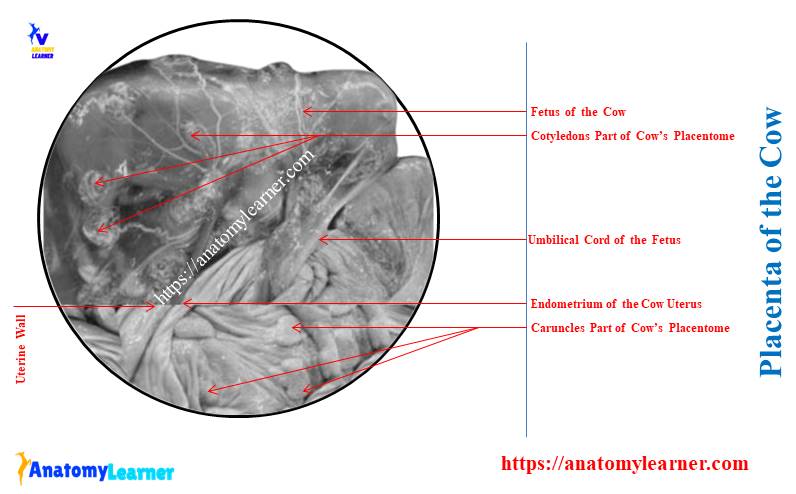
Here, the diagram shows the cow’s placenta with the fetal caruncles and maternal cotyledons. It also shows the umbilical cord, fetal membrane, and other structures of the bovine placenta.
Structure of a bovine placenta
To describe the full structure of a bovine placenta, you might cover the following –
- The extraembryonic membrane of the placenta and
- Various components of the placenta,
The extraembryonic membrane is formed around the fetus for protection. It will also help implant the fetus in the inner surface of the uterus (maternal endometrium).
The wall of the cow uterus has 3 coats – endometrium (inner), myometrium (middle), and perimetrium (outer). You may get the full guide on the cow uterus from the below-mentioned article –
The extraembryonic membrane is the fetal membrane of the placenta that consists of the following –
- Chorion – it is the outermost membrane that comes into contact with the maternal uterine endometrium,
- Chorioalanantois – is the fused structure of chorion and allantois,
- Allantois – is a continuous layer that encloses the allantoic cavity,
- Amnion – is the innermost membrane that closes to the fetus and
- Amniotic cavity – the cavity encompassed by the amnion and filled with the amniotic fluid. It provides the fluid environment in the placenta that protects the fetus.
The diagram shows the structures mentioned above from the fetal placenta. Here, the chorion surrounds the allantoic cavity, amnion, amniotic cavity, and fetus. Thus, this layer makes the fetal component of the bovine placenta.
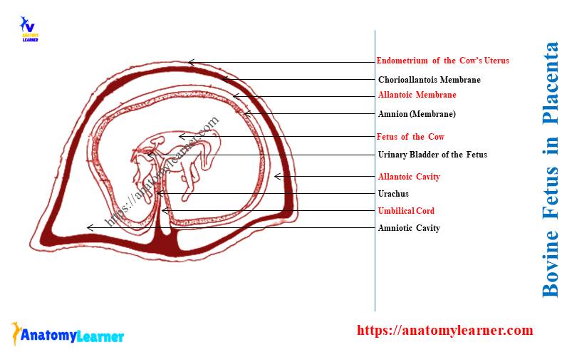
The allantois liens the inner side of the chorion and forms the chorioallantoic membrane. This allantois also lines the outer side of the amnion and forms the allantoamnion.
Thus, the allantois provide the vasculature to these two chorioallantoic and allantoiamnion membranes.
What are the water bags in the ruminant placenta?
The amnion is the fluid-filled cavity that contains the fetus. This amnion forms two cavities –
- Amnions fuse with an inner layer of allantois and form the allantoic cavity and
- An inner layer of the amnion contains fluid and forms the amniotic cavity, where you will find the fetus,
The allantoic cavity of the bovine placenta is known as the first water bag. This water bag continues with the cranial extremity of the fetal urinary bladder by the urachus. Finally, it passes through the umbilical cord.
Again, the fluid-filled amniotic cavity is also known as the second water bag of the fetal placenta. The first and second water bags typically refer to the fetal membrane during parturition.
So, the water bags of the ruminant placenta are –
- First water bag – allantoic cavity, and
- Second water bag – amniotic cavity,
During parturition, the allantoic sac is expelled first. Then, the amniotic sac is expelled out after the allantoic sac.
What type of placenta is found in cattle?
The cattle have the cotyledonary placenta or placentomes. This is one type of epitheliochorial placenta that contains 3 fetal and 3 maternal layers.
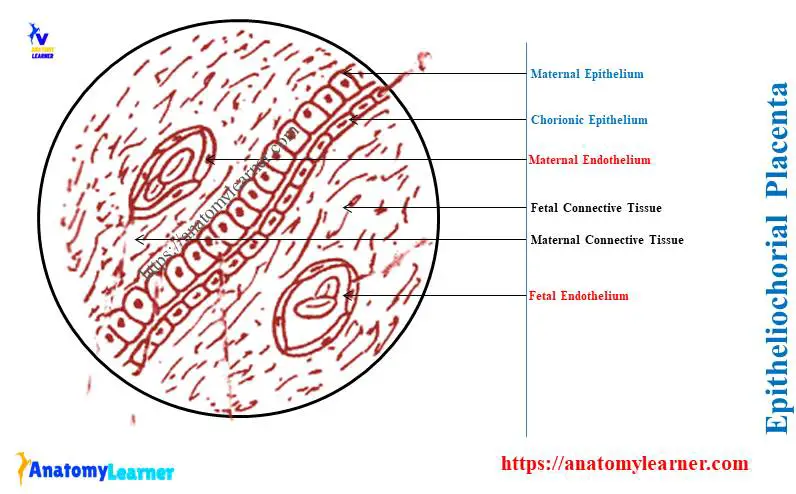
Here, the diagram shows the epitheliochorial placenta that may be found in horses, pigs, and ruminants. Again, the epitheliochorial placenta divides into two –
- Adeciduate placenta: the maternal component of the placenta does not slough during parturition. You will find adeciduate type placenta in horses and pigs.
- Partial deciduate placenta: the part of the maternal component of the placenta sloughed off during birth. You will find the particle deciduate type placenta in a cow, sheep, and goat.
What is an endotheliochorial placenta?
The maternal endothelium and connective tissue layers are absent in the endotheliochorial placenta. Here, the maternal endothelium begins in direct contact with the chorionic epithelium.
The endotheliochorial type of placenta is found in dogs. Here, in dogs, the maternal endothelium is lost during the birth.
Here, the diagram shows the deciduate endotheliochorial placenta from the dogs. It is also known as the zonary placenta in dogs.
What are the parts of the placenta of a cow?
The placenta of a cow consists of 2 parts – fetal and maternal parts. Here, the fetal component of the bovine placenta is formed by three layers of chorioallantoic membrane –
- Fetal endothelium: these endothelium line the allantoic vessels,
- Fetal connective tissue of the chorioalantoic membrane: it forms the bulk of the fetal part of the placenta. It is mesodermal origin and surrounds the fetal vessels.
- Chorionic epithelium: it is the surface epithelium layer of the chorioallantoic membrane,
The diagrams show the various parts of the bovine placenta with these six layers.
Again, the maternal part of the bovine placenta also consists of 3 layers of uterine endometrium –
- Maternal epithelium of the endometrium: it is the uterine lamina epithelium of the maternal part of the bovine placenta.
- Maternal connective tissue: this is the connective tissue that surrounds the vessels of the endometrium and
- Maternal epithelium: these are the epithelium that line the vessels of the maternal endometrium,
How many placentas do cows have?
The cows have only one placenta but multiple (60 – 75) placentomes. Here, the placentomes are the functional and discrete part of the bovine placenta.
A placentome of a cow consists of the connected cotyledon and caruncle. You will find this type of placentome only in the ruminant, like cows, sheep, and goats.
Here, the placentome of the cows (large ruminants) is dome-shaped or mushroom-shaped. The placentome of the ewes (small ruminants) is dish-shaped. They have a dent in the top.
What are cotyledons in cows?
Answer: the cotyledons are the fetal component of the cow placenta that forms the villous process. These cotyledons of the cows interdigitate with the maternal component of the placenta.
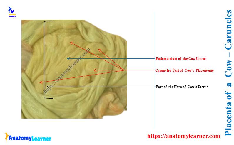
The diagram shows the oval button-like cotyledons from the cow’s placenta. Again, it shows the round and raised caruncles.
Here, the caruncles are the maternal component of the ruminant placenta. They consist of discrete units scattered over the endometrium of the ruminant uterus.
The caruncles are the proliferation of the connective tissue of uterine endometrium. You will also find these caruncles in the non-pregnant ruminants. But, these caruncles are absent in the horse and dog’s uterus.
The fetus of the ruminant pulled out of the interlocking caruncles and cotyledons.
Caruncles and cotyledons are seen on what animal?
Answer: the caruncles and cotyledons are the special structures found in the ruminant placenta. Thus, you will find the caruncles and cotyledons in cows, ewes, and does.
Thus, all ruminants have the cotyledonary type placental attachment in which exchanges occur at placentomes.
The caruncles project out from the surface of the ruminant uterus. It varies in diameter from half an inch to more than 3 inches.
As the pregnancy progresses in the ruminant, the size of the caruncles increases. The cranucles are larger in the gravid uterine horn than those in the nongravid uterus.
The surface epithelium of the ruminant caruncles is covered with the crypts. Within these crypts of the caruncles, the fetal placenta projects.
You will not find a free area between the caruncles. This area has no attachment between the maternal and fetal components.
Do goats have a placenta?
Answer: the goats have a placenta that looks like the cow’s. Anatomically, you will find similar structures in the goat placenta.
The number of the placenta may vary in the goat compared to the cows. You may find 1 – 3 (sometimes 4) placentas in the uterine horns of the goats (does).
What type of placenta do goats have?
Answer: the goats have the cotyledonary type placenta. Thus, you will also find the placentomes in the goat placenta, which consists of cotyledons and caruncles.
The shape of the goat’s caruncles differs from that of the cows. You will see the larger central depression on the caruncles of goats.
Within this central depression of the goat’s caruncles, the choronic villi attaches. Here, the diagram shows the convex cotyledonary placenta in the cows. Again, the diagram shows the concave cotyledonary placenta in ewe and does.
Conclusion
So, the placenta of a cow is a cotyledonary type that consists of special caruncles and cotyledons. The cotyledonary placenta of the ruminant possesses two components – fetal and maternal.
The goats and sheep also have the cotyledonary type placenta, like the cow. But, the shape and size of the caruncles in goats and sheep are a little different than those of the cows.