The chicken skeleton is unlike that of any other group of animals. A combination of reduction in the total number of bones and the fusion of many joints has resulted in a chicken or bird skeleton. Therefore, you will find a different skeletal modification in a chicken or bird. This article will show you the detailed anatomy of all bones from the chicken skeleton with a labeled diagram.
I will also enlist the peculiar anatomical features of every bone of chicken or bird with their short description. This might be a great resource if you want to know the details on chicken leg bone anatomy, chicken wing bone, and skull.
Chicken skeleton anatomy
First, I would like to inform you of the most interesting facts of chicken skeleton anatomy with a diagram. However, I have a short guide on avian osteology here as an anatomy learner. But, this article might help you to learn the details of bone anatomy from any birds. So, here, I will provide some interesting facts from the bones of a chicken.
- Most of the large bones of a chicken or bird possess air sacs (known as pneumatic bones) that connect with the respiratory system. These air sacs make the skeleton light compare to other mammals.
- There is a reduction in the total number of bones and fusion in many bones present in the chicken skeletal system.
- A lightweight beak is covering the mandible that replaces the teeth and reduces the weight of a skeleton. The bones of a chicken skull are mostly fused and possess a vast orbit.
- You will find a long and mobile neck in chicken containing more cervical vertebrae than mammals.
- The number of vertebrae reduces and fuses to form synsacrum (lumbar and sacrum), pygostyle (caudal) in chicken. You will find a uropygial or preen gland at the tip of the pygostyle of a chicken.
- There is no floor in the pelvic cavity of the chicken. This feature makes the pelvic more distensible to facilitate the passages of the eggs to the outside.
- The sternum of a chicken extends into a flattened lateral keel. This structure provides a large surface for the attachment of the primary flight muscles.
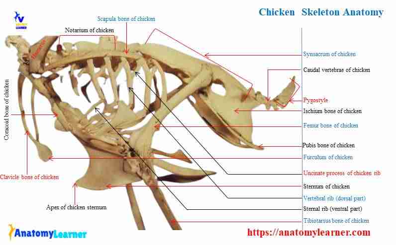
You will find two parts in each rib of a chicken. Except for the last rib, it possesses caudal extension, a unique feature of the chicken of bird skeleton.
Again, some of the bones from the leg are fused, forming a tibiotarsus and a tarsometatarsus in chicken.
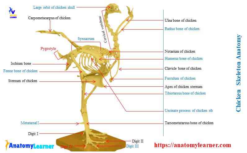
Chicken skeleton labeled diagram
Thanks for continuing to learn chicken skeleton with a labeled diagram. Here, I tried to show you every single bone from a chicken. Unlike mammals, you will find axial and appendicular structures in a chicken or a bird.
Now, it’s time to learn the peculiar features of every single bone of a chicken. Make sure you know the name of each bone from the axial and appendicular parts of chicken’s skeleton. You might see the following from a chicken –
- The skull bones with their peculiar features
- Chicken vertebrae in different region and their unique structures
- The bones of a wing with some peculiar characteristics
- Chicken leg bones and their special osteological features
In the labeled diagram, I showed you the skull bone, vertebrae (cervical to caudal), ribs, sternum, wing bones, and leg bones from a chicken. You may also get help from the video that I will add at the end of this article. That video might help you to identify all the bones of a chicken.
Chicken bone anatomy
In each bone from chicken skeleton anatomy, I will enlist the unique features first. Then I will go over the detailed anatomical features of each bone from a chicken.
“Structurally, the cortex of the bird’s bone is much thinner than that of mammalian. This is due to lightening their skeleton. As a result, many long bones are hollow and pneumatic in most of the bird.”
Okay, let’s start with the peculiar osteological features of the vertebrae of a chicken. You will find a lot of exceptional characteristics in the vertebrae of a chicken. Okay, let’s see what they are.
Vertebrae of a chicken skeleton
The segments of the vertebral column of a chicken are more challenging to different than that of the mammals. The following are some unique features that are present in the vertebrae of a chicken skeleton anatomy.
There are fourteen cervical, seven thoracics, fourteen fused lumbar and sacrum, and four to nine caudal vertebrae found in a chicken. As a whole, the vertebrae column is stiff due to the fusion of some vertebral segments.
The atlas of chicken is in the ring form that presents only one articular facet at its cranial end for the single occipital condyle.
Most of the cervical vertebrae of chicken possess styloid projection from transverse processes.
The last two thoracic vertebrae, along with lumbar and sacral vertebrae, fuses to form a unique structure (synsacrum).
The last few caudal vertebrae fuse to form a pointed bony projection, known as a pygostyle in chickens.
Great, now let’s move to the detailed anatomical facts of the chicken vertebrae with diagrams.
Cervical vertebrae of chicken
The cervical vertebrae of a chicken form an S-shaped structure that is more mobile and contains more vertebrae. Typically, you will find fourteen cervical vertebrae in the chicken, seventeen in a duck, and twelve in a pigeon.
The first cervical vertebrae are ring-shaped bones with a dorsal arch and ventral body. You will find an articular surface on the dorsal surface of the body for the dens of the axis. In addition, the second cervical vertebrae (axis) is larger than the first one (atlas).
The axis of a chicken has an elongated body that articulates with the third cervical vertebrae caudally. You will find an unpaired dorsal spinous process on the arch of the axis of a chicken.
On subsequent cervical vertebral bodies and the articular surface are saddle-shaped in chicken. There is a dorsoventrally convex and transversely concave cranial articular surface in the cervical vertebrae. Again, the caudal articular surface of these cervical vertebrae is shaped in an opposite, complementary fashion.
You will find the carotid processes ventrally from the mid-cervical region of a chicken skeleton. There is also a median ventral crest present in the cervical vertebrae of a chicken.
The transverse processes are perforating by the transverse foramen near their origin. Thus, the transverse foramina of cervical vertebrae give rise to a transverse canal similar to the foramen transversarium.
Thoracic vertebrae of chicken
In a chicken, you may find the first and sixth thoracic vertebrae as separate bones. The second to fifth thoracic vertebrae of a chicken fuse to form the notarium. Again, the last thoracic vertebrae of the chicken fuse with another unique structure – the synsacrum.
“The notarium is a rigid osseous unit that presents only in chicken and pigeon. But you will not find this structure in duck or goose. In duck and goose, the stabilization of the thoracic vertebrae is achieved by the ossification of tendon and ligaments.”
You will find an incomplete plate-like ventral crest in the notarium of a chicken. Again, the spinous process of notarium forms a dorsal spinal crest. The fusion of the transverse processes of notarium gives rise to a continuous plate known as the transverse lamina. In addition, you will find the articular surface for the ribs in the notarium or the thoracic vertebral segments of a chicken.
Lumbar and sacral vertebrae (synsacrum of chicken)
In a chicken skeleton anatomy, a total of fifteen to eighteen vertebrae (thoracic, lumbar, and sacral) contribute to form a synsacrum. But the number of bones may vary in different species of chicken. This is one of another exceptional osteological characteristics of chicken’s vertebrae than that of mammals.
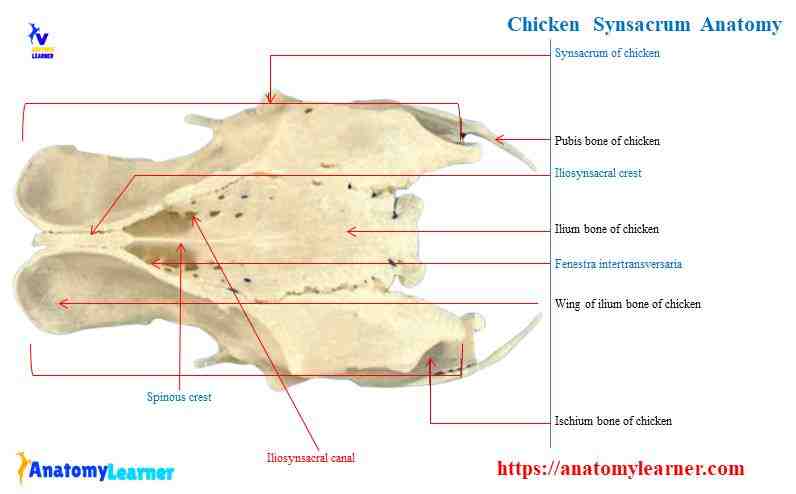
You will find a continuous ridge on the dorsal surface of the synsacrum that forms with the merge of the spinous processes. The name of this constant ridge is the spinous crest of chicken. You will also find the iliosynsacral crest at the dorsomedial edge of the iliac bones.
There is a solid bony connection between the ilium and the transverse and coastal processes of the synsacrum.
These features form a stable framework for the transmission of force during walking. In addition, there is a ventral crest present on the cranial surface of the synsacrum of a chicken.
Caudal vertebrae of chicken and pygostyle
The caudal vertebrae of the chicken skeleton may vary with different species. But, ideally, you will find four to nine caudal vertebrae in a chicken. In addition, there is a unique osteological feature present in the caudal vertebrae of a chicken than that of mammals. Some caudal vertebrae (last four to nine) fuse to form a pygostyle in a chicken.
The shape of this pygostyle of chicken is plate-like but may vary with species. You may also find triangular-shaped pygostyle in some species of chicken. There is a broad base of a pygostyle that articulates with the last free caudal vertebrae. The apex of the pygostyle is directed caudally. And the lateral parts of a pygostyle are known as the lamina.
But the other caudal vertebrae remain as individuals in chickens (first to third). The vertebral canal of the chicken’s caudal vertebrae is relatively wide compare to that of mammals. You will find the spinal cord that extends to the last segment of the vertebral column of the chicken.
The caudal vertebrae of chicken also possess a distinct transverse process and well-developed spinous processes.
Chicken skull bones anatomy
The bones of a chicken’s skull consist of thin plates formed either from connective tissue or from cartilaginous templates. I have a detailed guide on chicken skull anatomy where I showed the essential osteological features. Okay, let’s see what the most remarkable features present in chicken skull bones are.
- Most of the chicken bones are fused.
- You will find a large orbit that has a thin margin in a chicken skull.
- The upper jaw of a chicken is movable due to the presence of a nasal-frontal hinge.
- There is a single occipital condyle present in the skull of a chicken.
- You will not find any teeth in the upper and lower jaw of a chicken.
The occipital bone of a chicken skull forms the caudal wall of the brain. It also surrounds the foramen magnum of the skull of a chicken. In addition, there is a parabasal fossa in the occipital bone of a chicken that provides passage for blood vessels and nerves.
The frontal and parietal bones form the dorsal wall of the cranium of the chicken skull. There is a vast orbit that separates from the nasal cavity by the ectethmoid bone. The lacrimal bone forms the ventral and rostral continuation of the lateral orbital margin of the chicken.
The boney nostril bounds by the frontal and maxillary processes of the premaxillary bone. You will find a relatively small maxillary bone that articulates with premaxilla bone. Together they form a short, caudal terminal portion of the upper beak of a chicken.
You will find a rudimentary vomer bone in chicken. But in duck, this vomer bone completes the nasal septum. You will find a bar-shaped pterygoid bone in the skull of a chicken.
Thoracic limbs of a chicken skeleton
The thoracic limbs of a chicken skeleton consist of the bones of the pectoral girdle and the wing’s bones. A fully developed chicken pectoral girdle comprises the coracoid, scapula, and clavicle. Excellent, first lest’s discuss on bones of the pectoral girdle from a chicken.
Here, I will enlist some of the essential osteological features of the pectoral girdle’s bones of a chicken.
- The scapula of a chicken is elongated and located parallel to the vertebral column.
- There is a rod-shaped coracoid bone that connects the sternum’s cranial border with the chicken’s shoulder joint.
- The clavicle is the thin rod-shaped bones that form the furcula ventrally in chicken.
Now, let’s know some other osteological features of these bones of a chicken.
Scapula bone of a chicken
The scapula of a chicken is an elongated and blade-like structure. As you know, this bone runs parallel to the vertebral column of the chicken.
The cranial extremity of the scapula firmly binds with the coracoid bone of a chicken. You will find a long caudal extremity in the chicken’s scapula that is slightly curved. A neck separates the cranial extremity from the body of the scapula.
The scapula of a chicken joins with the clavicle craniomedially. You will find a supracoracoid groove that lies immediately lateral to this joint. There is also a glenoid process present at the lateral aspect of the chicken scapula. It completes the articular surface for the head of the chicken humerus.
The coracoid bone of chicken
The coracoid is the strongest and rod-shaped bone in the pectoral girdle of a chicken skeleton. You will find a body and two extremities in the coracoid of a chicken. There are also two surfaces – dorsal and ventral that merge at the lateral and medial margin of the coracoid.
The cranial extremity of the chicken coracoid bone possesses the following important osteological features-
- Acrocoracoid process of coracoid bone of chicken
- Supracoracoid groove of the coracoid bone
- Glenoid process of the coracoid bone
- The articular surface for humerus in coracoid bone and
- Articular surface for scapula in coracoid bone
The acrocoracoid process is a hook-like structure and the most prominent feature in the coracoid of a chicken. It extends medially and forms the supracoracoid groove for the tendon of the supracoracoideus muscle.
At the caudolateral aspect of the cranial extremity of the coracoid, you will find the glenoid process. The glenoid process of the coracoid forms the major component of the articular surface for the head of humerus bone. You will also find another articular surface for the scapula in the coracoid bone of a chicken.
The dorsal surface of a coracoid faces the trunk and posses a pneumatic foramen. This pneumatic foramen is for the outpouching of the clavicular air sac in chicken. In addition, the ventral surface of a coracoid bone merges smoothly with the intramuscular line.
Clavicle of chicken
You will find these two unique thin and rod-shaped structures (clavicle) in the chicken skeleton anatomy. The proximal extremity of the clavicle joins with the cranial extremity of coracoid and scapula bone-in chicken.
Distally, these two thin, curved rod clavicles join by the bony tissue and form wishbone or furcula. Again, the shape and the size of the clavicle may vary with the species. For example, it is blade-shaped in chicken and joins with sternal rostrum by fibrous connection. But the clavicles of parrots are reduced bones and do not form the furcula.
Wing bones from the chicken skeleton
The wing bones from a chicken skeleton consist of the humerus, radius, ulna, carpus, metacarpus, and digits. First, I will show you the unique osteological features from the wing bones of a chicken.
- First, let’s point out the essential features that you might know from the wing bones of a chicken –
- The body of the chicken’s humerus is twisted and possesses an oval head.
- Proximally the humerus of a chicken articulates with the coracoid and scapula bones.
- You will find a pneumatic foramen at the medial aspect below the head of the humerus.
- The radius and ulna bones of chicken’s wing are remaining in contact at two extremities. Therefore, the ulna bone is comparatively massive than that of the radius in chicken.
- You will find a vast interosseous space in between the radius and ulna bones of a chicken.
- There are two carpals at the proximal row (radial and ulnar) in the chicken. In addition, the distal row carpals fuse with the metacarpal bones.
- Three metacarpals fuse with the distal row of the carpus (actually known as carpometacarpus) in chicken. Thus, you will also find three digits in the wing of a chicken.
There are very little information about the wing bones of a chicken. If you want to learn more about the wing bones from a chicken, please continue this article.
Humerus of chicken
Unlike the mammal’s humerus, you will also find a body and two extremities in the humerus of a chicken. I tried to show you the essential osteological structures from the humerus of a chicken with a labeled diagram.
- At the proximal extremities of a chicken humerus, you will find the following vital structures –
- Head and transverse groove of the chicken humerus
- Dorsal and ventral tubercles of the chicken humerus
- Deltopectoral crest in a chicken humerus
- Intertuberal plane in the humerus of a chicken
- A bicipital ridge in the humerus of a chicken
You will find the structures mentioned above in the cranial surface of the proximal extremity of a chicken humerus. Again, you will find a pneumotricipital fossa with pneumatic foramen in the caudal surface of the proximal extremity of a humerus.
At the distal extremity of chicken humerus, you will find the following osteological features –
- Dorsal and ventral condyles and
- Dorsal and ventral epicondyles in the humeurs of a chicken
The head of the humerus of a chicken is ellipsoid and form the part of the shoulder joint. There is a transverse groove on the humerus for the acrocoracohumeral ligament. You will find a small dorsal tubercle that continues as a deltopectoral crest in the chicken humerus.
There is a sizeable ventral tubercle that merges with the bicipital crest in the humerus of a chicken. You will find a deep area that lies immediately adjacent to the ventral tubercle at the caudal surface of the humerus.
The distal extremity of a chicken humerus possesses barrel-like dorsal and ventral condyles. You will also find dorsal and ventral epicondyles just proximal to the respective condyles.
Radius and ulna of a chicken skeleton
You will find a variation in the radius and ulna bones of a chicken skeleton compared to mammals. These bones are approximately equal in length and lie parallel to one another in the chicken wing. But, the ulna bone of the chicken wing is more massive than that of the radius. Therefore, the smaller radius bone lies dorsal to the massive ulna in the chicken wing.
You will find a body and two defined extremities in both the radius and ulna bones of a chicken. At the proximal extremity of the radius bone of the chicken wing, you will find an articular surface for the humerus. You will also find an articular facet for the ulna bone of a chicken.
The body of the chicken radius bone is relatively straight and becomes thick at the distal part. Therefore, you will find the articular surfaces at the distal ends of the radius bone for ulna and radial carpal bone.
There is a less developed olecranon process at the proximal extremity of the ulna bone of a chicken. Therefore, you will find different articular surfaces for the ulna and scapula bones at the proximal ends of an ulna bone.
The body of the chicken ulna bone is slightly curved than that of the radius. At the distal extremity of the chicken ulna bone, you will find the pairs of condyles.
Carpal, metacarpal, and digits of chicken wing
There are two developed carpal bones (radial and ulnar) present in the chicken wing. These radial and ulnar carpal bones are arranged in the proximal row of the carpus. But the distal row of carpal bone fuses with the metacarpals to form carpometacarpus in chickens.
The radial carpal of the chicken is compact and possesses articular surfaces for radius, ulna, and carpometacarpus bones. On the other hand, the ulnar carpal is more oversized and distinctly angular in shape.
You will find carpus trochlea at the proximal end of each metacarpal (three in number) that articulates with the carpal bones. Again, at the distal extremity, each metacarpal bone of the chicken contains articular surfaces for phalanges.
You will find considerably reduced bone in the digits of a chicken wing. The first digit of the chicken wing contains only one cone-shaped phalanx, while the other two consist of two phalanges.
Chicken leg bone anatomy
The leg bones of a chicken skeleton consist of the pelvic girdle, femur, tibiotarsus, tarsometatarsus, and phalanges. Unlike the mammals, the pelvic girdle of a chicken consists of three bones – ilium, ischium, and pubis. But, in chicken, these pelvic bones are fused, forming os coxae and joins with the synsacrum.
There is a rigid connection between the pelvic bones and the vertebral column of a chicken. You will not find any pelvic floor in the chicken anatomy as there is no pelvis symphysis. So, the pelvis is open ventrally in the chicken and facilitates the egg passages.
Okay, now I will enlist some of the essential structures from the pelvic and leg bones of a chicken –
- The ilium, ischium, and pubis are joining with each other in chicken. Again, the ilium bones fuse with the synsacrum.
- You will find a thin elongated structure like pubis bone in the pelvic girdle of a chicken.
- There is an extra-large aperture in between the ilium and ischium, known as ilioischiatic foramen.
- The femur of chicken bents along the length and posses only the greater trochanter at its proximal ends.
- You will find a thin triangular patella which width is comparatively more than the length in chicken.
- The bones of the proximal row of the tarsus fuse with the distal end of the tibia and forms tibiotarsus (longest bone) in chickens.
- You will find a single long tarsometatarsus bone-in chicken that fuses with the distal row of tarsus bones.
Above these is very little information about the pelvic and leg bones of a chicken. If you want to know the detailed anatomical features of a chicken’s pelvic and leg bones, continue this article.
Ilium from the pelvic girdle of a chicken
The ilium is the most prominent bone in the pelvic girdle of a chicken. You will find three different parts in the ilium of a chicken – preacetabular wing, body, and postacetabular wing. Here, I will show you all the essential structures from the ilium bone of a chicken skeleton with a labeled diagram.
You might identify the following structures from the ilium bone of a chicken –
- Preacetabular wing of the ilium
- Iliosynsacral crest of chicken ilium bone
- Dorsal iliac crest of chicken ilium
- Lateral iliac crest of chicken ilium bone
- Body of the ilium bone of a chicken
- Preacetabular wing (dorsal and lateral surfaces)
- Dorsolateral iliac crest in preacetabular wing
- Preacetabular tubercles of ilium bone
- Iliosynsacral canal in ilium bone of a chicken
- Antitrochanter of ilium bone
You will find a dorsally concave preacetabular wing in the ilium of a chicken. Here is a preacetabular wing. You will find three crests – dorsal, lateral, and iliosynsacral.
The dorsal iliac crest bounds the ilium bone dorsally. Again, the lateral iliac crest forms by the cranial and lateral margin of the preacetabular wing of the chicken. The iliosynsacral crest forms with the fusion of the iliac ridge with the spinous process of the synsacrum.
You will also find some iliosacral canals that are enclosed by the medial surface of the preacetabular wing and the spinous and transverse process of a synsacrum. Thus, the body of the ilium contributes to forming the acetabulum and joins with the antitrochanter. In addition, you will find another structure (preacetabular tubercle) that projects cranioventrally from the ilium bone.
There are two surfaces (lateral and dorsal) in the postacetabular wing in a chicken. A dorsolateral iliac crest separates the dorsal surface from the lateral surface of the postacetabular wing.
This dorsolateral iliac crest continues as the dorsolateral iliac spine.
Ischium and pubis bones of chicken
The ischium of a chicken consists of a body and a wing. It contributes to forming the acetabular cavity caudally and the ventral part of the antitrochanter.
The wing of the ischium merges with the lateral surface of the ilium without any distinct boundary.
The pubis of a chicken is the most ventral part of the chicken pelvic girdle. This is a thin elongated bone that incompletely fuses with the ischium bone of the chicken. The body of the pubis bone helps to complete the ventral part of the acetabulum cavity.
The pubis bones of chickens are not fused by symphysis like mammals. Instead, you will find a thick, palpable apex in the caudal end of a pubis.
Femur of chicken skeleton
The femur of the chicken skeleton is a cylindrical shape that has a slight cranial curvature. You will find a well-developed body and two extremities in the femur of a chicken.
At the proximal extremity of the chicken femur, you will find a round femoral head. It possesses a small cavity for the ligament of the femoral head.
There is a femoral trochanter that projects laterally at the proximal end of the femur. In addition, you will find a body ridge (trochanteric crest) that extends distally from the trochanter. Finally, the neck of the chicken femur connects the head with the body of the femur.
You will find different muscular lines at the cranial and caudal surfaces of the body of a chicken femur. The body of a chicken femur is slightly curved.
At the distal extremity of the femur, there are lateral and medial condyles that separate by an intercondylar groove. The lateral surface of the lateral condyle possesses a fibular notch. You will also find the lateral and medial epicondyle in the respective condyles at the distal end of a chicken femur.
There is a patellar groove present in the cranial surface of the distal end of the chicken femur. It provides a gliding articular surface for the patella of a chicken.
Tibiotarsus of chciekn
The tibiotarsus (fusion of tibia and the proximal row of tarsus bones) is the most prominent bone in the chicken skeleton. You will find a long body and two extremities in the tibiotarsus of a chicken.
The proximal extremity of the chicken tibia is expanded and posses the following important osteological features –
- Small lateral and larger medial articular surfaces of the tibia
- Patellar crest in the tibia
- Lateral and cranial cnemial crest of the chicken tibia
- Intercnemial groove of the tibia
The interarticular area separates the lateral and medial articular surfaces of the chicken tibia. You will find a prominent cranial cnemial crest (tibial crest) in the proximal end of the chicken tibiotarsus. In addition, you will find the intercnemial groove in between the lateral and cranial tibial crests.
At the distal extremity of the chicken tibia, there are medial and lateral condyles that separate by the intercondylar groove. You will also find lateral and medial epicondyles proximal to the respective condyles of chicken tibiotarsus bone.
The fibula of the chicken is the rod, a slender structure that articulates with the tibiotarsus and the lateral condyle of the femur.
Tarsometatarsus bones of a chicken skeleton
The tarsometatarsus bones of the chicken skeleton form with the fusion of metatarsal with the distal end of tarsus bones. Only the first metatarsus bone is separate; the II to IV metatarsus fuses in chicken. The fifth metatarsal is absent in the chicken bone anatomy.
Let’s know what the essential osteological structures that you might identify from the tibiotarsus of a chicken are –
- Lateral and medial condyles at the proximal end of tibiotarsus bone
- The intercondyloid eminence of chicken tibiotarsus bone
- Hypotarsus of a chciekn
- Body of the metatarsal of chcicken
- Metatarsal I in chciekn leg
- Trochlea of metatarsal bones at the distal extremity
The intercondylar eminence separates the lateral and medial condyles of the chicken tibiotarsus. You will find an exception structure (hypotarsus) in chicken leg anatomy arising from the distal tarsal bones component.
The metatarsal bone I projects dorsomedially from its articulation with the tarsometatarsus of chicken. There is a calcaris process in chicken tibiotarsus where you will find a small fossa for the articulation with the first metatarsal bone.
Digits of chicken leg
There are three well-developed digits and one less developed digit present in the chicken leg. The number of the phalanges may vary with the number of digits. For example, you will find only two phalanges in the first digit of a chicken. Again, the fifth digit consists of five phalanges in chicken. But digit III is more extensive in the chicken leg.
The proximal and intermediate phalanges of the chicken leg possess a base, a body, and a capitulum. Again, the distal phalanges have a base, an apex, the base articulating with the penultimate phalanges.
Frequently asked questions on chicken bones
What is the skeletal system of a chicken?
The skeletal system of a chicken consists of the axial and appendicular skeleton as like other mammals. Therefore, you will find the skull bone, vertebrae, sternum, and ribs in the axial skeleton. In addition, you will find the wing bones and leg bones in the appendicular part of a chciekn’s skeleton.
How many bones are in a chicken skeleton?
There are almost one hundred and sixty-nine bones present in a chicken skeleton. You will find more than forty bones in the skull of a chicken. There are forty-two bones present in the legs (two) of a chicken. In addition, you will find twenty-five to twenty-seven bones in the wing of a chicken.
Do chickens have skeletons?
Yes, the chicken has some special osteological features in its maximum bones. You will find notarium, synsacrum, pygostyle, uncinate process, tibiotarsus-like structures in their skeleton.
How do you clean a chicken skeleton?
First, you might remove all the muscles from chicken’s skeleton. Then you might go through a long process that I have described in another article.
Do chickens have a bony skeleton?
Yes, chicken has a bony skeleton. But most of their bones possess some unique features to compare to that of mammals. For example, fusion and deletion are the main characteristics features of the chicken bones.
Do chickens have bones in their legs?
The leg bones of a chicken show the features of fusion and simplifications. For example, you will find the femur, longest tibiotarsus (fusion of tibia and the proximal row of tarsus), fibula, tarsometatarsus (fusion of metatarsal and a distal row of tarsus bones), and digits (three developed and one less designed) in chicken legs.
What are the 4 parts of the skeleton?
The four main parts of the skeleton are bones, cartilage, ligament, and joints. If you want to know the details anatomy of bones, cartilage, ligament, and joints from animals, go to the guide page and learn one by one.
Conclusion
So, you have got the variation in the different bones of a chicken skeleton. The most osteological variation is present in the bones of the skull, vertebrae, and chicken leg. All the chicken skeleton labeled diagrams might help you understand the variation in their osteological characteristics compared to mammals.
Now, it is time to learn more and practice with the actual chicken skeleton at your laboratory. You might take the help from all the labeled diagrams of a chicken skeletal system.