
The skeletal muscle fibers are elongated, cylindrical and multinucleated cells whose length may vary in different animals. In this short guide, you will get a basic concept of skeletal muscle histology from the real slide and labeled diagram.
You will also get the identification points of skeletal muscle histology slide with a little description here in this guide. I will show you the histological features from the real slide and skeletal muscle histology labeled diagram.
So, if you are interested to learn skeletal muscle histology slide identification with description, you may follow this guide.
Skeletal muscle histology
If you want to know the details of skeletal muscle, you might cover the light microscope structure, fine structure and the process of skeletal muscle contraction. But in this short guide, I will discuss only the identification points of skeletal muscle with a little description.
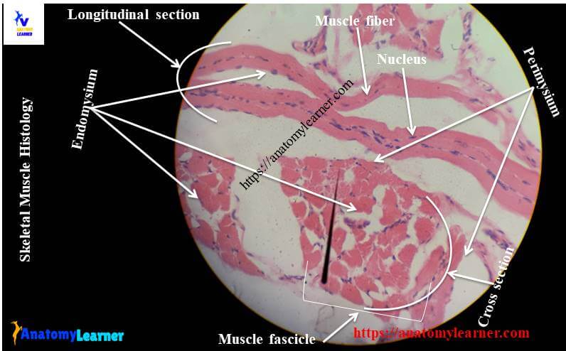
From the skeletal muscle histology slide, you might identify the following important structures under the light microscope. Please try to find out these structures from the skeletal muscle slide labeled images.
- #1. Longitudinal section of skeletal muscle
- #2. Cross-section of skeletal muscle
- #3. Skeletal muscle fibers of the longitudinal section
- #3. The nucleus of skeletal muscle fibers in longitudinal and cross-section
- #4. Cross striations of skeletal muscles
- #5. Endomysium of skeletal muscle fibers
- #6. Fibroblasts in endomysium
- #7. Perimysium in the cross-section part
- #8. Myofibril in cross-sectional part
- #9. Connective tissue in both longitudinal and cross-section parts
- #10. Blood vessels and other capillaries in both longitudinal and cross-sections of skeletal muscle
- #11. Cross striations of skeletal muscles
It is not easy to clearly show you all the structures from one slide picture, so please find the histological features from other slide pictures.
Skeletal muscle histology slide identification points
This is very important to know the characteristics features to identify the specific slide under the light microscope. Now, I will enlist the skeletal muscle histology slide identification points that will help you to identify this slide under the light microscope. Here, I tried to cover all the histological features (both in longitudinal and cross-sectional samples) from skeletal muscles.
Under the low power magnification, you will find the following identifying features of skeletal muscle in the longitudinal section –
#1. Presence of long, cylindrical and multinucleated muscle fibers without any branching.
#2. The nuclei (flatten in shaped) are present in the periphery in each skeletal muscle fibers.
#3. There are cross striation found in each skeletal muscle fibers of the provided sample.
#4. The endomysium is separating the skeletal muscle fibers bundles.
So, this is the longitudinal section of skeletal muscle fibers (skeletal muscle in tongue)
Again, you will find the following characteristics features of skeletal muscle under the microscope in cross-section –
#1. Presence of numerous grouped muscle fascicles in the cross section that covers with perimysium.
#2. Each muscle fibers shows granular appearances due to the cross section of myofibrils in the provided sample.
#3. Presence of ovoid peripheral nuclei in the muscle fascicles of the tissue sample.
#4. There are connective tissue, blood vessels and other capillaries in the sample tissue section.
So, this is the cross section of skeletal muscle (skeletal muscle of tongue).
Skeletal muscle histology description with labeled diagram
Well, let’s me describe the skeletal muscle histology with labeled diagram so that you might understand every single structure. I will show you the labeled diagram of both longitudinal and cross section of skeletal muscle.
The skeletal muscle fibers are supported by the connective tissue frameworks that are well appreciated in the cross section of muscles. The connective tissue of the muscle fibers provides support to the muscle fibers in the following manners –
#1. Epimysium of muscle fibers – a dense, irregular connective tissue layer surroundings the entire skeletal muscle fibers. This dense connective tissue sheath known as the epimysium of muscle fibers.
#2. Perimysium of muscle fibers – you know, a muscle fascicle is a small bundle of muscle fibers. A dense, irregular connective tissue also surrounds these small bundles of muscle fibers, which is perimysium of muscle.
#3. Endomysium of muscle fibers – loose connective tissue composed of reticular fibers supporting the individual muscle fibers. This reticular connective tissue fibers layers known as the endomysium of muscle. You will find more fibrocytes and blood vessels in the endomysium of muscle fibers.
You might also know the term sarcolemma, sarcoplasm, myofibrils and myofilaments. The sarcolemma is the thin plasma membrane of individual muscle cells.
Each muscle cell contains myofibrils which form the dot in the cross section of muscle at the light microscopic level. These are cylindrical structure and composed of thin and thick myofilaments that are responsible for the contraction.
Microscopic feature of skeletal muscle in longitudinal section
Each muscle fibers are the cylindrical and unbranched cylindrical cells under in longitudinal section (under the light microscope). You will find the elongated and flattened nuclei (multinucleated) in the muscle fibers just beneath the sarcolemma of muscle fiber.
Each muscle fibers is made of compact long cylindrical myofibrils in the sarcoplasm that arranged parallel to the long axis.
The thin myofibrils of skeletal muscle composed of actin, troponin and tropomyosin. Again the thick myofibrils of skeletal muscle composed of myosin. The arrangement of the actin, myosin, tropomyosin are very regular and form the distinct cross striation pattern in the skeletal muscle fibers. So, you will find the cross striation of alternative dark and light bands with Z lines.
Cross section of skeletal muscle
In the cross section of the skeletal muscle histology slide, you will find the numerous muscle fascicles (bundle of muscle fibers). These muscle fascicles are separated by the perimysium containing the blood vessels and connective tissue.
You will see the granular or dot appearance in the sarcoplasm of skeletal muscle. This occurred due to the cross section of each myofibrils of skeletal muscle.
Here you will easily identify the epimysium, perimysium and endomysium of skeletal muscle fibers. You will also find the ovoid nuclei in the skeletal muscle fascicles at the periphery.
Muscle slide images and videos
I think these labeled images and diagrams of skeletal muscle histology were helpful to understand the basics of this muscle structure. If you need more skeletal muscle images, let me know or follow anatomy learner on social media (for more updated images or notifications).
Don’t forget to read the other articles from anatomy learner (related to animal anatomy or histology) –
#1. Details guide and identification points of smooth muscle histology
#2. Dense connective tissue identification points with real slide images
#3. Loose connective tissue slide identification
Conclusion
This simple guide might help you to identify the skeletal muscle histology slide under the light microscope. If you think this guide is enough to learn the basics of skeletal muscle histology, share this with your friends interested in learning basic histology.
Thank you so much for staying with anatomy learner and learning the basics of animal anatomy and histology.