Cardiac muscle shows many structural and functional characteristics intermediate between skeletal and smooth muscles. Today, in this short article, I will show you the important histological features from the cardiac muscle histology slide.
You will get the basic guide to learn cardiac muscle histology with real slide images and labeled diagrams. I will also enlist the functions and identification points of cardiac muscle. After the end of the article, I will share the cardiac muscle histology drawing with you.
So, if you are interested to learn cardiac muscle histology, then continue this simple article.
Cardiac muscle histology
The cardiac muscle fibers are shorter than the skeletal muscle fibers and show the branching pattern. These muscle fibers have one or two centrally placed nuclei. From the cardiac muscle histology slide, you might identify the following important histological features.
I will try to show you these histological features from the real slide images and the diagram.
- #1. Longitudinal section of cardiac muscle
- #2. Cross section of cardiac muscle
- #3. Cardiac muscle fibers from both longitudinal and cross section
- #4. The nucleus of cardiac muscle fiber (single or binucleated)
- #5. Branching cardiac muscle fibers
- #6. Intercalated discs of cardiac muscle fibers
- #7. Cross striation of cardiac muscle fibers
- #8. Perinuclear sarcoplasm
- #9. Myofibrils of cardiac fibers
- #10. Endomysium, perimysium and epimysium of cardiac muscle
- #11. The connective tissue of cardiac muscle
Please find all these features of cardiac muscle from the slide images or labeled diagram.
Cardiac muscle histology slide identification
You might ask to identify the cardiac muscle slide under the light microscope at the laboratory.
I will enlist the most important identification points of cardiac muscle histology slide under the light microscope.
- #1. Presence of short cylindrical muscle fibers that shows branching (in longitudinal section of muscle fiber).
- #2. There is a centrally placed oval nucleus in each muscle fibers (sometimes, you may find two nuclei in some muscle fibers).
- #3. Presence of intercalated discs in the junction between the muscle fibers at their ends.
- #4. Muscle fibers show faint cross striation (less prominent than skeletal muscle fibers).
#5. Presence of muscle fascicles with centrally placed nucleus (in cross section of muscle fibers).
So, this is the slide of cardiac muscle histology (muscle of animal’s heart).
Cardiac muscle description
I will describe the histological features of cardiac muscle from both longitudinal section and cross section.
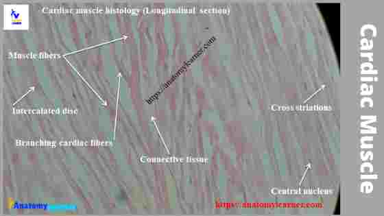
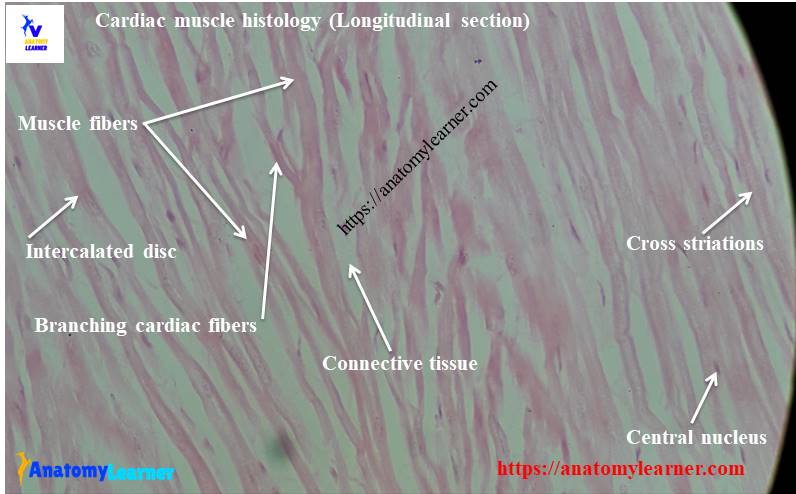
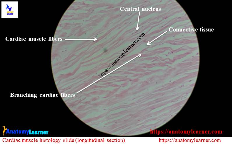
Longitudinal section of cardiac muscle
The cardiac muscle fibers are shorter than the skeletal muscle fibers and exhibits the branching pattern. There is one or two centrally placed oval nucleus in cardiac muscle fibers. The cytoplasm of these cardiac muscle fiber’s nuclei is acidophilic.
The most important identifying features of cardiac muscle histology are darkly stained transverse lines across the muscle fibers. These transverse lines are known as intercalated discs and located at the end to end junction of adjacent muscle cells.
Cardiac muscle cells have myofibrils similar to the myofibrils of skeletal muscle. The actin and myosin of the myofilaments (myofibrils) have a regular arrangement pattern like skeletal muscle. So, you will also find the cross striation in cardiac muscle fiber but less prominent than skeletal muscle’s cross striation. Surroundings of the nucleus of cardiac muscle fibers, you will find a prominent perinuclear sarcoplasm that is devoid of cross striation and myofibrils.
A network of fine reticular and collagenous fibers surrounds each cardiac muscle fibers. This network corresponds to the endomysium of the skeletal muscle fibers but is more irregular.
There is a vast blood supply found in the cardiac muscle. Well-developed capillary networks surround the individual cardiac muscle fibers.
Cross section of cardiac muscle fibers
In the cardiac muscle histology slide cross-section, you will find different muscle fascicles that exhibit granular appearances. In this section, you will find the oval centrally placed nucleus in the cardiac muscle fascicles.
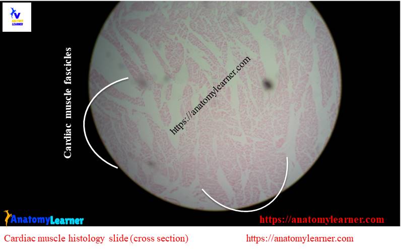
The connective tissue cells, fibrocytes and the fine reticular fibers of endomysium surround the cardiac muscle fascicles.
Location of cardiac tissue
You will find the cardiac muscle in animals’ hearts and the larger vessels attached to the heart.
#1. Heart wall
#2. Blood vessels attached to the heart
The function of cardiac muscle
The cardiac muscle is involuntary, and it is stimulated to contract by a mechanism similar to the skeletal muscle. This muscle helps to pump the heart by their involuntary movements and influence the rate of impulse conduction in the heart.
But the conduction system of the heart is made of modified cardiac muscle fibers which are thicker, larger and contain few myofilaments.
So the overview of cardiac muscle fibers are –
#1. Short branched muscle fibers
#2. Having uniform diameter
#3. One or two oval central nucleus
#4. Less cross striation
#5. Presence of intercalated discs
Cardiac muscle histology drawing
Now, I will share cardiac muscle histology drawing with you to understand every histological structure clearly.
This drawing of cardiac tissue is not perfect, but I hope it will help you get the basic concept of the arrangement of the muscle fibers. You might try to draw the better cardiac muscle.
If you need more drawing or real slide pictures of cardiac muscles, please let me inform or follow anatomy learners on social media. You will get a notification on social media when I update this article or images.
You might read the other article related to veterinary histology from anatomy learner –
#1. Smooth muscle histology with their identification points from slide pictures.
#2. Loose connective tissue identification from slide under light microscope
#3. Histology of connective tissue cells
#4. General features of a blood vessel with a labeled diagram.
I hope you could make the difference among three different types of muscles.
Conclusion
This was a short guide to learn the basics of cardiac muscle histology with slide images. If you think this guide helpful, you then share it with your friends who want to learn muscle tissue histology.
If you found any mistake on the cardiac muscle histology slide labeled diagram, please let me know. Don’t forget to check my newly published article here in anatomy learner.