The skin under a light microscope shows two distinct layers – epidermis and dermis. In the case of thin skin, the epidermis is very thin and lines with the keratinized stratified squamous epithelium. You will also find five different cells layers in the epidermis of both thick and thin skin under a light microscope.
The dermis of thick and thin skin comprises the superficial papillary layer and the deep reticular layer. Microscopically, you will find sweat glands, sebaceous glands, and hair follicles.
Here, I will show you the images of thick and thin skin under a light microscope with their identifying features. So, if you want to know the basics of the microscopic features of the skin, you may continue this article till the end.
Skin under microscope
Skin is the single heaviest organ covering the body surface and directly contacting the external environment. You will find two types of skin – thin or hairy skin and thick or non-hairy skin. The skin over the dorsal surface and on the lateral surface of the body is thick.
Again, the skin on the ventral surface of the body and the medial surface of the limbs are thin. You will find a thicker epidermis in the thick skin under a light microscope. In contrast, the thin skin contains a thin epidermis.

The microscopic features of both thick and thin skin consist of two layers –
A superficial epidermis layer that is made up of stratified keratinized squamous epithelium and five layers of cells, and
A deep dermis layer comprises connective tissue that contains sweat glands, sebaceous glands, and hair follicles.
The dermis layer of skin rests on the subcutaneous tissue, sometimes described as the third layer. So, you may tell there are three layers in the thick or thin skin – epidermis, dermis, hypodermis, or subcutaneous tissue.
You will learn the description of every single layer of skin in the next part of this article. Now, I will show the important microscopic features of thick and thin skin so that you may easily identify them.
Okay, let’s know the microscopic characteristics of the thick and thin skin with the labeled diagrams.
Microscopic features of thin skin
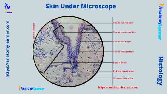
When you identify the thin skin, you will find the below-mentioned microscopic features. You will find both the epidermis and dermis layers under the microscope while viewing with 10x magnification. There is also a 40x magnification of thin skin where you will see the different types of cells.
- The sample tissue section shows that the thin epidermis comprises five layers of cells and lines with keratinized stratified squamous epithelium.
- There are two layers in the dermis of the thin skin – the superficial papillary layer and the deep reticular layer.
- You will find the dense connective tissue in the reticular layer of the dermis.
- Again, the thin skin’s dermis’s reticular layer contains sweat glands, sebaceous glands, hair follicles, and arrector pili muscles.
So, this is a thin skin microscope slide. You will find thin skin on most of the body except the palmar and plantar surfaces.
Microscopic features of thick skin
Under a light microscope, you will find the first difference between thick skin at the epidermis layer compared with thin skin. Again, you will find some structural differences in the reticular layer of thick and thin skin.
Here, I will show you the thick skin microscope image with 10x magnification (modified). Fine, let’s see the most important identifying features of a thick skin microscope slide.
- The tissue section shows the two distinct layers – the thick epidermis and dermis.
- Again, the thick epidermis consists of five layers of epidermal cells and lines with the keratinized stratified squamous epithelium.
- The reticular layer of the dermis does not show any hair follicles or sebaceous glands.
- You will find the papillae in the dermis of the provided sample and sweat glands under a light microscope.
So, this is a thick skin microscope slide. The epidermal cells (with 40x magnification) of the thick skin are shown in the next section of this article.
Identification of microscopic skin features
So, you could understand the important microscopic features in both thick and thin skin. From both the thick and thin skin microscope slide, you should identify the following features in common –
- Epidermis, dermis, and hypodermis (if present in the sample tissue) of thick and thin skin,
- Keratinized stratified squamous epithelium of thick and thin skin,
- Layers of epidermal cells of both thick and thin skin,
- Papillary and reticular layers of both thick and thin skin,
- Sweat gland and erector pili muscles of the skin,
- Hair follicles and sebaceous glands in the thin skin of an animal,
- Subcutaneous tissue or hypodermis of the thick and thin skin.
The different labeled diagrams already identified all these microscopic features from the thick and thin skin. You will also find more skin-labeled diagrams with their important microscope features on social media for anatomy learners.
Healthy skin under a microscope
So, in healthy skin, under a light microscope, you will find three distinct layers – epidermis, dermis, and hypodermis. The epidermis projects into the dermis as an epidermal ridge. In the epidermis lining in both thick and thin skin, you will find the keratinized stratified squamous epithelium.
The epidermis is ectodermal in origin, and it is avascular and nourished by diffusions. You may find the free nerve ending at the basal layer of the epidermis while viewing with the electron microscope.
Again, the microscopic view of a thick or thin skin shows five layers of cells. You will find four types of cells in the five layers of the epidermis.
Layers of the epidermis of thick or thin skin
You will find the following layers in the epidermis of thick or thin skin from its deep to the superficial surface –
- Stratum basale layer (basal layer),
- The second is the stratum spinosum layer or prickle cell layer,
- The third is the stratum granulosum layer,
- Stratum lucidum layer of skin, and
- Fifth is the stratum corneum layer of the skin.
Now, let’s discuss these epidermal layers from both the thick and thin skin of an animal.
Stratum basale of epidermis
In the stratum basale layer of thick and thin skin epidermis, you will find only a single layer of columnar or cuboidal cells. These cells rest in the basement membrane as found in the epithelial cell.
In the routine stain, you will see these stratum basale cells as basophilic. There is a large and oval nucleus in the stratum basale cells layer of the skin. So, the nucleus of the stratum basale occupies most of their cytoplasm.
In an electron microscope, you will see the desmosome (cell junction) between the stratum basale and stratum spinosum layers. The stratum basale attaches laterally to each other and overlays stratum spinosum cells by this desmosome. Again, they attach to the underlying basal lamina by hemidesmosome junction.
The mitotic figure in this stratum basale layer of the thick and thin skin microscope slide will also find the mitotic figure. The newly formed cells move towards the superficial layer.
There are also melanocytes and Merkel cells present in the stratum basale layer of the thick and thin skin.
A stratum spinosum or prickle cell layer of the skin
This stratum spinosum layer of the epidermis consists of several layers of irregular polyhedral cells. You will find the centrally placed nucleus in the stratum spinosum cells of the epidermis. The cytoplasm of the stratum spinosum cells layer contains the prominent tonofilaments (bundle of the keratine filaments).
There are numerous desmosomes in between the stratum spinosum cells. The tonofilaments of the stratum spinosum cells attach to the cell wall at the desmosome.
You may also see some mitotic figures in the stratum spinosum layer of the epidermis. Because of this fact, the stratum spinosum, along with the stratum basale layer, is called the stratum Malpighi or malpighian layer or germinative zone of the epidermis of thick and thin skin.
Stratum granulosum layer of epidermis
In the stratum spinosum, a few layers (two to five) of flattened or diamond-shaped cells are attached. A light microscope view shows the basophilic granules in the cytoplasm of the stratum granulosum cell layer.
Again, in the electron microscope view, you will find the irregular, membrane-bound, electron-dense keratohyalin granules and lamellated bodies.
The keratohyalin granules have a role in keratinization and barrier functions. They are the structural protein and precursor of filaggrin.
On the other hand, the lamellated bodies or membrane coating granules of the cytoplasm are the lamellar granules. These lamellar granules occur near the Golgi bodies and smooth endoplasmic reticulum. The size and number of the lamellar granules increase and move towards the cell membrane.
The lamellar granules of the stratum granulosum cell layer release lipid content by exocytosis in the intercellular space between the stratum granulosum and stratum corneum. Thus, this granulosum layer of the epidermis acts as a waterproof barrier.
Stratum lucidum of thick skin
Under a light microscope, you will find the stratum lucidum in the thick skin and hairless parts of the animal’s body. This is a thin, translucent, homogenous line between the epidermis’s stratum granulosum and stratum corneum.
A microscope image reveals several layers of fully keratinized, closely compact, dense cells present in the stratum lucidum of a thick skin. Normally, these dense cells are devoids of nuclei and cytoplasmic organelles. But, some microscopic figures of thick skin show there are some flattened nuclei may present in the stratum lucidum.
You will find the protein-bound phospholipid and eleidin in the cytoplasm of the stratum lucidum cells. Do you know what the eleidin is? They are the derivatives of the keratohyalin.
Stratum corneum of the epidermis of thick and thin skin
The stratum corneum is the most superficial layer of the epidermis, acellular. You will find flattened scale-like elements (keratinized squamous cells) containing keratin filaments embedded in protein. So, in the cytoplasm of these squamous cells, you will find the keratin filaments.
Again, the plasma membrane of these cells is thickened. These cells are held together by glue-like substances containing lipid and carbohydrates. These elements of the stratum corneum cells provide support to the cells.
The presence of lipid content makes this layer highly resistant to water permeability. In addition, these elements allow the cell to resist invasion by microorganisms and destruction by an environmental agent.
The thickness of the stratum corneum of a thick skin varies in different animal bodies and species areas. You will find more thickness in the palmar and plantar surfaces of the limbs.
The superficial layer of the thick skin epidermis is constantly shed off and is replaced by the proliferation of cells in the deeper layer.
Skin cells under a microscope
Within the epidermis of a skin, you will find squamous, diamond-shaped, and polyhedral cells under the light microscope. But, an electron microscope shows two different types of cells present in the epidermis layer of a thick or thin skin – keratinocytes and non-keratinocytes.
The keratinocytes are the more predominant cells in the epidermis of thick and thin skin. Again, you will find three different types of non-keratinocytes in the epidermal layer of the skin –
- Melanocytes of the epidermis,
- Langerhans cells (dendritic cells of Langerhans), and
- Merkel cells of the epidermis.
Now, I will provide the histological features of these epidermal cells with their identifying points and functions.
So, let’s start with the keratinocytes of the epidermis.
Keratinocytes of epidermis
These are the most abundant cells types in the thick and thin skin that forms the epidermal layer. But, do you know how these keratinocytes are formed? Well, they are formed from the stem cells present in the basal layer of the epidermis.
From the basal layer, the keratinocytes enter the stratum spinosum layer, and some of them go further through mitotic division. So, these keratinocytes (at the stratum spinosum layer) are known as intermediate stem cells. But, when they leave the spinosum layer, keratinocytes do not show any further mitotic division.
The keratinocytes of the epidermis produce the tough complex scleroprotein known as the keratin. Again, the keratin comprises the amorphous protein (from keratohyalin granules) and fibrilar protein (from tonofibrils).
The keratinocytes of the epidermis migrate from the stratum basale toward the surface and begin to undergo keratinization. I hope you would like to learn about the short keratinization process in the epidermis of thick or thin skin.
After entering the stratum spinosum, keratinocytes lose their mitotic activities and start to synthesize keratin. The plasma membrane of the keratinocytes thickens, and disintegration of nuclei and organelles occurs.
Thus the cornification and desquamation of the keratinocytes cells occur and form the keratin substances on the surface of the epidermis. This is a very short process of keratinization, but if you want to learn the details of the process, you may read the following.
Steps in keratin formation
You will find numerous intermediate filaments in the basal epidermal cells. These are the tonofibrils or cytokeratin filaments of the basal cells. The basal cells move to the stratum spinosum layer of the epidermis, and the tonofibrils change. Thus the tonofibrils convert them to keratin filaments.
The keratinocytes pass the stratum spinosum and enter the stratum granulosum layer. In this layer, the keratinocytes lose their mitotic activities and synthesize the keratohyalin granules.
These keratohyalin granules contain specialized proteins that are rich in sulfur-containing amino acids. So, the keratin contains the keratin filament embedded in the keratohyalin. Cells of the superficial layer of the stratum granulosum are packed with keratin.
The plasma membrane of the cell thickens and loses its nuclei. Thus there forms an acellular layer of thin flakes.
Melanocytes of epidermis
The epidermis melanocytes are derived from the melanoblast that arises from the neural crest. These melanocytes are responsible for the synthesis of melanin.
You will find the melanocytes in the stratum basale layer of the thick and thin skin. They are also found in the hair matrix of the hair follicles, external root sheath, and ducts of sweat and sebaceous glands.
Under the electron microscope, you may find several dendritic processes in the melanocytes of epidermal skin. These dendritic processes either extend between the adjacent keratinocytes or run parallel to the dermal surfaces.
The dendritic process of the melanocytes joins the cells of the germinative zone. You will find a spherical nucleus and some typical organelles in the skin’s melanocytes. Again, the cytoplasm of the melanocytes is clear except for the melanosome.
The melanocytes lack tonofilaments and desmosomes. They need tyrosinase enzyme to produce the melanin with the process of cytokine secretion. You will not find tyrosine in the white-colored animal.
On the other hand, the melanosome possesses pigment-containing ovoid granules that impart color to the skin and hair of the animals. The pigment granules are randomly distributed within the cytoplasm of the melanosome. But, they often become localized over the nucleus and form a cap-like structure. This structure protects the nucleus from ultra-violet radiation.
Langerhans cells of epidermal skin
In the stratum spinosum (2nd) layer of thick and thin skin epidermis, you will also find Langerhans’ dendritic cells and the melanocytes. But, their function is quite different than those of the melanocytes.
You may also find these Langerhans cells in the stratified squamous epithelium of the upper digestive tract, female genital tract, sheep rumen, lymph node, and dermis layer of skin.
Let’s see what the Langerhans cells look like. They are star-shaped cells and have an identical nucleus and cytoplasm containing typical organelles. But, you will not find the tonofilaments and desmosome in the cytoplasm of the Langerhans cells of the skin.
They also possess the long dendritic process that transverse the intercellular space and reaches up to the granular cell layer. In the cytoplasm of a Langerhans cell, you will find the Langerhans or Birbeck cell granules.
Electron microscopic view of the Langerhans cells reveals that these Bribeck are the distinctive rod or rocket-shaped granules in the cytoplasm.
The Langerhans cells play an important role in protecting the skin against viral and other infections. It is believed that the cells take up the antigens in the skin and transport them to the lymphoid tissue, where the antigen stimulates T-lymphocytes.
Again, the Langerhans cells play an important role in controlling the rate of cell division in the epidermis of the thick and thin skin.
Merkel cells of the skin
You will find the Merkel cells in the stratum basale of the epidermis of an animal’s thick and thin skin. There is an irregular and lobulated nucleus in the cytoplasm of a Merkel cell of the skin.
The cytoplasm of the Merkel cells is clear and lacks tonofilaments. There is also vacuolated cytoplasm in the Merkel cell that contains electron-dense spherical granules.
Sensory nerve endings are present with these Merkel cells. Merkel cells join with the axon and form the Merkel cell nutrient complex. This complex is the tactile hair discs in the stratum basale layer of the epidermis.
The Merkel cells believe in functioning as slow adapting mechanoreceptors for the touch.
The dermis layer of a skin
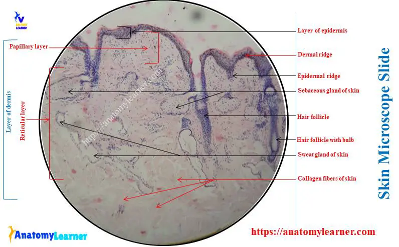
Before describing the dermis layer of thin and thick skin, let’s talk about the dermal-epidermal junction. Under the light microscope, you will see the junction between the epidermis and dermis of the skin, called the dermal ridge or papillae.
Again, the dermal ridge interdigitates with the skin’s epidermis and forms the epidermal ridge. The microscope view of the skin slide clearly shows two layers in the dermis – one is the papillary layer, and another one is the reticular layer.
The dermis (2 layers) of the thick and thin skin is made of vascular connective tissue. You will find the numerous fibrocytes, mast, macrophages, plasma cells, fat cells, and chromatophores in the dermis layer of the skin.
Let’s talk about the papillary and reticular layers of an animal’s thick and thin skin. The papillary layer is superficial, whereas the deep layer is a reticular layer.
A papillary layer of the dermis
This is the superficial and thinnest layer of the dermis of the thick and thin skin. It consists of loose connective tissue in contact with the epidermis layer of the skin.
The papillary layer of the dermis may protrude into the epidermis and give rise to the dermal papillae. Again, these dermal papillae contain the blood capillaries and Meissner’s corpuscles (tactile corpuscles). You will also find the anchoring fibril in the papillary layer of the dermis.
So, from the papillary layer of the dermis, you might identify the following histological features –
- The loose connective tissue of the papillary layer,
- Dermal papillae in the dermis, and
- Epidermal ridge or papillae.
Fine, now, let’s talk about the microscopic features of the reticular layer of the dermis from both the thick and thin skin of an animal.
Reticular layer of the dermis
The reticular layer is the thickest and deep layer of the dermis. This layer consists of dense irregular collagenous connective tissue (few cells). It also contains a considerable number of elastic fibers. You will also find the fatty tissue between the thick collagen bundle fibers.
The reticular layer of the dermis rests on the superficial fascia that attaches to the deeper structures. There are smooth muscle fibers located near the hair follicles in the reticular layer of the dermis. These smooth muscle fibers of the dermis layer are the erector pili muscles.
There are also sweat and sebaceous glands present in the reticular layer of the dermis. This reticular layer of the dermis is also rich in blood and lymph vessels.
Again, at the deeper aspect of the reticular layer, you will find numerous receptors like the Pacinian corpuscles (pressure receptors) and Krause end-bulbs (cold receptors).
Hypodermis or subcutaneous tissue layer of skin
There is a loose arrangement of the collagen and elastin fibers in the hypodermis of an animal’s thick and thin skin. This hypodermis allows skin flexibility and free movement over the underlying structure.
You will also find the fatty tissue in the hypodermis of the thick and thin skin. These fatty tissues may form small clusters of cells or large masses. Thus, they create a cushion or pad for the feet of the animal.
Blood and nerve supply to the skin
There are blood vessels in the skin derived from the number of arterial plexuses. You will find three major plexuses in both an animal’s thick and thin skin. The deepest plexus is present over the deep fascia, whereas you will find other plexus just below the dermis.
Below the level of the dermal papillae, you will also find another arterial plexus. You will not find any blood vessels in the epidermis of the skin. The skin’s epidermis derives nutrition entirely by diffusion from the capillaries in the dermal papillae.
Numerous arteriovenous anastomoses in the skin regulate blood flow through the capillaries bed. Thus, these arteriovenous anastomoses regulate body temperatures.
The skin of an animal is richly supplied with sensory nerves. You will find the dense network of nerve fibers in the superficial part of the dermis of thick and thin skin.
Again, the nerve fiber of the skin does not penetrate the deeper part of the epidermis of the skin. The skin also receives the autonomic nerve that supplies the smooth muscle in the wall of blood vessels and arrector pili muscles. They also provide the secretomotor supply to the sweat glands of the skin.
Dry face skin under a microscope
The dry face skin under a light microscope also shows similar histological features. So, dry face skin comprises two layers – the epidermis and the dermis. The epidermis is the superficial layer, whereas the dermis is located directly below the skin’s epidermis.
In the face skin, you will find a very thin epidermis lined by the keratinized stratified squamous epithelium. Like the normal skin histology, you will also find the five different layers and four different types of cells in the epidermis of dry face skin.
Again, the dermis of dry face skin consists of two distinct layers – the superficial papillary layer and the deeper reticular layer. The papillary layer of the dry skin indents the base of the epidermis and forms the dermal papillae.
In addition, the deeper reticular layer of the dry face skin forms the bulk of the dermis and contains dense irregular connective tissue. You will also find the sweat and sebaceous glands, hair follicles, and arrector pili muscles in the reticular layer of the dry face skin.
The hair follicles of the reticular layer of the dry skin show the hair bulb at their base. Again, you will find the dermal papillae at the base of the hair follicle in the reticular layer. Here the thin strip of smooth muscle (the arrector pili muscle) attaches to the hair follicles.
But, how will you differentiate the sweat glands from the sebaceous gland from the reticular layer of a dry face skin slide? Well, the sebaceous glands are associated with the hair follicles of the skin. You will also find a small portion of the hypodermis containing fatty tissue in the dry face skin.
Functions of skin in animals
The skin of an animal provides mechanical protection to underlying tissue. So, you will find the thickest skin in the area where the friction is more in an animal body. The skin also acts as a physical barrier against the entry of microorganisms and other harmful substances.
Fine, now, let’s look at some of the important functions of the animal’s skin –
Protection: it protects against mechanical trauma, invasion of microorganisms, evaporation, and ultra-violet ray. It also offers protection against damage to tissue by chemical, heat, and osmotic influences.
Sensory reception: it contains many receptors for pain, touch, temperature, and pressure. The presence of relatively sparse and short hair over most skin increases its sensitivity.
Synthesis of vitamin – D: the epidermis of the skin involves the synthesis of vitamin – D. Some degree of exposure to sunlight is essential for the synthesis of vitamin D. The ultraviolet light converts 7 – dehydrocholesterol to vitamin D.
Thermoregulation: the thermoregulation is performed by the glands, blood vessels, and fatty tissue of the skin. Numerous arteriovenous anastomoses can control the blood flow through the capillaries of the thick and thin skin.
So, blood flow through capillaries is kept to a minimum to prevent heat loss in cold conditions. Again, in warm condition, the blood flow increase to promote cooling.
Excretion: it helps to excrete the catabolic nitrogen waste products and water—the sweat glands of the skin act as an excretory organ.
Absorption: it absorbs certain lipid-soluble substances, drugs, and others.
Storage: it stores glycogen and cholesterol.
Besides these functions, skin also plays other different functions in an animal.
The skin under the microscope labeled.
Now, I will finally show you the different labeled skin diagrams under a light microscope. The thin skin labeled diagram shows the keratinized stratified squamous epithelium in its epidermis. Again, this diagram shows the five layers (identified) of the epidermis with four different types of cells.
The dermis of the thin skin labeled diagram shows the two distinct layers – papillary and reticular. Here, the papillary layer of the dermis shows the loose connective tissue fibers, cells, and ground substances.
Again, the reticular layer of the thin skin’s dermis shows different important microscopic features. It shows both the longitudinal and transverse sections of the hair follicles with the hair bulb and epidermal ridges.
The diagram also shows the sebaceous and sweat glands, arrector pili muscles, ducts of sweat glands, and adipose tissues. I will describe how you differentiate the sebaceous glands from the sweat glands under the light microscope.
The thin skin labeled diagram shows a thicker epidermis that lines with the keratinized stratified squamous epithelium. But, the diagram did not show any sebaceous glands and the hair follicles in the dermis part.
Again, the thin skin labeled diagram shows the sweat glands in its dermis layer. Both the thin and thick skin shows the keratin substances at the upper surface of the epidermis.
Here, in both thick and thin skin diagrams, the different epidermis layers are identified so clearly. You may easily understand the microscopic features of the cells of the different layers of the epidermis.
Hair follicles of the skin under microscope
Hair is present on the animal’s skin, covering almost the whole body. In an animal hair, you will mainly find three parts – shaft, root, and bulb. The visible part of the animal hair is the shaft, whereas the embedded part of the hair into the skin is the root.
The root of the animal hair has an expanded lower end called the bulb. Again, the root of each hair is surrounded by a tubular sheath called the hair follicle. The hair follicles are made up of several layers of cells derived from the skin layers.
The shaft of the animal hair consists of three layers – cuticle, cortex, and medulla. Again, the wall of the hair follicle comprises three main layers –
- The inner root sheet is present only in the lower part of the follicle,
- An outer root sheet that is continuous with the stratum spinosum layer of the epidermis, and
- The connective tissue sheet is derived from the dermis.
In the inner root of the follicle, you will find some important structures like the cuticle, Huxley’s layers, and Henle’s layer.
Arrector pili muscles of the skin
The skin under the light microscope shows some smooth muscle band that attaches at one end to the dermis. These are the arrector pili muscle that passes obliquely from the lower part of the hair follicles towards the junction of the epidermis and dermis.
A sebaceous gland lies at the angle between the hair follicles and the arrector pili muscles. The contraction of the arrector pili muscle affects the sebaceous secretion.
You may learn more about the different appendages of the skin from another article by an anatomy learner.
Suggested reading for you: Microscope features of hair follicles and glands of the thick and thin skin with the labeled diagrams.
How do you differentiate sebaceous glands from sweat glands?
The skin labeled diagram shows the sebaceous glands in the dermis that are closely associated with the hair follicles. Each gland comprises several alveoli connected to a broad duct that opens into hair follicles.
Each alveolus is pear-shaped and consists of a solid mass of polyhedral cells. The diagram shows the difference in the size of the inner and outer cells of the sebaceous glands. It shows the larger, rounded lipid content cells in the inner part of the sebaceous gland. In contrast, the outermost cells of the sebaceous glands are small and rest on the basement membrane.
The secretion of the sebaceous gland is oily sebum that keeps the skin and hair soft. It also prevents the dryness of the skin and makes it resistant to moisture.
There are two types of sweat glands (typical and atypical) in the animal body that produces sweat. The skin labeled diagram shows a transverse section of the typical sweat glands of the skin. All these sweat glands are located on the thick and thin skin (reticular layer).
A microscopic view of the sweat glands shows the oval (polyhedral) nucleus. It also shows the myoepithelial cells that surround each sweat gland of the animal skin.
Get more skin-labeled diagrams on social media for anatomy learners.
Frequently asked questions on skin under microscope
So, let me provide the short answers to the commonly asked questions on the skin under the microscope. Here, you may find microscopic features of the thick and thin skin of different types of animals.
What is the human skin look like under a microscope?
You will see the same microscopic figure of human skin as the animals. In humans, you will also find thick or thin skin in different body regions. The thin skin of humans also possesses three distinct layers – epidermis, dermis, and hypodermis.
The keratinized stratified squamous epithelium also lines the epidermis of the human skin. And the epidermis of a human skin also comprises five different layers like the animal skin.
Again, the dermis of the human skin comprises two-layer – papillary and reticular layers. You will find similar microscopic features in the dermis of the human skin.
What does a pimple look like under a microscope?
What does the skin look like after a needle?
Have you ever seen skin in a microscope after inserting the needle? I will show you what the skin looks like after needle insertion.
Conclusion
The skin under the light microscope shows three distinct layers – epidermis, dermis, and hypodermis. You will find almost similar microscopic features in the epidermis of animals’ thick and thin skin. But, there is a great variation in the microscopic features of the dermis layer in between the thick and thin skin.
The dry face and human skin will also show the same microscopic figure under the light microscope. It is very important to differentiate the sebaceous glands of skin from the sweat gland.