
The urinary bladder is a muscular sac-like structure that temporarily stores urine and discharges out prediocally through the urethra. In the urinary bladder histology, you will find three or four defined coats like the tubular organs. Here, I will show you all the layers from the urinary bladder histology slide with microscopic images.
You will also get information on the variation of different layers of the urinary bladder in different species. But, first, I will introduce you to the important histological features with the appropriate identifying points of the urinary bladder slide. Then I will go with the details histological features of the different layers of the urinary bladder with their special features in different animals.
Urinary bladder histology
In urinary bladder histology, you will find almost all the features of a typical hollow organ. It consists of a tunica mucosa, thick tunica muscle coat, and tunica serosa. But, in some animal species, you may find the distinguished lamina muscularis layer and tunica submucosa.
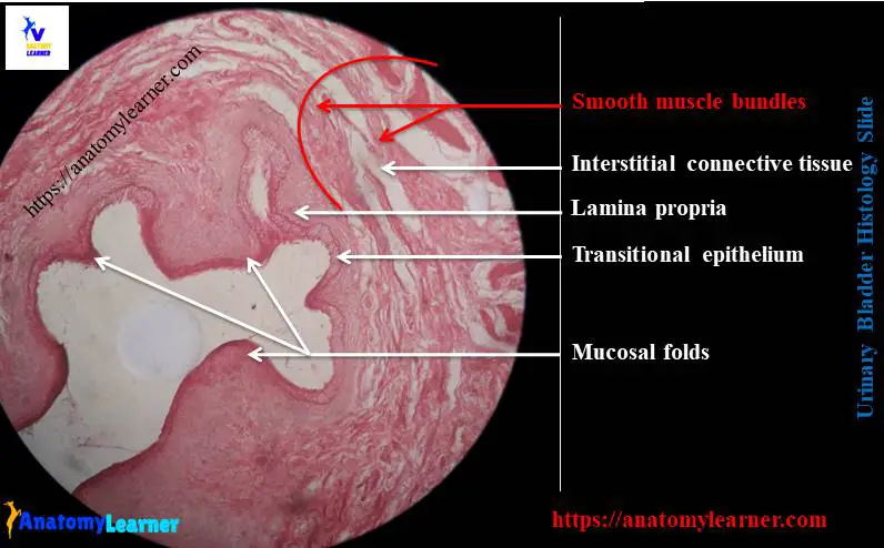
The tunica mucosa of the urinary bladder exhibits numerous mucosal folds that line with the transitional epithelium. In the lamina propria, fine connective tissue fibers, numerous fibroblasts, and blood vessels will be found. Again, in a few animal species, you may find the distinct lamina muscularis layer that divides the lamina propria from the tunica submucosa.
“For your kind information, the exceptional features of different layers of urinary bladder slide in different animal species will discuss later. For that, you may read the full article to get these exceptional features of urinary bladder histology.”
The tunica muscle layer of the urinary bladder slide is thickest and consists of two or three indistinct layers of smooth muscles. You will find a great variation in the pattern of this muscle layer of the bladder in different species.
There is interstitial connective tissue present in between the smooth muscles bundles of the tunica muscularis layer. This interstitial connective tissue merges with the connective tissue of the tunica serosa of the urinary bladder.
In the urinary bladder histology slide, you may find tunica serosa or tunica adventitia layer. The superficial surface of the urinary bladder lines with the tunica serosa. But, the inferior surface of the urinary bladder possesses the connective tissue layer without any mesothelium (tunica adventitia). This connective tissue adventitia of the bladder merges with the connective tissue of adjacent structures.
Urinary bladder histology slide identification points
This part of the article might help you to identify the urinary bladder histology slide so quickly. First, I will enlist some of the important structures from the urinary bladder slide. Then, I will provide the identification points for the urinary bladder slide under the light microscope.
Let’s try to identify the following histological features from the urinary bladder slide –
- The mucosal folds and the star-shaped lumen of the urinary bladder.
- A transitional epithelium that covers the mucosal fold of the bladder.
- The lamina propria and lamina muscularis layer (in a few species) of the bladder.
- Two or three ill-defined smooth muscles layers with the interstitial connective tissue
- The tunica serosa or adventitia of the bladder (based on the superior or inferior surfaces)
Now, you may write the following identification points for identifying the urinary bladder histology slide under the light microscope.
- The tissue section shows three distinct layers – tunica mucosa, tunica muscularis, and tunica serosa.
- Presence of numerous mucosal folds that line with the transitional epithelium
- The tunica muscle layer possesses three ill-defined smooth muscle bundles and interstitial connective tissue.
- Presence of thin layer of loose connective tissue with mesothelium lining in the tunica serosa layer.
So, this is a urinary bladder slide. But, there may be few changes in identifying characteristics based on the animal and the superior or inferior surface of the bladder.
Tissue layers of the urinary bladder
Normally, the urinary bladder wall consists of three layers – tunica mucosa, tunica muscularis, and tunica serosa. But, in the ruminant, horse, dog, and pig, you will find the tunica submucosa layer in the urinary bladder histology slide.
Here, I will discuss the detailed histological characteristics of different layers of a urinary bladder. The three (or four) tissue layers of a urinary bladder are –
- Tunica mucosa of the bladder wall
- The tunica submucosa layer of the bladder wall (not present in humans and cats)
- A tunica muscularis layer (consist of three or two ill-defined smooth muscle layers) and
- The tunica serosa or adventitia of bladder wall
“If you want to memorize the basic histology of the four different layers of a tubular or hollow organ, you may read the article related to hollow organ histology.”
The major histological difference will find in the lamina propria or tunica submucosa of a bladder in different species. There will also find a difference in the pattern and layers of smooth muscle bundles of the tunica muscle layer of a bladder in different species.
Tunica mucosa and lining epithelium of the urinary bladder
The tunica mucosa of the empty and contracted urinary bladder shows numerous mucosal folds. But these mucosal folds will disappear during bladder distension. Here in the tunica mucosa of the urinary bladder histology slide, you will find the below-mentioned characteristics.
It consists of the transitional epithelial lining and underlying connective tissue (lamina propria). The transitional epithelium of the bladder is thicker and consists of five or six-cell layers. You will find low cuboidal, columnar, and dome-shaped cells in the superficial layer of the transitional lining. These cells often contain two nuclei or even are polyploids.
Again, the plasma membrane of the superficial cell layer is thickened and prominent. In addition, you will find the rounded and more columnar basal cells in the deep layer of the transitional epithelium of a bladder.
The lamina propria of the urinary bladder slide consists of connective tissue fibers, numerous fibroblasts, and blood vessels (venules and arterioles).
A lamina muscularis of a small, isolated bundle of smooth muscle is present in the horse, ruminant, dog, and pig. But, you will not find any distinct lamina muscularis layer in the urinary bladder of cats and humans.
So, the presence of this lamina muscularis layer divides the loose connective tissue layer into an inner lamina propria and an outer tunica submucosa in the urinary bladder of a ruminant, horse, dog, and pig.
You may find some mucosal gland in the tunica mucosa of the urinary bladder, especially near the internal urethral opening.
When the urinary bladder distends with urine, the lining epithelium becomes thinner. This results from the ability of the epithelial cells to change their shape and shift over one another.
The tunica muscularis layer of the urinary bladder histology slide
The tunica muscle layer of the urinary bladder histology slide is thick, and it remains a form of smooth muscle meshwork. There is a great variation in this muscle layer in different animal species. In most species, like a ruminant horse, this tunica muscularis is not well layered. It comprises irregular-shaped interweaving bundles.
But in some species like a cat, you will find three layers of smooth muscle in the tunica muscularis. The internal and external muscle fibers of this layer tend to be longitudinal. In between the longitudinal layers, you will find thicker circularly arranged smooth muscle bundles.
Do you know the functions of these three layers of smooth muscle bundles of a urinary bladder? Well, the contraction of these muscle layers is responsible for the emptying of the bladder. That is why these muscles are known as detrusors.
In between the smooth muscle layer, there present visible interstitial connective tissue. You will find a thicker circular smooth muscle fiber just above the junction of the urinary bladder with the urethra.
Tunica adventitia and serosa layer of the bladder
In the tunica adventitia of the urinary bladder (inferior surface), fibroelastic connective tissue with numerous blood vessels, nerves, and lymphatics. But the superior surface of the urinary bladder covers by a mesothelium of the peritoneum that forms the tunica serosa layer.
Urinary bladder anatomy
This part of the article is not important if you have a good piece of knowledge on the urinary bladder anatomy. Here, I will provide the little anatomical facts of a urinary bladder with the diagram. But, if you wish to know the detailed anatomical facts of the urinary bladder of different animals, you may read the articles from the gross anatomy section.
Grossly, the urinary bladder of any animal is a thicked, walled muscular sac that serves as a reservoir for urine. The shape and the size depending upon the amount of urine it contains. In the empty stage, it remains completely in a contracted condition and lies on the floor of the pelvic cavity.
Again, in distended condition, it becomes oval and gets projected to the abdomen of the animal. But in a horse, it does not extend up to the abdominal cavity even in distended conditions.
The urinary bladder divides into three parts – vertex, body, and neck. The cranial blind end of the bladder is the vertex that bears a cicatricial tissue on its middle. Again, the body of the urinary bladder is rounded and dorsoventrally compressed.
The dorsal surface of the bladder is convex, and the ventral surface is flat that related to the floor of the pelvic cavity. You will find a peritoneum that covers the dorsal or superior surface of the bladder. But, the bladder of a dog is covered by the peritoneum.
The caudal narrow tubular part of the bladder is the neck. It is thick-walled and is continuous with the urethra. Three ligaments hold the urinary bladder in its position – the lateral ligament, ventral ligament, and the round ligament.
Urinary bladder histology slide diagram
Now, I will try to show you all the features from the urinary bladder histology with actual microscopic slide images and the labeled diagram. Here, you will find the contracted and the stretched mucosa of the urinary bladder.
I tried to show you the superficial transitional epithelium (low cuboidal, columnar, or dome-shaped cells). There may present binucleated cells in the superficial layer of the urinary bladder.
Here, the deeper cells of the epithelium are rounded, and the basal cells are more columnar. The lamina propria of this microscopic slide image consists of connective tissue fibers, fibroblast cells, and blood vessels.
The tunica muscularis of this slide image shows the ill-defined smooth bundle layers (interweaving pattern). Again, the urinary bladder microscopic image shows the interstitial connective tissue in between the smooth muscle bundles.
The stretched condition of a urinary bladder shows the reduction of the number of cells layer. Again, the superficial cells of the stretched urinary mucosa show the flattened epithelium (squamous). The thickness of stretched urinary mucosa maybe three to four cell layers.
In some regions of the stretched urinary bladder, it resembles stratified squamous epithelium. The mucosa membrane of the bladder will not show any folds. Other histological features from the different layers of a stretched bladder are almost similar to the contracted bladder.
I think the above-mentioned urinary bladder microscopic diagrams might be helpful for you to understand every single structure so clearly. In addition, if you need more diagrams on the microscopic urinary slide, you may join anatomy learner on social media.
How to draw the urinary bladder microscopic slide image?
So, this part of the article might help you to draw the urinary bladder histology slide image. First, you should draw the mucosa membrane (folded) of the bladder. Now, you should try to draw the transitional epithelium on the mucosa of the bladder. Please provide five to six layers of the cell on the urinary mucosa.
Let’s draw the lamina propria of the bladder (provide loose connective tissue along with the cells, blood vessels). Now, you might try to draw the lamina muscularis layer of the urinary bladder. For that, you may provide inner longitudinal, middle circular, and outer longitudinal layers of smooth muscle fibers (if you want to draw the muscle coat of a cat or human’s bladder).
Or you may provide ill-defined layers of smooth muscles fiber (in interweaving fashion) if you want to draw the urinary bladder (muscle coat) of ruminant, horse, and pig. Don’t forget to draw the interstitial connective tissue in between the smooth muscle bundles of fibers.
Now you should draw the tunica adventitia or serosa layer of the urinary bladder.
Frequently asked questions on a bladder microscopic slide
Fine, in this section of the article, I will try to solve the common inquiries on the urinary bladder histology slide. I hope you will find your desired question on the urinary bladder microscopic slide image with its answer.
What is the histology of the urinary system?
You might know the histology of the kidney, ureter, urinary bladder, and urethra from the urinary system. You will find the detailed guide on the kidney, ureter, urethra here in anatomy learner (histology learning section).
In the kidney histology slide, you might identify the renal cortex and renal medulla. Then you might also identify the nephron (renal corpuscles, ascending and descending convoluted tubules, a loop of Henle) and others. All the structures are shown here in this article (kidney histology slide).
Again, in the ureter histology slide, you might identify the star-shaped lumen that lines with the transitional epithelium. The tunica muscularis layer of the ureter might be identified under a light microscope. Here you will find all the details guide and slide identification points for the ureter histology slide.
Again, from the urethra (both male and female), you might identify the lining epithelium and surrounding structures (depends on male or female).
What is the epithelium of the urinary bladder?
The epithelium of the urinary bladder is the transitional epithelium that consists of superficial columnar or cuboidal cells and deeper rounded or columnar cells. The layer of the transitional epithelium of a urinary bladder may change with the contracted and stretched conditions.
There are five to six layers of cells (consists of superficial dome or columnar type cells and deeper rounded type cells). The plasma membrane of the superficial cells layer is so prominent in the urinary mucosa.
Again, in the stretched urinary bladder, the transitional epithelium changes its shape. The thickness of the epithelium layer reduces into three to four layers. Again, the superficial cell layer becomes squamous-type cells.
What are the 3 tissue layers of the bladder?
Normally, in the urinary bladder microscopic slide, you will find three distinguished tissue layers (but it depends on the animal species). The three tissue layers of a bladder are the tunica mucosa, tunica muscle layer, and the tunica adventitia or serosa layer.
But, in some animal species, you will find the tunica submucosa layer that contains loose connective tissue with connective tissue cells and blood vessels.
If you love to learn details about the three or four different layers of the urinary bladder, you may read this article from the start till the end.
What is the urinary bladder?
The urinary bladder is a muscular sac-like structure that is responsible for reservoirs of urine. The reservoir urine from the urinary bladder of any animal will discharge periodically through the urethra.
This urinary bladder is located on the animal’s pelvic floor (when empty), extending into the abdominal cavity when it fills with urine.
There are three different parts of the urinary bladder – the vertex, the body, and the neck. The vertex of the bladder is the caudal blind sac-like part. Again, the body of the urinary bladder is dorsoventrally compressed in most animals.
There are three ligaments found in the urinary bladder that help to hold in position. These ligaments of the urinary bladder are the ventral ligament, lateral ligament, and round ligament.
How is the bladder adapted to its functions?
Which structures connect the kidney to the urinary bladder?
The ureter connects the kidney to the urinary bladder. There are two ureters present in the urinary system of any animal. These are the tube-like structure that originates from the hilus of the kidney and ends on the cranial end of the urinary bladder.
The length of the right ureter is larger than that of the left ureter of an animal. You may learn more about the gross anatomy of a ureter and the histological features of a ureter from an anatomy learner.
What is the structure of the urinary bladder?
The urinary bladder is a muscular bag-like structure, and its wall consists of three or four coats. I have already described every single coat of the urinary bladder with the microscopic slide and diagram. If you reach this part first, I will highly recommend you to read the full article.
What type of tissue is the bladder?
In the urinary bladder histology slide, you will find three types of tissues – epithelium, connective, and muscular (smooth muscle). The epithelium tissue lines the tunica mucosa layer of the urinary bladder. Again, you will find the numerous connective tissue fibers and cells in the lamina propria and tunica submucosa.
Again, in the tunica muscle layer, you will find ill-defined three or two layers of smooth muscle bundles. In addition, there are loose connective tissue and simple squamous lining epithelium present in the tunica serosa layer of the urinary bladder.
Conclusion
I think this simple guide might help you to learn the basics of urinary bladder histology. The microscopic images of the urinary bladder and the labeled diagram might help you easily identify the slide under the light microscope. Most characteristic features of the urinary bladder slide are found in the tunica mucosa layer, lining epithelium, and tunica muscularis layer.
But, there are variations in the lamina muscular layer in some species. You will find isolated smooth muscle bundles in the lamina muscularis layer of a few animal’s urinary bladder histology slides.