The blood is a specialized connective tissue that is fluid and circulates through the vascular channel. In the blood histology slide, you will find different types of cells with their specific features. This might be a short article where I will show you all the cells from the blood microscope slide with a labeled diagram and actual pictures.
First, I will share some general information on the blood and histology of blood cells with their identification points. Then I will also share how you may make a blood slide to evaluate different types of cells (erythrocytes and leukocytes). Finally, I will summarize the hematopoiesis process so that you may get the essential information so quickly.
Blood histology
The main goal is to provide the basic and essential identifying points of blood cells histology. But before going into the details, I would like to share some basic information on animal blood. The total volume of the circulating blood of animals may vary with the species.
You will find eight to ten percentage blood in large domestic animals of their body weight. Again, in laboratory animals, you will find six to seven percent of blood to body weight. You know, there are several vital functions of blood as well as they involve crying nutrients to cells and carrying waste products to excretory organs.
In the blood, you will find cellular components and plasma-protein riched fluid. The cells of the blood are three major types – erythrocytes (Red Blood Cells), leukocytes (White Blood Cells), and Platelets (Thrombocytes).
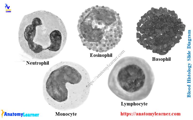
In the blood histology slide (smear), all of the cells mentioned earlier with their useful identifying features. There are five different types of leukocytes in blood – neutrophils, eosinophils, basophils, monocytes, and lymphocytes. The neutrophils, eosinophils, and basophils are known as the polymorphonuclear granulocytes. Again, the monocytes and lymphocytes are known as the mononuclear agranulocytes.
In addition, you will find ninety-one to ninety-two percentage water and eight to nine percentage solutes (protein, lipid, electrolytes) in blood.
Blood collection and slide preparation
If you want to study all the blood cells under a light microscope, you should collect blood and make a thin smear. You may collect blood from the jugular vein, femoral vein, brachial or saphenous vein of the animal.
To study the blood cells, you may follow any of the following methods. If you collect blood into a vial containing anticoagulants (Ethylenediaminacetic acid), you may go with this process –
The collected blood from an animal must be centrifuged, and it will show the three distinct layers. In the lower layer, you will find the erythrocytes called the packed cell volume (PCV; 45% of the total blood volume).
There is a thin gray-white middle layer that is known as the Buffy coat. In the upper part of the buffy coat, you will find the platelets and leukocytes in the lower part. You may evaluate these cells by differential leukocytes count method (DLC).
Again, in the uppermost layer, you will find the plasma portion of the blood. If you don’t want to follow this method, you may go with the simple procedure below.
After collecting the blood, you may take a small drop of blood and prepare a thin blood film. You might air dry and fix it in the methanol. Then the blood slide might be stained with the RomanVsky’s dye (Wright or Giemsa).
The Wright dye is methylene blue (basic dye) and eosin (acidic dye). Here, the methylene blue will attract the acidic nuclei that provide the deep blue color to it. Again, the eosin (acidic dye) will attract alkaline cytoplasmic constituents and give a pink tint to them.
Histology of blood cells
Now, it’s time to evaluate the blood cells from the blood histology slide under the light microscope. Make sure you use the oil immersion to examine erythrocytes, leukocytes, and platelets. Using the wright stain, you will find the following color changes in the different types of blood cells.
- The cytoplasm of the erythrocyte will stain in pink color.
- Neutrophil nuclei stain dark blue.
- The nuclei of eosinophil stain dark blue, and the granule will stain bright pink color.
- Again, the basophil nuclei stain dark blue or purple, cytoplasm pale blue, and granule stain deep purple.
- Monocyte cytoplasm stain pale blue, and the nucleus stains medium blue.
- The nuclei of the lymphocyte will stain dark-purple blue with pale blue cytoplasm.
- Finally, the platelet stain with light blue.
Fine, let’s find all these blood cells under the light microscope.
Erythrocytes histology
The erythrocyte is a non-nucleated biconcave disc that varies with animals. You will find the typical biconcave appearance in the erythrocytes of dogs, cats, and sheep. Again, you will find the shallow concavity in the erythrocyte of horses and cats.
In addition, you will find the flat discs in the erythrocyte of goat and pig blood. In the blood cell histology slide, you may identify the erythrocyte by the following points –
- The presence of a central pale area surrounds bu the orange cytoplasm
- Having the biconcave discs (non-nucleated)
The size of the erythrocyte may also vary with the species. You will find the largest erythrocyte in the dog and the smallest erythrocyte in the goat blood.
The immature anuclear erythrocyte is known as the reticulocytes. These reticulocytes appear polychromatic (pinkish-blue) on wright stain. Sometimes, you may find the Heinz body that appears as a pale area within the cytoplasm of the Red Blood Cells of the dog and cat. This results from the oxidation of the hemoglobin.
Abnormalities of erythrocyte blood cells
You may find variations in the size and shape of the erythrocyte. If there is any variation in the size of the erythrocyte, this is known as anisocytosis. Again, if you find any variation in the shape of the erythrocyte ten, it is known as poikilocytosis.
But, you may find the spindle, rod, pear, triangle-shaped erythrocyte in goats. Again, in camel and llama, you will find the elliptical-shaped erythrocytes.
Functions of erythrocytes
The erythrocytes contain hemoglobin that is essential to mammalian life. It helps to carry oxygen to the tissue of the animal body.
White blood cells histology (leukocytes)
The number of white blood cells is fewer than the erythrocytes. Again, the count of the leukocytes in a blood histology slide may vary with the animal species. You will find numerous neutrophils in the blood film of a dog and cat. Again, the lymphocytes are predominant in the ruminant, rat, and mice blood.
As I told you before, there are two types of leukocytes in blood – polymorphonuclear granulocytes and mononuclear agranulocytes. The polymorphonuclear granulocytes possess specific granules in their cytoplasm.
- Neutrophil cells – possess neutral granules,
- Eosinophils – contains the pronounced acidophilic granules, and
- Basophil – contains the distinct basophilic granules.
In addition, the mononuclear agranulocytes of the white blood cells are monocytes and lymphocytes. Let’s try to identify all the leukocytes from the blood film one by one.
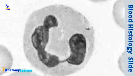
Neutrophil blood cells
The neutrophils are produced and released from the bone marrow and circulate for six to ten hours. Then neutrophils will migrate to the tissue. You will find forty to seventy percentage neutrophils of the total leukocytes counts.
You will find the following features in the neutrophil histology –
- It contains heterochromatic (clumped chromatin) segmented nuclei with three to five lobes. But, occasionally, you may find an extra chromatin lobe like a drumstick.
- The cytoplasm of the neutrophils contains a pale grayish-blue color that has a moderate number of fine pinkish or pale granules.
“Drum-stick like extra chromatin lobe will normally present in the female blood. These extra chromatin are known as the barr body in female.”
You will find two types of granules (azurophilic and specific granules) in the cytoplasm of the neutrophil. They contain a different chemical that plays a significant role in regulating neutrophil production and defending the body against bacterial infection.
The granules of the neutrophils are larger and reddish in rabbits, guineapig. Again, in chicken, this neutrophil is known as the heterophils.
Functions of neutrophils
Neutrophil forms the first line of defense against microbial infection in the animal body. It has an essential role in bacterial phagocytosis and in modulating the inflammatory process and subsequent tissue damage. In addition, the granules of the neutrophil work together to destroy the phagocytic bacteria.
Eosinophil blood cell histology
In the blood histology slide, you will find two to eight percentage eosinophils of the total leukocytes. The main characteristics of eosinophil under the light microscope are listed below.
- Eosinophil contains polymorphic nuclei that are less condensed and segmented (two).
- The presence of specific granules in the cytoplasm of eosinophil
- These granules are loosely packed in the cytoplasm of the eosinophil.
Again, you will find the Golgi body, mitochondria, ribosome, rough endoplasmic reticulum in the cytoplasm of the eosinophil. The granules of the eosinophils may vary in different animal species. You will find numerous uniform, spherical granules in the eosinophil of sheep, goats, cows, and pigs. They take the bright orange stain that nearly fills the cells.
Again, in a horse, you will find the largest granule in the eosinophil that takes the bright orange stain. In addition, the cytoplasm of the dog eosinophil contains pinkish or light orange granules.
Functions of eosinophils
Eosinophils play a role in allergic and anaphylactic reactions and infestation caused by a parasite. They also have a limited phagocytic and bactericidal capacity.
Basophils blood cell
In a blood smear, you may rarely find the basophil in the animal. You may identify the basophils by the below-mentioned identifying features.
Presence of segmented or irregular-shaped heterochromatic nuclei that possess two or three lobes (but less pronounced than eosinophils)
The cytoplasm of the basophils contains homogenous metachromatic and prominent (coarse) granules.
Granules of the basophils are water-soluble, and degranulation may occur during the staining. It will take the reddish-violet stain and invariably fill the cytoplasm of the basophils.
Again, the granules of the basophil’s blood cells may vary in different species. You will find larger and fewer granules in the dog basophils than that of the cow and horse. There are rod-shaped, orange gray granules present in the cytoplasm of cat basophils.
In addition, you will find the more prominent and spherical or oval reddish violet granules in the other domestic animals.
How do you differentiate basophils blood cells from connective tissue mast cells? Well, I may help you to distinguish the basophils blood cells from the connective tissue mast cells.
The basophil is a smaller cell, but the mast cell is more significant in shape. You will find the segmented or spherical nuclei in basophils, but in mast cells, you will find the irregular or spherical nuclei. Again, you will find fewer granules in basophils blood cells that are evenly distributed. But, in mast cells, you will discover numerous granules that are eccentrically distributed.
Functions of basophils
The basophil has a significant role in mediating inflammatory reactions, lipid metabolism, blood coagulation. They also have a limited phagocytic and bactericidal activity like eosinophils.
Monocytes blood cell histology
In the blood histology slide, you will find the largest leukocytes – monocytes. They are about three to eight percent of total leukocytes. The monocyte is also considered as the precursor of tissue macrophage.
You will find the following identifying features in the blood smear for monocytes –
Presence of highly pleomorphic nuclei (nucleus may appear in elongated, irregularly contoured folded, intended, horse-shoe-shaped).
The cytoplasm of the monocytes contains large grayish blue color and often appears foamy or vacuolations.
The cow monocytes are very difficult to differentiate from the large lymphocytes.
Functions of monocytes
The blood monocytes transform into the tissue macrophage that acts as phagocytic cells. Again, the blood monocytes and the tissue macrophage are the mononuclear phagocytic system (MPS). In addition, monocytes produce several substances of biological importance like colony-stimulating factors (CSFs), cytokines (IL I, IL III), tumor necrosis factor (TNF), and lysosomal enzymes.
Lymphocytes blood cells
The number of lymphocytes in the circulating blood varies with the animals. You will find twenty to forty percentage lymphocytes of total blood cells in dogs, cats, and horses. Again in ruminant and rat, you will discover sixty to seventy percentage lymphocytes in total leukocytes.
There are two types of lymphocytes – small and large lymphocytes. Under the blood histology slide, you will find the following figures –
For small lymphocytes of animals –
- Presence of round, dense or slightly indented heterochromatic nuclei
- The cytoplasm of the small lymphocyte is light and pale blue.
- Presence of a few azurophilic granules in the small lymphocytes
Again, for the large lymphocytes –
The presence of a few indented nuclei and cytoplasm possess more abundant, homogenously blue-stained granules.
Both the small and large lymphocytes are found in cows, sheep, and goats.
Functions of lymphocytes
The lymphocyte plays a role in cell-mediated immunity (T – lymphocytes), and antibodies mediate immunity or humoral immunity (B – lymphocytes).
T – lymphocytes also have a role in regulating hematopoiesis and differentiation of B-lymphocytes into plasma cells. The lymphocytes are functionally grouped into three types of cells – T –lymphocytes, B- lymphocytes, and Natural Killer cells.
Platelets blood cells histology
In the blood histology slide, you will find discoidal or spherical or elongated cells (individually or in small to large clusters) known as platelets. They are the short-living blood cells in animals (life span is about nine to twelve days).
The cytoplasm of the platelets contains microfilaments, microtubules, glycogen, and heterogeneous granules. Do you know the functions of the microfilaments and microtubules of platelets? Well, they maintain the typical shape of the platelet.
The platelets’ thin exterior coat is the platelet’s external membrane that covers by amorphous materials. It is responsible for platelet adhesive properties.
Functions of platelets
The platelet maintains the integrity of the blood vessel. It also plays a significant role in blood coagulation and clot reactions. Again, the granular contents form a large secondary hemostatic plug. In addition, platelets contain proteins that have procoagulants, antiheparin, inflammatory, and growth-promoting activities.
Get more blood cells microscope pictures on social media of anatomy learner.
Hematopoiesis – Blood cell formation
The bone marrow is the primary production site for all blood cell –lines called the pluripotential hemopoietic stem cells. The blood cell formation is a very complex process (known as hematopoiesis). So, I will show you only the short process of blood cell formation (erythrocytes and granulocytes).
You will find two types of bone marrow in the animal – the red bone marrow and yellow bone marrow. The red bone marrow is hematopitically active, whereas the yellow bone marrow contains fat and is hematopitically inactive.
The prenatal hematopoiesis begins in the wall of the yolk sac in mammals. In addition, postnatal hematopoiesis starts from the bone marrow in late gestation and parturition. But a few weeks after the birth, hematopoiesis begins in the liver and spleen.
You might learn the following process from hematopoiesis –
- Erythropoiesis (formation of erythrocytes)
- Granulopoiesis (formation process of granulocytes)
- Monocytopoiesis (formation of monocytes)
- Lymphopoiesis (formation process of lymphocytes) and
- Thrombopoiesis (formation process of thrombocytes)
Now, I will share the erythropoiesis and granulopoiesis in a short form. If you want to learn the other different types of hematopoiesis, you may read the other articles from the blood histology section.
Erythropoiesis (formation process of Red Blood cells)
This is the development process that leads to the formation of mature erythrocytes. It begins with multipotent stem cells that differentiate into unrecognizable erythroid progenitors (burst forming and colony-forming units).
The whole process of erythropoiesis will occur in the following steps –
- Formation of erythroblasts
- Proerythoblast formation
- Formation of basophilic erythroblast
- Polychromatic erythroblast formation
- Formation of normochromic erythroblast
- Orthochromatic erythroblast formation
- Reticulocytes or polychromatophilic erythroblast formation and
- Finally, the formation of erythrocytes
You will find the following changes in the complete steps of erythropoiesis –
- The cell size will decreases
- Nuclear chromatic – condensed
- Hemoglobin accumulates (increased acidophilia)
- Ribosome – decrease (decrease basophilia)
- Nucleus – ejection
Now you may learn in detail what happens in every step of erythropoiesis.
Erythroblast is the most considerable cell that contains a spherical euchromatic nucleus with one to three nucleoli. It possesses the deep blue cytoplasm (basophilic). The proerythroblast does not contain any nucleoli, and the mitotic division occurs.
In the basophilic erythroblast, you will find clumped chromatin in a radial pattern, and the cytoplasm will take the deep blue stain. Again, in polychromatic erythroblast, you will see the dark condensed nucleus with prominent clumped chromatic. The cytoplasm of the polychromatic erythroblast is grayish orange. In addition, the hemoglobin begins to accumulate in the polychromatic erythroblast step.
In the normochromic erythroblast, the dense nucleus and reddish cytoplasm will be found. This is because of the accumulation of more hemoglobin in the cytoplasm of the normochromic erythroblast. Again, you will find the pyknotic nucleus in the orthochromatic erythrocyte step. The cytoplasm of orthochromatic erythrocytes contains slightly polychromatic to normochromic features.
On the other hand, in reticulocytes, the cytoplasm becomes eosinophilic, and the nucleus will eject, and it becomes the erythrocyte.
Granulopoiesis – formation of granulocytes
This is the development process of three granulocytes (neutrophil, eosinophil, and basophils). Here, I will also share a short process of these three blood cells formation.
You will find the following steps in the granulopoiesis process –
- Myeloblast formation
- Formation of promyelocytes
- Myelocytes formation
- Formation of metamyelocytes
- Band formation and
- Formation of mature granulocytes
Let’s discuss the formation of a neutrophil blood cell. You know, you will find C or S or V-shaped nucleus, pale blue cytoplasm, and neutral granule in the neutrophil blood cell histology.
The myeloblast is an oval or spherical-shaped cell containing spherical euchromatic nuclei with three to five nucleoli. The cytoplasm of the myeloblast is light blue (basophilic).
Again, the promyelocytes are giant cells containing an oval or spherical nucleus and lighter blue cytoplasm. In addition, the myelocytes include the spherical to slightly indented nuclei with condensed chromatin. You will not find any nucleoli in the nucleus of the myelocytes.
The cytoplasm of the myelocytes becomes pale blue, and granules appear. In the metamyelocytes, you will find kidney-shaped heterochromatic nuclei. The cytoplasm of the metamyelocytes contains specific granules.
In the next step of granulopoiesis, C, S, or V-shaped nuclei (band) will form. The cytoplasm becomes paler blue and contains neutral granules, and finally, it becomes the mature granulocytes (neutrophils).
Conclusion
I hope you got the basic idea on all cells of the blood histology slide. All the blood cell histology labeled diagrams might be helpful for you to identify them so quickly. The neutrophil contains heterochromatic segmented nuclei with three to five lobes. Again, the eosinophil contains the polymorphic segmented nuclei.
So, all the blood cells possess specific features in their cytoplasm and nuclei. Now, you may practice and identify the blood cells from the blood histology slide.