Dog Ovary Anatomy – Shape, Size, Location, and Ligament with Diagram

Dog Ovary Anatomy - Location, Shape , and Ligaments

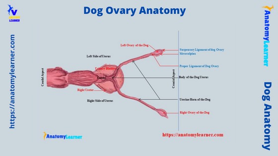
The dog ovary anatomy shows distinct surfaces, borders, extremities, and ligaments. You will also see the defined medulla and cortex in the ovary of a dog which possesses different structures and features. But, the location and structure of the dog’s ovary may vary with cows. The location of the canine ovary is significant as you … Read more

Cardiac Muscle Under Microscope with Labeled Diagram

Cardiac Muscle Under a Microscope Labeled 400X Diagram

The cardiac muscle under a microscope shows a short cylindrical fiber with a centrally placed oval nucleus. You will find some unique features in cardiac muscle that will help you to differentiate it from skeletal and smooth muscles. Quick answer: the cardiac muscle microscope slide shows the cylindrical fiber with 1 or 2 nuclei. The … Read more

Lymphocytes Under Microscope with Labeled Diagram

Lymphocytes Under a Microscope with Labeled

Lymphocytes under a microscope show a round to slightly indented nucleus with clumped heterochromatin. You know these lymphocytes are agranulocytes and form the second-largest population of white blood cells. This article will show you the structure (morphology) of different types of lymphocytes (small, large, B, and T) under light and electron microscopes. So that after … Read more

Adipose Tissue Under Microscope with Labeled Diagram

Adipose Tissue Under Microscope Labeled

The adipose tissue under a microscope shows an aggregation of fat cells or adipocytes. You will find this adipose tissue subcutaneously throughout the body except over the eyelid, auricle, and other parts of the animal’s body. The adipose tissue is considered the specialized connective tissue which plays an essential role in energy homeostasis. Microscopically, you … Read more