During studying the osteological features of the animal’s bone, you will find different types of articular surfaces. Here, you will know the answer to the question – ‘what are the types of articular surfaces in bone’?
Quick answer: facet, fovea, condyle, and trochlea are the significant types of articular surface in the animal’s bones. These articular surfaces are different in terms of their external appearances.
Here, I will show the major differences of these various types of articular surfaces from the animal’s bones. So, with a diagram, let’s learn the features of the different types of articular surfaces from the animal’s bones.
What are the types of articular surfaces in animal bones?
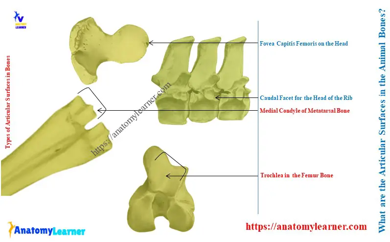
Explanation of the types of articular surfaces: the bones of the animal skeletons have different types of articular surfaces. The major types of articular surfaces on the bones are –
- Facet: small articular surface,
- Fovea: small pit-like articular surface on the bone,
- Condyle: knuckled-shaped paired articular surface of the bone and
- Trochlea: the pully-like grooved articular surface of the bone.
Let’s see the essential features of these four main types of articular surfaces from the bone in Table 1 –
| Types of articular surfaces in the bone | Typical features of the surfaces |
| Facet | Facet is a comparatively small and smooth articular surface of any animal skeleton bone. |
| Fovea | The fovea is the small cavity-like articular surface of the bones. |
| Condyle | Condyle is the paired articular surface of any bones of the animal skeleton. |
| Trochlea | Trochlea is the large, pully-like grooved articular surface of the animal’s bone. |
Facets of the animal bones
There are different facets in the various bones of the animal skeleton. From them, I am going to enlist some of the typical examples of facets –
- Cranial and caudal articular facet of the ox’s thoracic vertebrae,
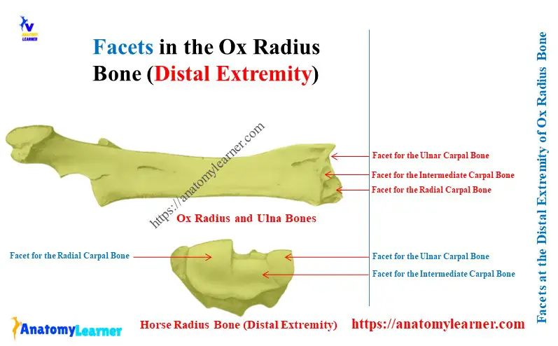
- Articular facets for the carpal bones at the distal extremity of the ox radius bone,
- Caudal articular facets on the transverse process of the horse’s last lumbar vertebra,
I have previously described the features of the facets from the animal bone. You may read the below-mentioned article to get a complete idea of the features of the facet –
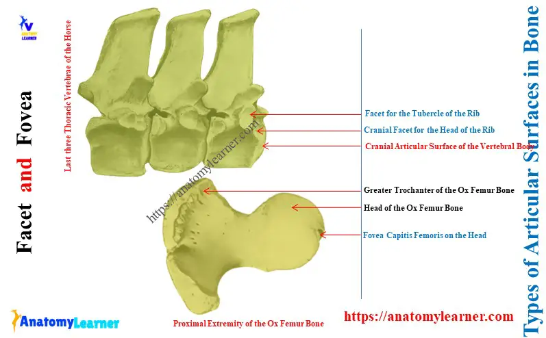
Here, the diagram shows the small articular facets from the cranial and caudal aspects of the body of the animal vertebrae. These articular facets are small compared to the articular surfaces of the body of vertebrae.
The distal end of the animal’s radius-ulna bones show 3 small articular surfaces for the proximal row’s carpal bones. Here, the medial one is the facet for the radial carpal; the middle one is the facet for the intermediate carpal bones. Finally, the lateral facet is for the ulnar and accessory carpal bones.
Fovea in the animal bone
The best example of the fovea from the animal bone is the fovea capitis femoris. At the proximal end of the animal, the humerus bone shows a clear, deep, and cavity-articular surface.
The round ligament attaches with this fovea capitis femoris. This type of small and cavity-like articular surface is the fovea.
Here, the diagram shows the fovea from the proximal end of the horse femur bone.
Condyle in the animal bone
You will find typical features of the condyles in different bones of the ox or horse skeletons. Let’s see some of the examples of the condyle of different bones –
- Medial and lateral condyles of the ox metacarpal bone,
- Medial and lateral condyles of the ox metatarsal bone,
- Lateral and medial condyles at the distal extremity of the horse or ox humerus bones,
- Lateral and medial condyles at the caudo-distal extremity of the ox femur bone,
The article might help you to get a complete guide on the features of the condyle and epicondyle of animal bones –
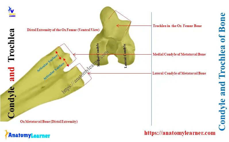
Here, the diagram shows the lateral and medial condyles from the distal part of the ox metatarsal bone. Each of the condyle at the distal extremity shows the paired articular surfaces.
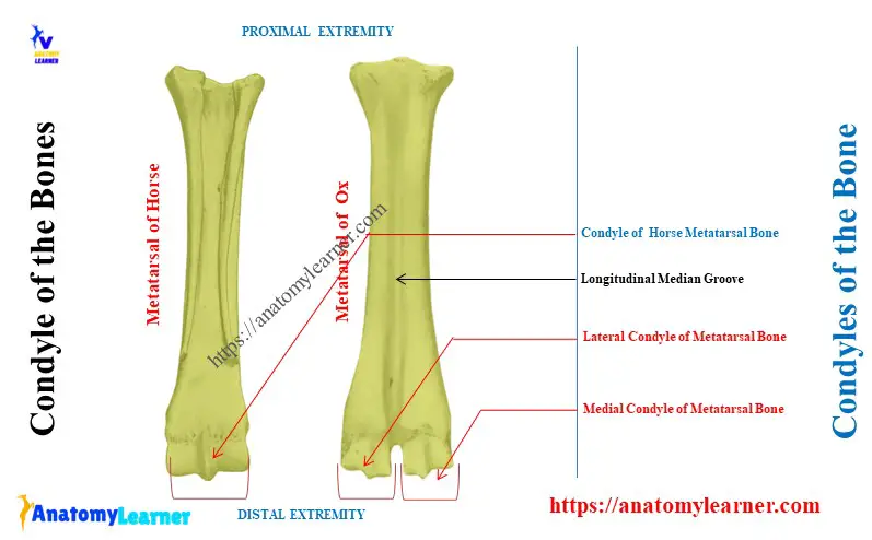
Again, the metacarpal and metatarsal of the horse show only one condyle at their distal extremity. It also represents two articular surfaces divided by a median ridge.
Trochlea in the animal bone
Examples of the trochlea in the animal bones –
- Trochlea at the cranio-distal extremity of the horse or ox femur bone,
- Trochlea at the distal extremity of the horse or ox radius bones,
Here, the diagram shows the cranio-distal extremity of the ox femur bone. It shows a grooved surface where the patella articulates.
This articular surface is pully-like, where the patella runs upward and downward. So, this is a trochlea of the ox femur bone.
The trochlea from the horse femur bone is more profound than the ox humerus bone.
Conclusion
So, facet, fovea, condyle, and trochlea are four significant types of articular surfaces in the bones. All these articular surfaces possess distinct, unique features and are termed by their name in different bones.