Transitional epithelium is a pseudostratified type with a wide variety of appearances. Here, you will know the answer to the question – ‘Where is transitional epithelium found?’
Quick answer: transitional epithelium is found in the renal pelvis and calyces, ureter, urinary bladder, and urethra. This epithelium may also be seen in the palpebral conjunctiva, larynx, and nasopharynx.
I will show the microscopic appearance of transitional epithelium from different parts of the animal body. So, to know the location and identifying features of transitional epithelium under a light microscope, let’s continue this article.
Where is transitional epithelium found?
Transitional epithelium lines most of the hollow organs of the urinary system and is capable of considerable distension. Following are the organs of the urinary and other systems of animals where you will find transitional epithelium –
- In the lining of the renal pelvis and calyces,
- Ureter,
- Urinary bladder,
- The lining of the urethra,
- In the palpebral conjunctiva and
- The lining of the larynx and nasopharynx,
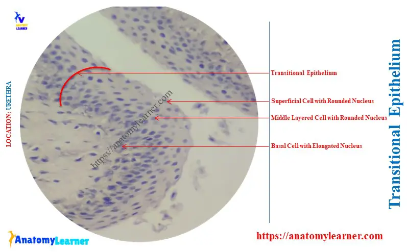
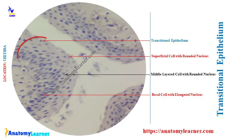
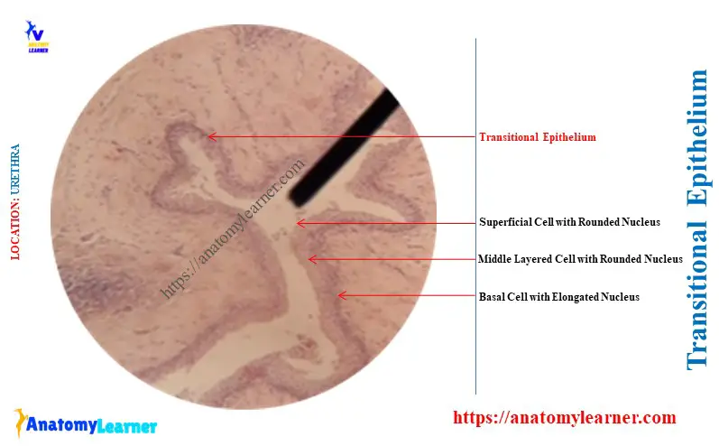
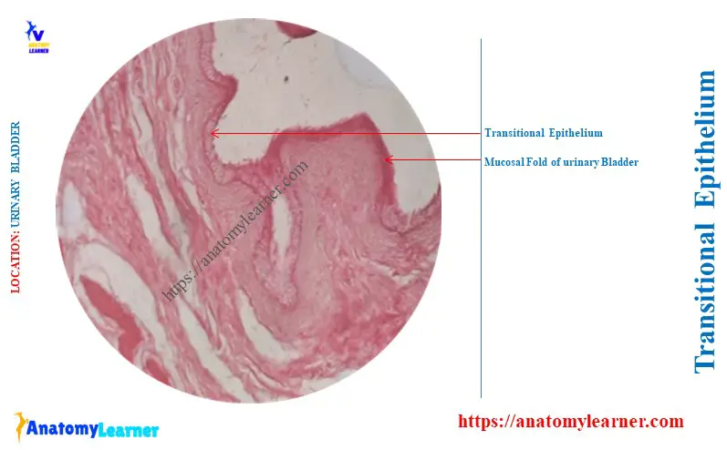
The cells of the transitional epithelium increase in size from the basal layer to the superficial layer. Here, the diagram shows the transitional epithelium from the mucosal fold of the urethra.
What does transitional epithelium appear as?
Transitional epithelium is a multilayered epithelium that contains 4 – 6 cells thick. This type of epithelium differs from the stratified epithelium.
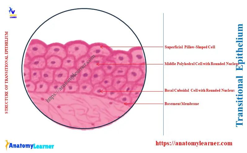
The shape of the transitional epithelium cells depends on the degree of organ distension. The surface cells are large and “pillow-shaped” when the epithelium is under little tension.
The middle layer is made up of polyhedral or pear-shaped cells. Again, the deeper cell layers are small and irregular.
When the transitional epithelium is stretched, the cells become flattened and elongated. In this condition, the total height of the epithelium increases.
Under the light microscope, the luminal cells of transitional epithelium appear relatively smooth compared to pseudostratified. Again, you will find the thicker plasmalemma (plaques) under the electron microscope.
During organ contraction, these plaques fold together much like a hinge. They produce typical transitional epithelium on the cell surface. Again, upon the distension, these plaques unfold, allowing expansion of the luminal surface.
The cells of the transitional epithelium are firmly united to one another by numerous desmosomes. Thus, these cells retain a relative position when the epithelium is stretched or relaxed.
Identification points of transitional epithelium
Following are the identification points for the transitional epithelium under the light microscope –
- This type of epithelium shows several layers of cells (4 – 6) with various–shaped nuclei,
- The superficial cells of the transitional epithelium show a “pillow-shaped” appearance,
- Sometimes, the nuclei of the transitional epithelium show the mitotic figures,
Here, the diagram shows the transitional epithelium cells from the different layers with their nuclei.
Where would you most likely find transitional epithelium?
Answer: the typical transitional epithelium is most likely found in the urinary bladder and parts of the urethra. Cells of the transitional epithelium of the urinary bladder can be stretched considerably without losing their integrity.
When the bladder is stretched, it appears thinner, and the superficial cells become flattened. Here, the diagram shows the typical transitional epithelium from the urinary bladder.
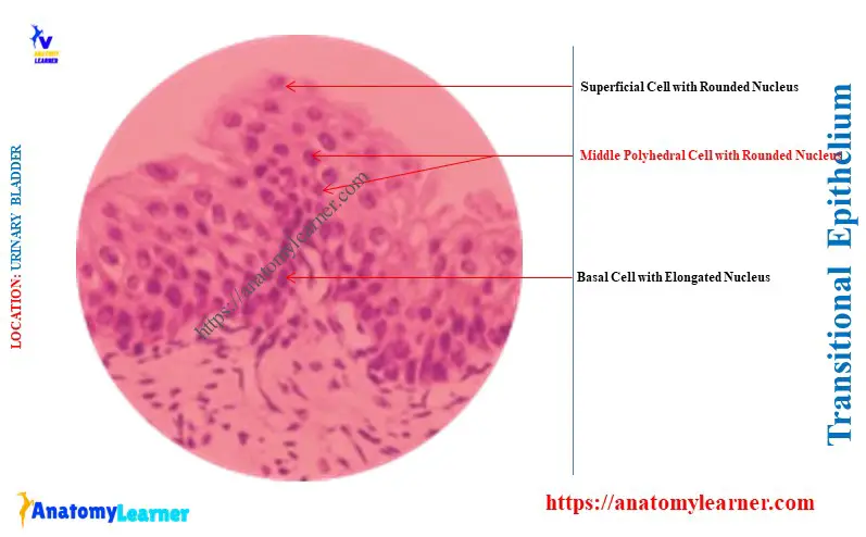
This diagram also shows the below-mentioned microscopic features –
The epithelium of the relaxed urinary bladder is made up of 4 – 6 layers of cells,
It shows the pillow or rounded-shaped superficial cells. They change their shape according to the degree of distension of the urinary bladder.
Sometimes, these cells of the transitional epithelium show double nuclei.
The plasma membrane of the superficial cells of the urinary bladder is thickened at the luminal surface. They form the cuticle responsible for the osmotic barrier between the urine and tissue fluid.
The transitional epithelium of a distended urinary bladder is made of 3 or 4 layers of cells. The superficial cell layer of this condition becomes flattened.
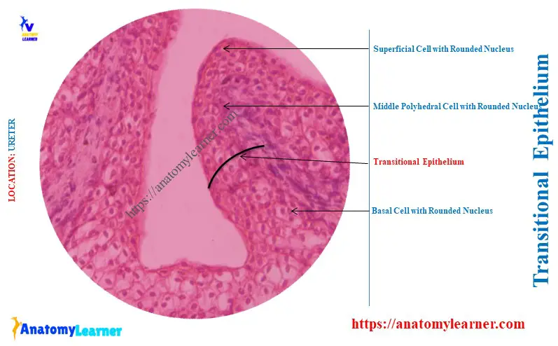
Transitional epithelium in the animal ureter
Here, the diagram shows the transitional epithelium from the mucosal fold of the animal ureter. The epithelium of the ureter shows several layers of rounded nuclei.
The superficial cells of the ureter lining are not flattened, as the nuclei are also rounded. These superficial nuclei of the cells show mitotic figures under the light microscope.
What is the difference between transitional and stratified epithelium?
Answer: the differences between transitional and various types of stratified epithelium are given below –
Transitional vs stratified squamous epithelium: transitional differs from stratified squamous in that the cells of the superficial layer are not squamous. Again, the cell layers are less in the stratified squamous than the transitional epithelium.
The stratified squamous epithelium may present the keratin layer on the superficial surface. But, you will not find the keratinization in the transitional epithelium.
Transitional vs stratified cuboidal epithelium: the stratified cuboidal shows two layers of cells, whereas the transitional epithelium presents 4 to 6 layers of cells. Again, the stratified cuboidal shows two layers of rounded nuclei under the light microscope. However, the transitional epithelium shows multiple layers of polyhedral nuclei.
Transitional vs stratified columnar epithelium: the stratified columnar shows two layers of cells – basal cuboidal and upper columnar. Sometimes, you may find the goblet cells interposed between the stratified columnar cells.
However, the transitional epithelium doesn’t show such microscopic features as the stratified columnar. Again, you will not find any goblet cells in the structure of the transitional epithelium.
Conclusion
So, the concise answer to the question – ‘Where is transitional epithelium found?’ was revealed. The best examples of transitional epithelium lining organs are the renal pelvis, calyces, ureter, urinary bladder, and urethra. However, the palpebral conjunctiva, larynx, and nasopharynx also show this transitional epithelium lining on their tunica mucosa layer.