The stratified cuboidal epithelium has a limited distribution in the animal body. This guide will help answer the question: Where is stratified cuboidal epithelium found?
Quick answer: the stratified cuboidal epithelium is found in the excretory ducts of salivary glands, sweat glands, and developing ovarian follicles. This type of cuboidal epithelium is also found in the excretory duct of the animal’s pancreas.
I will show you how these stratified cuboidal shapes look in different organs under the light microscope. I will also provide some key features for identifying this stratified cuboidal epithelium.
Where is stratified cuboidal epithelium found?
This type of epithelium consists of two or more layers of cuboidal cells. Like other epithelium, it is not widely distributed in the animal body.
You will find the stratified cuboidal epithelium in the following organs –
- Larger excretory ducts of the salivary glands,
- Large and small ducts of the sweat glands,
- Primary and secondary ovarian follicles (developing ovarian follicles), and
- Larger excretory ducts of the pancreas,
But you may also find the stratified columnar epithelium in these larger excretory ducts of the pancreas, salivary, and sweat glands.
What does stratified cuboidal epithelium look like?
The stratified cuboidal cells are arranged in two or multiple layers. Here, the superficial cells have the same length, breadth, and height. Thus, they look like the typical cuboidal cells.
Under the light microscope, these cuboidal cells show centrally placed rounded nuclei. The typical stratified cuboidal features may be seen in the excretory ducts of the salivary gland.
It shows two layered cuboidal cells with a central rounded nucleus. The basal layer cuboidal cells rest on the thin basement membrane.
So, the typical features of the stratified cuboidal epithelium are –
- Presence of the two-layered cuboidal cells (sometimes multiple layers),
- Cells are look like cuboidal (equal length, breadth, and height),
- The presence of a deeply-stained rounded central nucleus in both two layers (typical),
- Shows the pink–stained cytoplasm of the cuboidal cells,
Under the light microscope, the cell boundary of the stratified cuboidal is not visible. Thus, you will only see the two-layered rounded nuclei in the ducts or ovarian follicles.
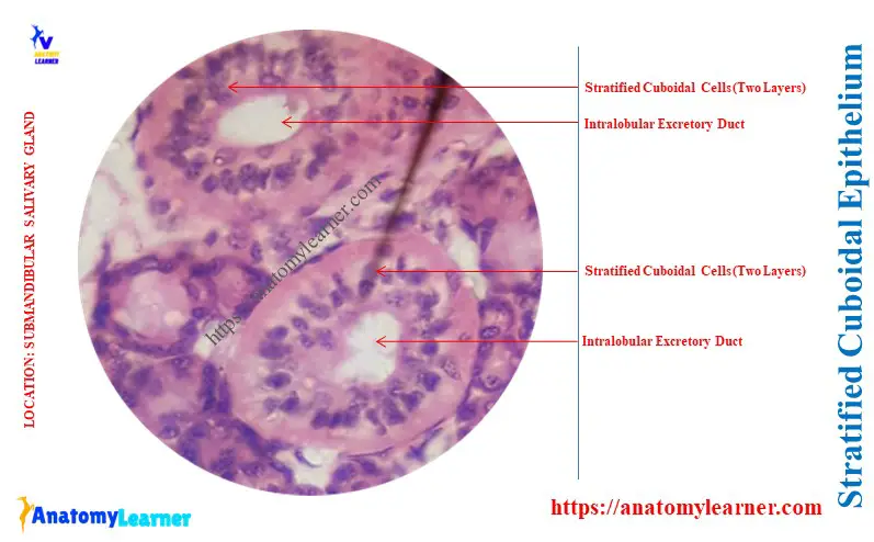
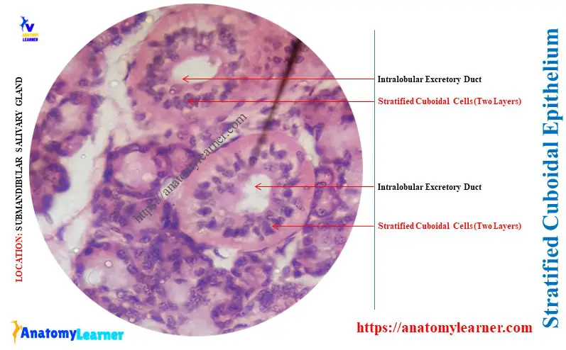
Stratified cuboidal in the excretory ducts of the salivary gland
The major salivary glands, like the parotid, submandibular, and sublingual present different excretory ducts. The excretory ducts of these salivary glands are lined by the stratified cuboidal.
- Related article: salivary glands histology slide (parotid, mandibular, and sublingual glands),
The diagram shows the intralobular excretory ducts of the animal’s parotid salivary gland. It is clearly lined by the two layers of cuboidal cells (stratified cuboidal). These intralobular excretory ducts are located within the lobules of the animal’s parotid salivary glands.
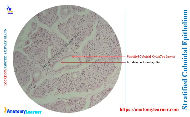
The intralobular ducts join with the larger ducts in the connective tissue septa of the parotid salivary gland. Now, the intralobular ducts become larger and the lumen wider.
The epithelium of the intralobular ducts becomes taller and can increase from cuboidal to columnar. Thus, you may see the stratified columnar epithelium in the lining of the larger excretory ducts of the parotid salivary gland.
Again, the epithelium of the excretory ducts can increase from the columnar to the pseudostratified.
Similar features of the lining epithelium may be found in the intralobular excretory duct of the submandibular gland. Here, the diagram shows the stratified cuboidal epithelium from the excretory ducts of the submandibular gland.
The lining epithelium of the intralobular excretory ducts of sublingual varies from low columnar to pseudostratified. Thus, you will not find the stratified cuboidal here in the excretory ducts of the sublingual gland.
Stratified cuboidal in the excretory ducts of the sweat gland
The dermis of the thick and thin animal skin presents the sweat glands. Here, the excretory ducts of these sweat glands are lined by the stratified squamous epithelium.
Here, the higher magnification of the sweat gland with the excretory duct shows the stratified cuboidal epithelium. Again, the diagram represents the myoepithelial cells that surround the excretory ducts of the sweat glands.
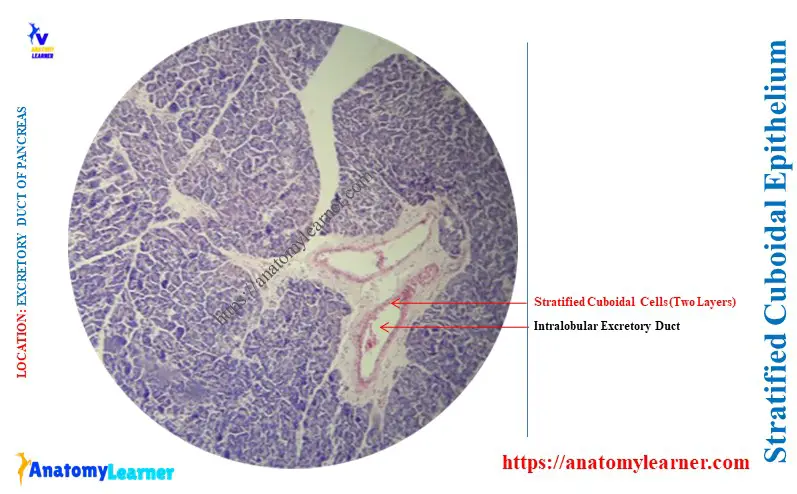
The lining of the excretory ducts of the pancreas
The interlobular ducts of the animal’s pancreas also show the stratified cuboidal epithelium. Sometimes, you may also find the simple cuboidal epithelium lining in the lumen of the excretory ducts of the pancreas.
Here, the diagram shows the stratified cuboidal epithelium from the interlobular ducts of the pancreas.
You may know more about the histological features of the animal pancreas from the below-mentioned article –
Stratified cuboidal in ovarian follicles
The developing ovarian follicles, like the primary and secondary, show the stratified cuboidal epithelium. These cuboidal cells surround the primary oocyte, and the outer layer rests on the basement membrane.
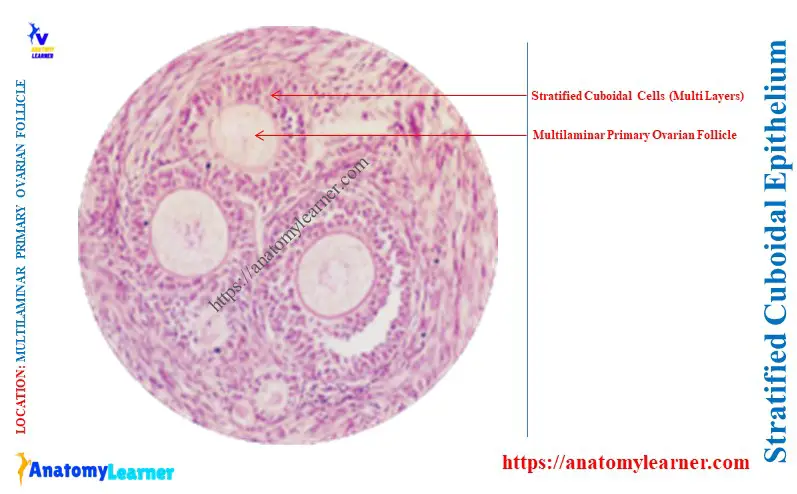
Here, the diagram shows the multilaminar primary and secondary oocytes with stratified cuboidal. The tertiary ovarian follicles also show stratified cuboidal cells (granulosa cells).
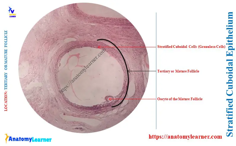
Both the developing follicles of the ovary are surrounded by the stromal cells. The inner layer of the stromal cells converts into the theca external cells in the corpus luteum.
The granulosa cells of the tertiary follicle become a polyhedral shape in the later stage of development. You may know more about the histological features of the ovarian follicles from the animal ovary microscope slide.
Conclusion
So, the concise answer to the question – ‘Where is stratified cuboidal epithelium found?” was revealed. Typical features of the stratified cuboidal cells are found in the excretory ducts of sweat glands, salivary glands, and ovarian follicles.
Cuboidal shaped two layered cells with a central rounded nucleus are the key identifying features of stratified cuboidal. I hope you will now identify this cuboidal epithelium from the excretory ducts and ovarian follicles under the light microscope.