In adrenal gland histology you will find an outer cortex and inner medulla in animals. Adrenal gland are paired and located near the upper pole of kidney embedding in adipose tissue.
Hi anatomy learner, if you are looking for the best guide to learn adrenal gland histology with different labeled slide and diagram then this article is for you. Fine, in this article I am going to share adrenal glands cortex and medulla histology in easy way so that you might understand.
I am sure that after reading this article you will able to identify the adrenal gland under light microscope with their identifying histological features. You will also find the adrenal glands slide histology drawing in this article.
I will also share the information of adrenal gland functions and adrenal gland hormones with you. So, if you are interest to learn details histology of adrenal glands then I would like to invite you to read this full article.
Adrenal gland histology
In adrenal gland histology, you might identify the following important histological features under the light microscope.
#1. Capsule of adrenal gland of animals
#2. Cell and structure of zona glomerulosa (cortex of adrenal gland)
#3. Cells and structure of zona fasciculate (cortex of adrenal)
#4. Cells and structure of zoan reticularis (cortex of adrenal gland)
#5. Two types of chromaffin cells and structure of adrenal medulla in animal adrenal gland
#6. Zona intermedia in few animals
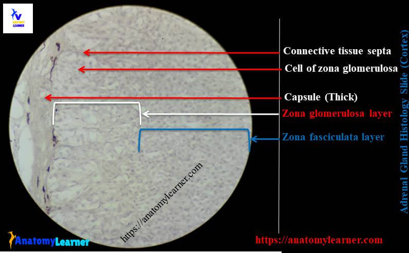
Histologically, there is a thin layer of capsule that usually made with dense irregular connective tissue along with few smooth muscle fibers. The trabeculae originated from this thin capsule but not enter into the medulla of adrenal gland.
You will find the following two concentric layers in adrenal gland histology in different animals –
#1. Adrenal cortex – that is generally yellow in color at this is the peripheral zone of adrenal gland
#2. Adrenal medulla – this is the central layer of adrenal gland and reddish brown in color
I am going to discuss these two different concentric layers of adrenal gland separately here in this article.
Adrenal gland cortex histology
The cortex of adrenal gland is subdivided into three distinct zones in different animals. You will find the following three main zones in adrenal gland histology in most of the animals –
#1. Zona glomerulosa – this is the outermost zona in adrenal glands cortex histology
#2. Zona fasciculate – this is the largest and intermediate zone in between the zona glomerulosa and zona reticularis of adrenal cortex
#3. Zona reticularis – this is innermost zone of adrenal cortex and adjacent to the adrenal medulla
But you will find the zona intermedia of adrenal cortex in few animals like horse, dog, and cat even in ruminant (occasionally). You might identify these different zones from adrenal gland cortex histology under light microscope.
Zona glomerulosa of adrenal glands cortex histology
You will find this layer just beneath of the thin capsule of adrenal gland. The cell in this layer varies from tall columnar to spherical in different species. You will find tall columnar cell in horse, donkey’s adrenal gland cortex histology whereas spherical shaped in other domestic animals.
These cells of zona glomerulosa generally arranged in closely packed or cluster or in arcs in different animals. Generally in ruminant, you will find irregular clusters and cords of cells. But in dog, cat, pig and horse these cells are arranged in arcs; so this zone is also called zona arcuata in these animals.
You may find few acidophilic granules in the cell of bovine zona glomerulosa layer.
Zona fasciculate of adrenal gland cortex histology
This zona fasciculata is the largest zone of adrenal gland cortex in animal. You will find polyhydral cells (cuboidal or columnar cells) that arranged in straight cords in adrenal cortex.
Most of the cells in zona fasciculata of adrenal gland cortex histology have large numbers of lipid droplet. During routine tissue processing, these lipid droplets are dissolved and results the vacuolated cells. These vacuolated cells of zona fasciculata are also known as spongiocytes.
Zona reticularis of adrenal cortex in animal
This is the innermost layer of adrenal gland cortex histology in animals. You will find an irregular network of anastomosing cell cords in zona reticularis of adrenal glands cortex histology. These cells are polyhedral and the other features are same as the previous layers cells.
You may find few lipid droplet and pigment granules in these cells of zona reticularis.
Adrenal gland medulla histology
The medulla of adrenal gland histology of animals is composed with clump or cords of chromaffin cells that are separated by dense network of sinusoidal cappiliaries. These chromaffin cells are polyhedral or columnar in shaped in different animals.
There are two distinguished type of chromaffin cells in medulla of adrenal glands histology in animals and they are –
#1. Epinephrine cells of adrenal medulla and
#2. Norepinephrine cells of adrenal medulla
Norepinephrine cells have larger granules and have strong chromaffin reaction; epinephrine cells have smaller granule and have less affinity to chromium slat.
In the adrenal cortex of cow, pig, horse; medulla mainly subdivided into clear zones – an outer zone of large epinephrine cells and an inner zone of clusters of small polyhedral norepinephrine cells.
Adrenal gland histology slide labeled diagram
I am going to show you the different layers of adrenal glands cortex histology and medulla with real slide and labeled diagram. I hope this adrenal gland histology slide labeled diagram will help you to understand the every single structures and cells in different layers of adrenal gland cortex and medulla easily.
If you need more adrenal glands slide pictures and labeled diagram of adrenal gland histology then you follow anatomy learner at here in social media. Hope you will get your desire adrenal gland slide pictures.
Adrenal gland histology drawing
Fine, I am also interest to share the adrenal gland histology drawing with you; you might follow and try to draw the better diagram of adrenal gland in animal.
Adrenal gland functions
Do you want to know the functions of adrenal gland in animals? I am not going to describe the details functions of adrenal gland of different animals. Rather I prefer to enlist the main functions of adrenal gland cortex and medulla in different animals.
#1. Zona glomerulosa of adrenal cortex produce primarly aldosterone and deoxycorticosterone that regulate electrolyte level in extracellular body fluid
#2. The cells of zona fasciculata are involved in the production of cortisol and corticosterone
#3. Zona reticularis cells produce mainly dehydroepiandrosterone
#4. Epinephrine and norepinephrine acts on different androgenic receptors of animal’s body
That’s great; If you want to know more about the functions of adrenal glands in animals then please go through the histology of adrenal gland pdf book.
Conclusion
I tried to make simple histology of adrenal glands for you. Hope you could understand the adrenal gland histology with labeled diagram.
If you like this guide then you may share this article with your friends who want to learn adrenal histology. Again, you might read other different histological features of other gland like ovary, thyroid, pituitary and other from anatomy learner at here.
Thank for staying with anatomy learner; get update on very single post at here in social media.