
In ovary histology, you will find an outer cortex and inner medulla in most of the animal except horse. Ovary is an ovoid structure and is a combined exocrine and endocrine gland in female. It produces both the ova and ovarian hormones – estrogen and progesterone.
Hey, do you looking for the best guide to learn ovary histology from different animals? Fine, in this article I am going to discuss different follicles of ovary histology of animals.
In this single article you will get all the identifying characteristics of ovarian cortex, ovarian medulla and different follicles with real ovary histology slide pictures and labeled diagram. You will also know the different characteristics of ovarian follicles in different animals.
I am so excited to provide you a normal ovary histology pdf and ppt at the end of this article; this will really help you to learn and practice.
Okay, let’s get into today’s article – histology of normal ovary with slide pictures and labeled diagram.
Ovary histology
In this article I will focus only the identification of ovarian cortex and contains of ovarian cortex (different follicles). You will learn how you could identify different types of ovarian follicles under light microscope. I will try to make it so simple so that you might learn and identify the follicles from real ovary histology slide.
Followings are the structures or follicles you might identify under the light microscope from ovary histology at laboratory –
#1. Cortex of animal ovary
#2. Different stages of follicles from ovarian cortex (primordial, primary, secondary, tertiary or grafian follicles)
#3. Different parts of a mature or grafian follicle
#4. Histological characteristics of follicular atresia
#5. Histological features of corpus luteum from ovary
#6. Ovulation process (shortly)
Please find all these structures or follicles from the labeled diagram below; hope you will get the basic idea about the histology of animal ovary.
Identification of ovarian cortex from ovary histology slide
You might like – identification of ovarian cortex from ovary histology real slide. Followings are the characteristics that might help you to identify the ovarian cortex under light microscope –
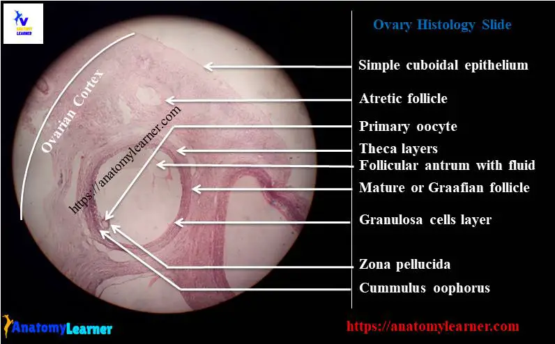
#1. The surface of the ovary is covered by simple cuboidal epithelium. You will find a thick connective tissue layer just beneath the surface of the epithelium
#2. Presence of growing ovarian follicles at various stages of development (primordial, primary, secondary, tertiary or mature follicles; you will find the identifying characteristics of these different follicles at below)
#3. There are follicular atresia and intestinal endocrine cells on ovary cortex (you will learn the characteristics of this follicular atresia at below)
#4. Sometime you will find corpus luteum and corpus albican at the cortex of ovary
Normal ovary histology – ovarian follicles and follicular atresia
I will describe how you will identify the following different growing ovarian follicles from normal ovary histology slide.
#1. Primordial follicles of ovary
#2. Primary follicles of ovary
#3. Secondary follicles of ovary and
#4. Tertiary or mature or graafian follicles of ovary
First I would like to discuss on the different parts or structures of a mature follicles from animal ovary histology.
Structure of a mature follicle of ovarian cortex
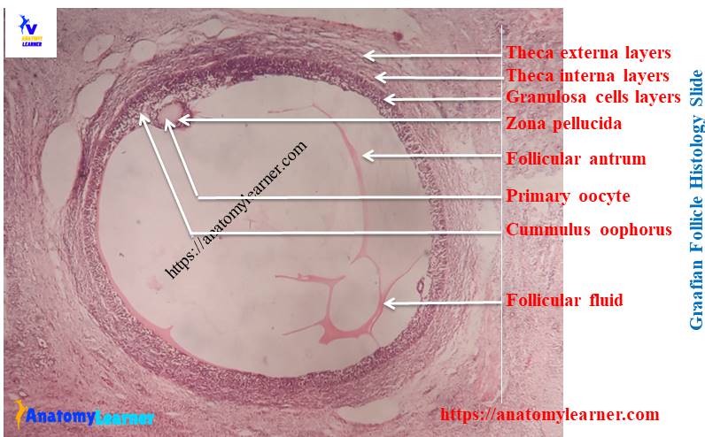
You will find the following structures or parts or cells in a mature or graafian follicle of ovarian cortex. Please try to find these following structures or cells from the labeled pictures of ovary –
#1. Primary oocyte or secondary oocyte of mature ovarian follicle
#2. Granulosa cells of mature follicle
#3. Cummulus oophorus of follicle
#4. Corona radiate of oocyte
#5. Zona pellucida of primary oocyte
#5. Follicular antrum of ovarian follicle
#6. Theca layer – theca externa and theca interna
Fine, now learn the details histology of mature or graafian follicle from animal ovary.
Tertiary follicle or graafian follicle
In tertiary follicle you will find primary or secondary oocyte which is surrounded by the stratified epithelium (known as granulosa cells and layer is known as stratum granulosum). This tertiary follicle also known as the vesicular or graafian follicle. But just before ovulation this tertiary follicle is known as the mature follicle.
The stratum granulosum of tertiary follicle is surrounded by theca layers – inner vascular theca interna and outer supportive theca externa layer.
In tertiary follicle, the theca interna cells are spindle shaped and located in fine reticular fiber; you will find extensive blood and lymph cappilaries in this theca interna layer. But in mature follicle the cells of theca interna become polyhedral and increased in size. Again, the theca interna consists of thin layer of loose connective tissue with fibrocyte in tertiary follicle.
A fluid filled cavity is formed among the granulosa cells of tertiary follicle; this cavity is known as antrum and the fluid that fills antrum is known as liquor folliculi. Antrum enlarges due to more accumulation of liquor folliculi and oocyte displaced eccentrically.
The granulosa cells that attached to the oocyte are called cumulus oophorus. The oocyte is covered by a glycoprotein layer which is known as zona pellucida (around the plasma membrane of oocyte). The granulosa cells that immediately surround the oocyte became columnar and these cells known as corona radiata. These cells (corona radiate) provide nutrient support to oocytes of tertiary or graafian follicle of ovary.
Okay, now learn how to differentiate other different growing follicle from tertiary follicle of ovary histology normal slide.
Primordial, primary and secondary follicles of ovarian cortex
In primordial follicle you will find a primary oocyte that surrounds by a simple squamous epithelium of follicular cell (granulosa cells).
The primordial follicle of ovary histology are located at the outer cortex and evenly distributed in ruminant and sow. But in carnivorous you will find primordial follicle in a cluster form in cortex of ovary.
The primary follicles of ovary are composed of a primary oocyte that surrounded by simple cuboidal epithelium of follicular cells (granulosa cells). Again, the secondary follicles of ovarian cortex are composed of a primary oocyte surrounded by a stratified epithelium of polyhedral follicular cells. You will find small fluid filled clefts among the follicular cells (granulosa cells) of follicle.
Hope you will able to differentiate different types of follicles from ovary cortex histology.
Atretic follicles of ovary histology
During follicular development, most of the follicles of ovary regress; only small percentage of oocytes are ovulated. This regression of follicle is known as atresia of follicle. How you identify this atretic follicle under light microscope from ovary histology?
Well, you will find the following hostological features for atretic follicle of ovary –
#1. The basal lamina of granulosa layer became folded and thickens
#2. Granulosa and theca layers became atrophoid and fibrotic or hyalinez (glassy membrane appearance) around the antrum of follicle
Corpus luteum of ovary
Do you want to know about the corpus luteum of ovary and want to identify under light microscope? Hope, this part will help you to identify corpus luteum from ovary histology.
Follicles rupture after the ovulation and then collapse and shrink; so the folds of follicular wall become more extensive. This rupture of the follicle also known as corpus hemorrhagicum.
Theca externa form poorly defined capsule around the developing corpus luteum. The stratum granulosum layer becomes more vascularized by extensive capillaries from theca interna.
Granulosa cells become enlarge, luteinize and form large luteal cells of corpus luteum of ovary. Again, the theca interna cells become the small luteal cell of corpus luteum of ovary.
So in corpus luteum from ovary histology, you will find the following two types of luteal cells –
#1. Large luteal cells (granulosa cells) and
#2. Small luteal cells (theca interna cells)
How you will identify these lutein cells under light microscope from ovary histology? Well, the large luteal cells are polygonal and have large spherical vesicular nucleus; they also contain numerous lipid inclusions. Again, small luteal cells have more lipid inclusions. Both the large and small luteal cells produces progesterone hormone in animal.
How ovulation occurs?
I am not going to details on ovulation process in animal; rather I prefer to provide a short description of ovulation process in animal.
You know, ovulation is the process of release of oocyte from matures follicles. During the development process of tertiary follicle, it is surrounded by more blood and lymph vessels. These help to increase the secretion of liquor folliculi on the antrum of follicle.
The increased accumulation of liquor folliculi causes the follicle to swell; small hemorrhage occurs in the follicular wall. The follicular wall becomes thin and transparent where the stigma formed (site of follicular rupture); this rupture caused by the release of collagenases. Oocyte release from this ruptured follicular wall.
If you want to know the details process of ovulation then let me inform. Hope I will provide a complete guide for ovulation here in anatomy learner histology section.
Ovary histology slide labeled diagram and drawing pictures
I am so happy to share ovary histology real slide picture, labeled diagram and drawing pictures with you. Hope these resources will help you lots to learn ovary histology. If you need more ovary real slide pictures then you may follow anatomy learner at here.
Fallopian tube histology
You might learn the next organ histology from female genital system of animals – fallopian tube histology. If you want to learn other histological features of different organs from different body system then you may follow anatomy learner histology section.
Conclusion
Hope you learn the basic histology of ovary with anatomy learner. Now you will able to identify the normal ovary histology slide under light microscope.
If you got value from ovary histology labeled diagram and drawing picture then share this article with your friends who want to learn ovary histology.
Thanks for staying with anatomy learner and learn different organs histology with me.