The animal trachea is the major component of the respiratory system that extends larynx to the root of the lungs. In this article / guide, I will explain the details of animal trachea anatomy with labeled diagrams and real pictures.
Quick answer: Animal trachea anatomy is a cylindrical structure that consists of 50 – 60 incomplete hyaline cartilage rings and divides into cervical and thoracic segments.
Thus, we need to know the extension, anatomical composition, and special features of the two segments of the animal trachea.
Animal trachea anatomy
First, you should know the extension of the animal trachea (also known as the windpipe). It is almost cylindrical and extends from the larynx to the level of the base of the heart, where it divides into the right and left bronchi. These bronchi enter the corresponding lungs of the specific animals.
The animal trachea is made of incomplete hyaline cartilage rings, membrane, and smooth muscle. The number of cartilage rings varies in different animals. In a ruminant, you will find about 50 cartilaginous rings, whereas a horse has 50 – 60 rings. Again, the sheep and goat possess 40 – 50 flexible, membranous, and cartilaginous rings.
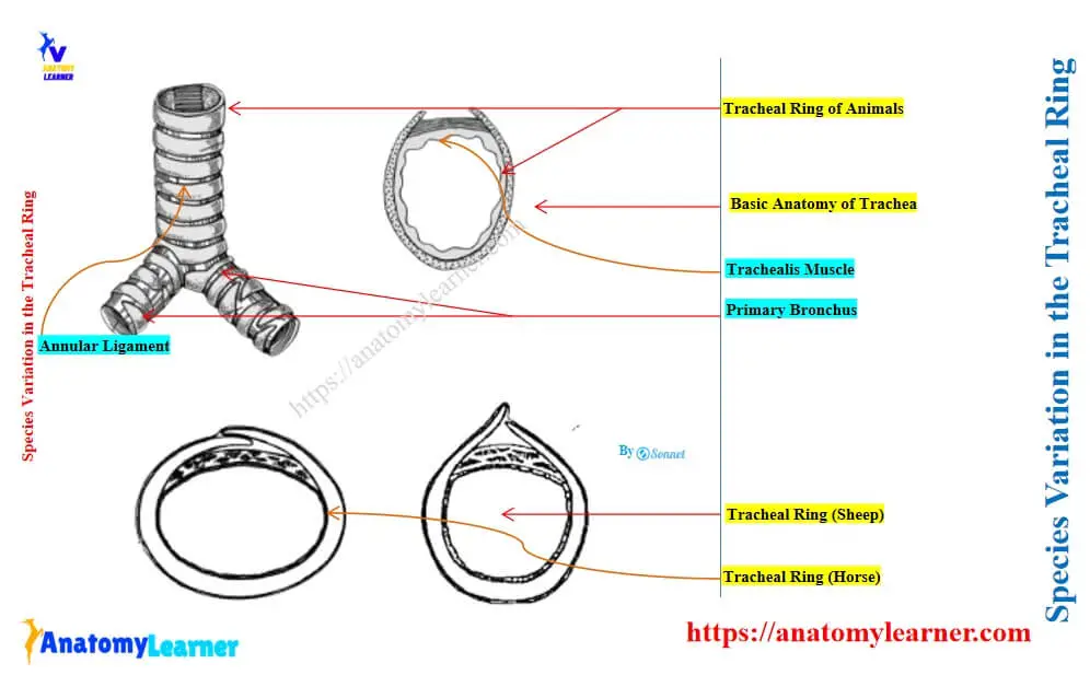
These cartilaginous rings form the wall of the animal trachea and remain open on its dorsal aspect. However, the open end of the trachea is completed with the connective tissue membrane and tracheal smooth muscle fibers.
The trachea occupies a median position in most animals, except near its termination (at the level of the fifth or sixth intercostal space). At this point, the trachea is pushed a little to the right by the arch of the aorta.
Length and caliber of the tracheal ring in different animal species
The length, diameter, and caliber of a tracheal ring vary in different animal species. In a horse, the length is about 70 to 80 centimeters, whereas it is about 65 centimeters in an ox, and 25 centimeters in a goat and a sheep.
The average caliber is about 2 – 2.5 inches (5 – 6 centimeters) in the horse trachea. But the caliber of the trachea is relatively small in cows compared to horses. Its width is about 4 centimeters, and its height is about 5 centimeters in an average-sized cow. The sheep and goat have have small caliber, which is less than 2 centimeters.
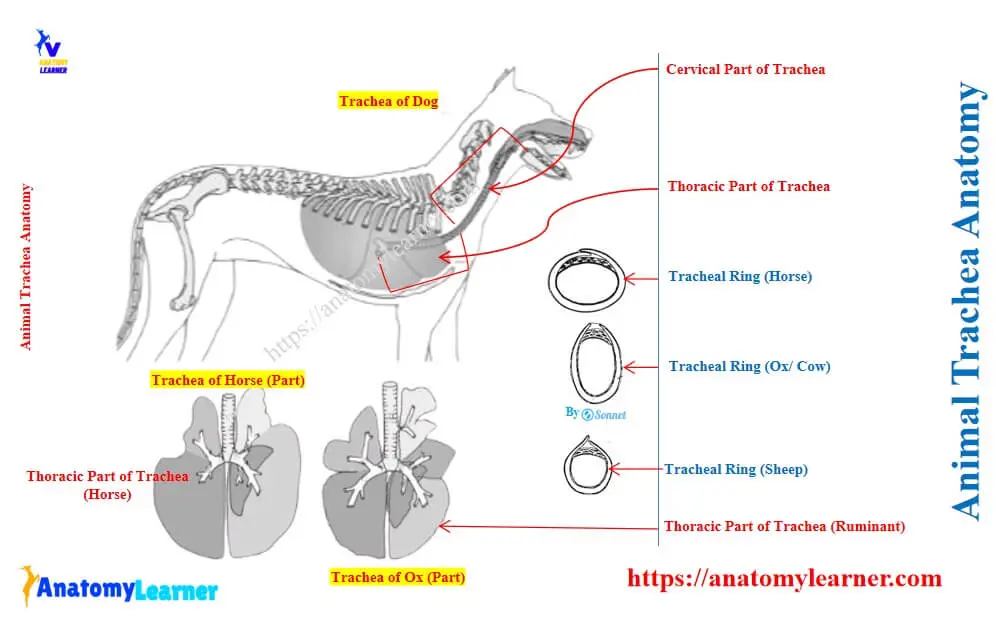
Parts of the animal trachea
The parts of the animal trachea that are situated in the neck and the thoracic cavity are called the cervical part and the thoracic part, respectively. The trachea is longer in horses compared to the large ruminant.
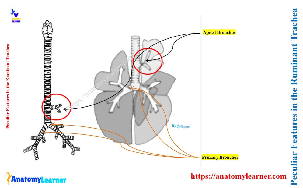
Again, the small ruminant possesses a shorter trachea than that of the large ruminant. The thoracic segment of the ruminant trachea shows peculiar features (apical bronchus), whereas it is absent in the horse and dog.
Let’s discuss the detail anatomical features of the cervical and thoracic parts of the animal trachea.
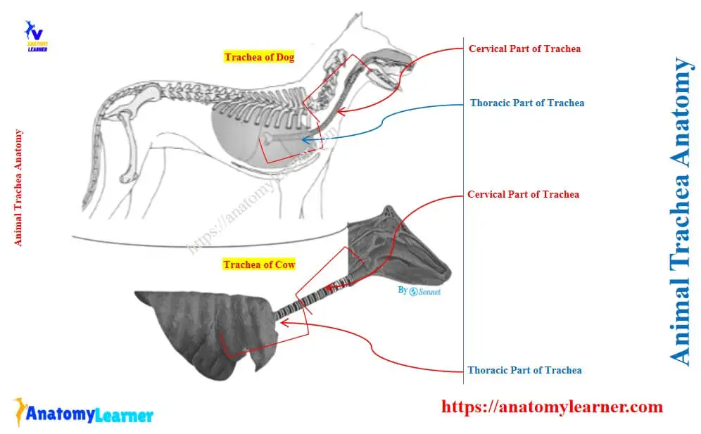
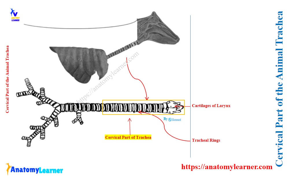
The cervical part of the animal trachea
The cervical part of the animal trachea extends from the larynx (at the level of the first or second cervical vertebra) to the level of the thoracic inlet. This part of the trachea is related to different organs or structures on its dorsal, ventral, and lateral aspects.
Dorsally, the cervical part of the animal trachea is related to the esophagus in the cranial half of the neck and to the longus colli muscles in the caudal half of the neck.
Ventrally, this segment of the trachea is related to sternothyroideus and sternohyoideus muscles. But the cranial extremity is related only to the sternohyoideus muscle.
Laterally, the cervical part of the trachea is related to sternocephalicus, omohyoideus, and sternothyroideus near the larynx. Again, it is also related to the scaleni muscles near the thoracic inlet.
The cervical segment of the animal trachea is also related to the deep cervical lymph nodes and tracheal lymphatic duct. However, it is also related to the jugular vein and phrenic nerve on the right lateral aspect near the thoracic inlet.
On the dorsolateral aspect of the cervical part of the animal trachea, you will find different organs and structures. Thus, it is essential to describe the relationship of different organs that are related dorsolateral to the cervical part of the trachea separately.
Organs related to the dorsolateral aspect of the cervical part of the animal trachea
In the middle of the neck, the esophagus lies on the dorsolateral aspect of the trachea. But, in the caudal part of the neck, it is related to the left lateral aspect of the trachea.
Again, the common carotid artery, vagosymphathetic trunk, and recurrent laryngeal nerve lie on the dorso-lateral aspect of the cervical part of the trachea. But, near the thoracic inlet, these structures lie on its lateral aspect.
The thyroid glands lie on the dorsolateral aspect of the first three or four cartilaginous rings of the cervical part of the animal trachea. Again, the isthmus of the thyroid gland lies ventrally to the cervical part of the trachea.
In young animals, the thymus may lie on the ventral and ventrolateral aspect of the trachea.
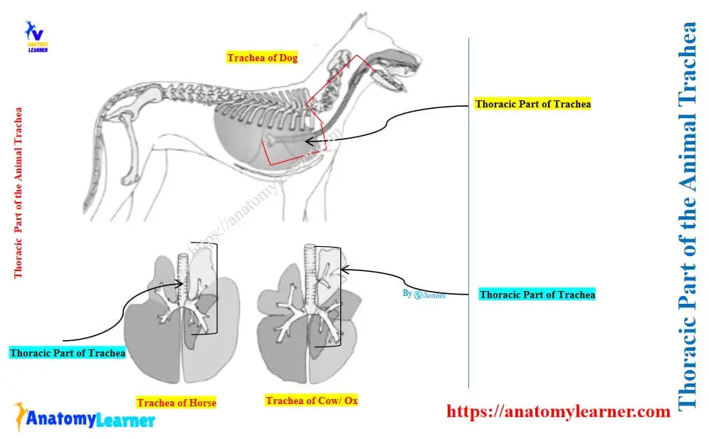
Thoracic part of the animal trachea
The thoracic part of the animal trachea is located at the dorsal aspect of the cranial and middle parts of the mediastinum. Again, different organs and structures lie on the dorsal, ventral, right lateral, and left lateral to the animal’s trachea.
Dorsal aspect: The longus colli muscle and esophagus lie on the dorsal aspect of the cervical segment of the trachea. But, actually, the esophagus lies on the dorsal aspect of the trachea at the level of tracheal bifurcation.
Left lateral aspect: From the thoracic inlet to about the level of the third rib, the esophagus lies on its left lateral aspect. Again, the thoracic duct, left axillary, left cardiosymphatheic nerves, left lung, and the aortic arch close to the tracheal bifurcation are also related to the left lateral aspect of the trachea.
Right lateral aspect: The vagus, right cardiosymphatic nerves, costocervical and vertebral vessels, and the sympathetic trunk between the caudal and middle cervical ganglia lie on the right lateral aspect of the thoracic part of the animal trachea.
Ventral aspect: The brachiochephalic trunk, cranial vena cava, common carotid, right pulmonary arteries, mediastinal lymph nodes, and left recurrent laryngeal nerves lie on the ventral aspect of the trachea.
The bifurcation of the animal trachea occurs opposite to the fifth or sixth ribs and about 4 – 5 inches ventral to the vertebral column.
Peculiar features in the ruminant trachea
The thoracic segment of the ruminant trachea (ox, sheep, goat) gives off an extra bronchus on its right side at the level of the third intercostal space. This is the apical bronchus in ruminants that ventilates the apical lobe (shown in the figure) of the right lung. But you will not find such peculiar features in the trachea of horses and dogs.
Species variation in the tracheal ring
In the fresh state, the tracheal rings of different animals show little variation, so that you may easily differentiate the species. The cross-section of the tracheal rings from different animal species shows different shapes and helps in their identification.
Tracheal ring in a horse
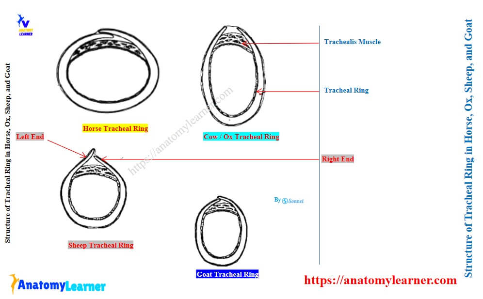
There are 50 – 60 flexible, membranous, and cartilaginous tracheal rings in the horse. These rings are bent to form incomplete hoops that open dorsally. The free extremity of the cartilaginous ring overlaps specifically at the cervical region, with the right end overlying the left (shown in diagram).
But the free extremity of the tracheal ring in the thoracic region does not meet dorsally. Here, the trachealis muscles consist of smooth muscle fibers. They extend transversely across the dorsal part of the tracheal wall. This muscle also attaches to the inner surface of the cartilaginous tracheal rings.
The cross-section of a horse’s tracheal ring is almost circular. The average diameter is about 6 centimeters and soon becomes flattened dorsoventrally. The lumen of the horse tracheal ring may vary considerably in size due to the action of tracheal muscles.
Tracheal rings in large and small ruminants
The large ruminant, like the ox, has 48 – 60 cartilaginous rings in their trachea. These rings are bent so that their free dorsal ends almost touch, but never overlap (shown in the figure).
The cross-sectional of the ox trachea shows an ellipse outline (C-shaped) with its greatest diameter in the sagittal plane. Again, the free dorsal end of the ox and cow tracheal rings form a dorsal crest.
In the case of a goat, the tracheal rings are bent and form a U-shaped outline. The wall of the trachea between the open ends of the rings is flat and entirely membranous.
Again, the cross-section of the sheep trachea differs from one region to another. It is almost cylindrical at the larynx region with a low dorsal crest. In the middle third, the cross-sectional outline of the sheep trachea is U-shaped (like that of a goat).
At the caudal third of the sheep trachea, the dorsal free ends of the rings extend further, and the left ends overlap the right end.
Vessels and nerves of the animal trachea
Branches of the common carotid and broncho-esophageal arteries supply the wall of the animal trachea. Again, the veins of the trachea are tributaries of the jugular veins and the broncho-esophageal veins.
The lymphatic vessels of the animal trachea drain into the deep cervical, costocervical, cranial, and middle mediastinal and tracheobroncheal lymph nodes.
Finally, the parasymphathetic and sensory nerve fibers from the recurrent laryngeal nerve and sympathetic nerve fibers from the sympathetic nerve trunk innervate the wall of the animal trachea.
Structure of the animal trachea
The wall of the animal trachea consists of 4 layers –
- The inner mucous membrane,
- Second layer: submucosa,
- A musculo-cartilaginous layer, and
- Adventitia layer of trachea,
Now, let’s know the details of these 4 different layers of the animal trachea.
Mucosa layer of the animal trachea
The mucosa of the animal trachea is thrown into numerous low longitudinal folds. Histologically, you will find the pseudostratified columnar ciliated epithelium lining the mucosa. This lining epithelium contains numerous goblet cells, which rest on the basement membrane. The cilia of the epithelium beat cranially and carry the mucous secretions and entrapped practical of foreign materials.
The lamina propria of the animal trachea is a relatively thin fibrous layer. It is separated from the submucosa layer by a fibroelastic membrane. This fibro-elastic membrane consists of longitudinal elastic fibers within a collagenous framework. You will also find the true lymph nodules and the aggregation of lymphocytes in the lamina propria of the animal trachea.
Submucosa of the animal trachea
This layer of animal trachea is rich in elastic fibers and also contains fat. Microscopically, you will find numerous tracheal glands that open into the lumen of the trachea.
These glands may also be found in the deeper layers of the lamina propria and between the cartilaginous rings in the musculocartilaginous layer. These types of glands are numerous in the ventral and lateral walls of the animal trachea.
Musculocartilaginous layer of the animal trachea
This layer of animal trachea consists of cartilaginous rings or plates, fibroelastic tissue, and tracheal muscle. Here, the cartilaginous plates are composed of hyaline cartilage surrounded by the perichondrium.
The cartilaginous plates bent so that they are roughly horseshoe in shape with the opening dorsally. Though the exact shape of the cartilaginous rings or plates varies in different species (described before).
The dorsal opening in each plate is filled with connective tissue and tracheal muscle. Here, the tracheal muscle of the animal trachea is smooth in nature that arranged in a circular fashion. This area of the cartilage is called the membranous part of the animal trachea.
Typically, the tracheal muscle attaches to the inside of the cartilage rings in most animal species. But, in the dog and cat, this muscle attaches to the outside of the plates or rings.
The cartilage plates are held together by a fibroelastic tissue along their cranial and caudal border. They form the anular tracheal ligament in the animal trachea. In animals, the first cartilage plate attaches to the cricoid cartilage (CC) of the larynx by the cricotracheal ligament.
“The trachea must function as a rigid tube, or it would collapse when the lungs expand. The rigidity is provided by the cartilaginous rings or plates.”
Adventitia of the animal trachea
It is a connective tissue layer that blends with the musculocartilaginous layer. Together, they surround the whole trachea.
Why is the animal trachea capable of expansion?
The animal trachea must be capable of expansion so that it can accommodate any increase in the volume of air passing from the lungs. It is capable of this expansion because –
- Hyaline cartilage of the animal trachea has a certain inherent flexibility.
- The cartilaginous plates or rings are incomplete dorsally, so that they can expand,
- Its mucous membrane layer is thrown into longitudinal folds, and
- There is a considerable amount of elastic tissue in the submucosa layer,
Conclusion
The animal trachea anatomy consists of 50 – 60 incomplete cartilaginous plates, a fibroelastic membrane, a muscular layer, and inner mucosa. It is a flexible, membranous, and cylindrical tube that occupies a median position and extends from the larynx to the dorsal base of the heart.
The trachea possesses two defined segments (cervical and thoracic), which relate to different organs in respective areas. Species variation occurs in the shape of the cartilaginous rings of the trachea in cross-section. Due to the presence of cartilage rings, longitudinal folds, and elastic fibers in the mucosa, it is capable of expansion when necessary.