
Cartilage is specialized for the supportive role in the animal’s body that contains chondrocytes, a supportive framework of collagen or elastic fibres and firm but pliable ground substances. You know, there are three different types of cartilage based on the structural basis of matrix – hyaline, elastic and fibroelastic cartilage. This post will describe the basic histology of hyaline cartilage with slide images and labeled diagram.
Hey there, do you want to learn cartilage histology in details? Well, you will find the details guide of three different types of cartilage here in the anatomy learner blog.
But here in this post, you will learn the essential histology and identification points of hyaline cartilage with slide labeled images. You will also know the components of cartilage histology structure like the cells, fibers and ground substances.
If you want to learn cartilage histology in details, you might start with this post. After completing this post, you will understand the histological features of hyaline cartilage histology slide under the light microscope.
Hyaline cartilage histology
First, I would like to point out the essential histological features from the hyaline cartilage histology slide under the light microscope. These will help you to understand every single component of the hyaline cartilage structure.
- #1. Perichondrium of hyaline cartilage structure
- #2. Inner chondrogenic layers of hyaline structure
- #3. Differentiating chondroblasts (if possible)
- #4. Fibroblasts in the perichondrium of hyaline cartilage
- #5. Pericellular matrix of the hyaline structure
- #6. Territorial matrix of hyaline cartilage
- #7. Interterritorial matrix of the hyaline structure
- #8. Isogenous chondrocytes in the lacunae
Fine, now you might find out these structures from the hyaline cartilage histology slide image.
Hyaline cartilage histology identification points
So, do you want to identify the hyaline cartilage histology slide under a light microscope with proper identification points? Well, here I am going to enlist the identifying characteristics of hyaline cartilage slide under the light microscope.
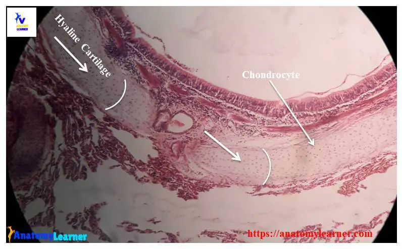
#1. The provided sample tissue shows the homogenous and glassy basophilic matrix (both the territorial and interterritorial matrix).
#2. Presence of isogenous groups of chondrocytes (cell nest) within the basophilic matrix of the sample tissue.
#3. There is a perichondrium that covers the sample tissue peripherally.
#4. The number of chondrocytes increase in size from the periphery to the centre of the sample tissue
#5. Presence of numerous fibroblasts within the perichondrium.
So, this is a hyaline cartilage slide. You may also enlist other relevant histological features of hyaline cartilage structure if you want.
Description of hyaline cartilage structure
If you are interested to know the details histology of hyaline cartilage, then you may continue this article. Now, I will describe the histological features of hyaline cartilage with slide images and a labeled diagram.
But, before to start I would like to inform you of the fundamental component of cartilage histology. In cartilage histology, you will find three different components – cartilage cells (chondroblasts, chondrocytes), fibers (hyaline or elastic fibers) and ground substances.
Cells of cartilage histology
You will find two different types of cartilage cells – chondroblasts and chondrocytes. The chondroblast is an oval-shaped cell with a spherical nucleus that actively forms the matrix of cartilage. Usually, these chondroblasts are found in the growing cartilage of the animal’s body.
Again, the chondrocyte is the mature but less active cell in the cartilage matrix (after complete formation of cartilage matrix). The chondrocyte’s shape may vary from elongate to spherical and depends on the location within the cartilage. They are found in lacunae and also responsible for the production of fibres and ground substance.
Fibers of cartilage structure
You know, the matrix of a cartilage histology structure contains the fibers and ground substance as found in other different connective tissue of the body. You will find mostly collagen fiber (primarily type II and type I) and elastic fibers in the cartilage matrix structure.
These collagen or elastic fibers form the framework of the matrix of cartilage histology. If you want to know more about collagen fibers and elastic fibers, you may read articles in the connective tissue section.
The ground substance of cartilage structure
The ground substance of the cartilage structure composed of chondroitin sulphate, keratine sulphate and hyaluronic acid.
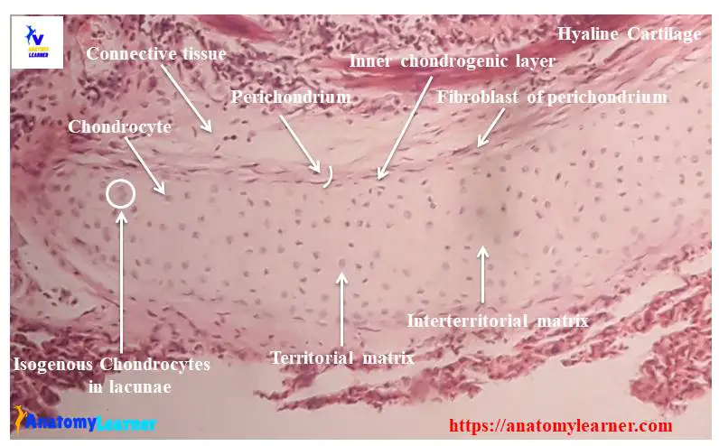
You will find two different types of matrix in the cartilage structure – territorial matrix and interterritorial matrix.
The territorial matrix is the newly formed matrix without the fibers that immediately surrounds the chondrocytes. Again, the territorial matrix is the old matrix of the cartilage structure with fibers located among the chondrocytes. The territorial matrix is more basophilic, whereas the interterritorial matrix is less basophilic.
Histological Features of hyaline cartilage
Hyaline cartilage is the most common type of cartilage in the animal’s body that serves as a skeletal model for most of the bone.
#1. Location of hyaline cartilage
#2. Cells of hyaline cartilage
#3. Matrix of hyaline cartilage histology (ground substance and fibers)
Location of hyaline cartilage
First, you might know where you will find hyaline cartilage in an animal’s body. You will find hyaline cartilage in the following organs or structures of animal’s body –
#1. Tracheal ring of animal
#2. Thyroid cartilage of animal
#3. Cricoid cartilage of different animals
#4. Costal cartilage
#5. Articular cartilage of different bones
#6. Epiphyseal plate
#7. Bronchi of lung structure
#8. In the nasal septum of animals
#9. In the larynx histology structure
Cells of hyaline cartilage structure
In the mature hyaline cartilage histology slide, you will find various size chondrocytes. Near the hyaline cartilage surface, these chondrocytes are small in size, and the lacunae are elliptical. But in the deep hyaline cartilage structure, the chondrocyte becomes larger and more polyhedral shaped.
Lacunae are the oval or elliptical space like cavity in the matrix of hyaline cartilage. Some lacunae contain only one chondrocyte, and some others include two, three or four chondrocytes. These type of multicellular lacunae are also known as cell nest or isogenous cells groups.
Matrix of hyaline structure
The extracellular matrix of hyaline cartilage histology is homogenous and glassy in appearance. The amorphous ground substance of a hyaline structure is generally firm and contains a collagen fiber network (type II).
You will find the pericellular, territorial matrix and interterritorial matrix in the hyaline cartilage structure. A thin layer of matrix is found around each chondrocyte which lacks collagen fibers but contains proteoglycans. This type of matrix refers to the pericellular matrix of the hyaline structure.
The territorial matrix surrounds the pericellular matrix composed of a network of fine collagen fibers and ground substances.
Again, the interterritorial matrix lies outside the territorial matrix that fills the space among the matrix structure’s lacunae or chondrocytes.
Perichondrium of hyaline cartilage
You will find a vascular connective tissue layer that surrounds the hyaline cartilage structure peripherally (except the articular surface). This connective tissue layer of hyaline cartilage is known as perichondrium.
Perichondrium of hyaline cartilage histology consists of two distinct layers – the inner cellular and outer fibrous layers.
The cellular layer immediately adjacent to the hyaline cartilage matrix that contains chondroblasts and networks of small blood vessels. You will find irregularly arranged collagen fibres and fibroblasts in the outer fibrous layer of the perichondrium.
Hyaline cartilage slide images
Do you need more hyaline cartilage histology slide images? Great, you may follow anatomy learner on social media to get more updated cartilage histology slide images.
Now, I will provide you with the drawing tutorial of hyaline cartilage slide images. If you want, you may follow this simple cartilage drawing tutorial.
You might also read other articles like elastic cartilage histology and fibrocartilage histology from the anatomy leaner blog.
#1. Histological features of loose connective tissue with labeled images
#2. Bone histology – the compact bone and spongy bone histological features with labeled pictures
Conclusion
This is the best guide to learn hyaline cartilage histology with labeled diagram and real slide pictures. Now you will able to identify the hyaline cartilage histology slide under a light microscope with proper identification points.
If you think this article helps learn cartilage structure, share it with your friends who want to start learning connective tissue structure. Again, if you want to get more articles like this cartilage structure, then follow anatomy learner