The cow liver anatomy comprises lobes, surfaces, borders, vessels, and nerves. It is the largest solid gland in the cow’s body.
I will discuss and identify the main anatomical facts from the cow or ruminant liver with a diagram.
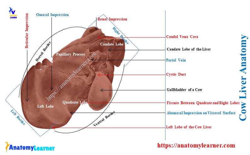
Quick overview: the cow liver locates to the right of the median plane and possesses 4 lobes and 2 surfaces. Here, the caudate lobe possesses 2 processes from the renal impression with the right lobe. You will find various impressions of the cow’s internal organs on the visceral surface of the liver.
Here, you will also find (see) the description of all the cow liver lobes, surfaces, and borders. I will show how the ruminant liver attaches to the abdominal cavity with various ligaments.
Finally, you may also compare the anatomical facts of the liver between cows and horses.
Let’s get started to know the important anatomical facts of the cow liver with a diagram.
Cow liver anatomy
The liver is the larger solid gland in a cow and is considered the extramular digestive organ. First, I would like to show (ID) you the important features of the different lobes, surfaces, and borders of the cow liver.
If you want to know (learn) the list of primary organs from the cow digestive system, the below-mentioned article might help you –
Here, I will identify the below-mentioned features from the cow liver anatomy –
- Four lobes of the cow liver (specify the name in the diagram),
- Caudate and papillary processes of the caudate lobe,
- Renal impression between the caudate and right dorsal lobes,
- Diaphragmatic and visceral surfaces of the cow liver,
- Impression of ribs on the diaphragmatic surface,
- Falciform and round ligaments from the diaphragmatic surface,
- Serous covering of the cow liver (Glisson capsule),
- Porta hepatis from the visceral surface of the cow liver,
- Reticulum, omasal, and abomasal impression on the visceral surface of the cow liver,
- Borders of the cow liver (right, left, dorsal, and ventral),
- Gall bladder fossa on the ventral surface of a cow liver,
- Wide groove on the dorsal border of the liver, and
- The thin left border of the cow liver,
The liver diagram identifies all these anatomical facts from the cow’s liver. You will also find other features of the cow liver in other diagrams.
Unique features of the cow or ruminant liver
Now, you may easily tell the unique anatomical facts from the cow liver. Here, I will enlist some of the unique features of the cow liver structure –
- It is the largest gland located right of the median plane and in the abdominal cavity,
- Indistinct fissure divides the cow liver into 4 different lobes – right, left, caudate, and quadrate,
- The cow liver represents diaphragmatic and visceral surfaces with 4 various borders,
- The internal surface of the various lobes represents the impression of different visceral organs,
- You will see the deepest impression on the caudo-dorsal area between the caudate and right dorsal lobes (renal impression),
- A gallbladder is present on the visceral surface of the cow liver (between the right and quadrate lobes),
- The dorsal border is thick that possesses the caudal vena cava, and other borders are comparatively thin,
- A bile duct comes from the liver and gall bladder that open into the duodenum at its second bend,
Here, I will also provide the summary of the cow or ruminant liver structure in Table 1 –
| Cow liver anatomy | Features |
| Color | Reddish brown |
| Weight | 4.5 – 5.6 kilograms |
| Location | Right of the median plane (in the abdominal cavity) |
| Lobes | 4; right, left, caudate, and quadrate |
| Surfaces | 2; diaphragmatic and visceral |
| Borders | 4; right, left, dorsal, and ventral |
| Impressions | Renal, reticulum, Omasal, abomasal, and Cystic |
| Ligaments | Round, falciform, Right and left triangular, Coronary ligament and two others (let’s see from the attachment of the liver) |
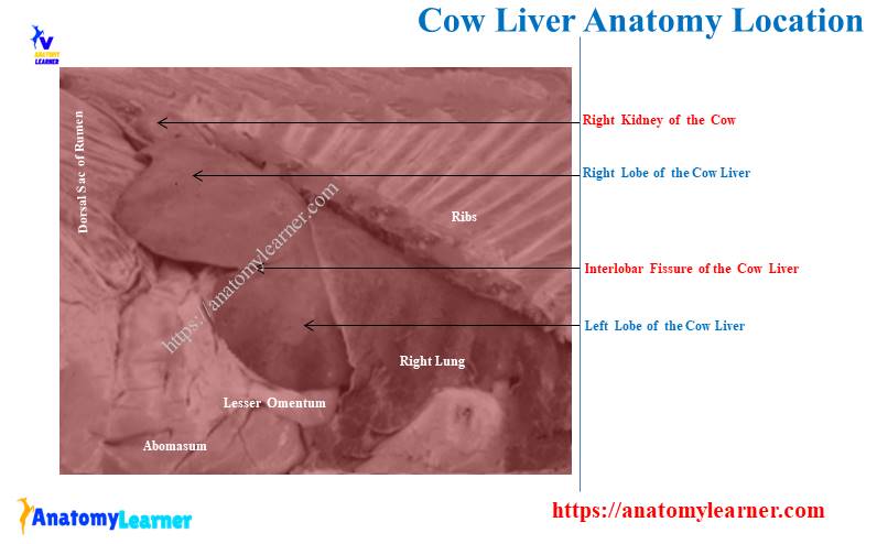
Location of the liver in cattle or cow
The cattle or cow liver lobes are almost entirely to the right (RT) of the median plane. It is placed in the abdominal cavity where the long axis directs cranioventrally (downward and forward direction).
Due to its oblique direction in the abdominal cavity, you will find the right lobe dorsally. In contrast, the left lobe of the cow liver directs ventrally.
Surface anatomy of the ruminant liver: it extends from the right kidney (RK) at the last rib (13th) to the plane of the ventral third of the sixth intercostal (6th) space.
I hope the provided diagram might help you understand the cow liver extension within the abdominal cavity.
There are other different organs in the thoracic part of the abdominal cavity. You may get an idea of these organs that locates in the cow abdomen from the below-mentioned article –
Body cavities and organs with labeled diagram – the major and minor body cavities of the animals,
How big is a cow’s liver?
Answer: you will see a larger liver in cows than in other animals. The medium-sized cow shows a liver weighing 4.5 – 5.6 kg (kilograms).
Whereas medium-sized sheep and goat’s liver weighs 550 – 750 grams. On the other hand, the horse has extensive liver and weighs about 4 to 5 kg (kilograms).
Colour of the cow liver: in fresh condition, you will find a reddish-brown colour in the cow’s liver.
Liver of ox anatomy description
In this section, I will describe the followings from the ox liver anatomy –
Anatomical features from the ox or cow liver lobes –
- Caudate lobe (medial to the right lobe),
- Right lobe (right in position),
- Quadrate lobe (between right and left lobes), and
- Left lobe (left in position),
Anatomical facts from the surfaces –
- Diaphragmatic surfaces (parietal or outer surfaces), and
- Visceral surface (an inner surface that has an attachment with the surrounding organs),
Identification of 4 borders with their important facts –
- The right border of the cow liver,
- Left border of the cow liver,
- The ventral border of the liver, and
- Dorsal border of the liver (important),
And finally, you might also describe the vessels and ducts from the cow liver structure. Let’s learn the details and facts from the cow liver.
Cow live lobes
There are 4 lobes in the structure of a cow liver, but they are not clearly separated from each other (except caudate). This is because (reason) you will not find any fissure in the cow liver structure that divides the lobes.
Again, the lobes are distinct in sheep or goats than the cow or ox.
Caudate lobe: here, the caudate lobe of the cow liver lies between the vena cava and the left branch of the portal vein. You will find two processes in the structure of the caudate lobe –
- Larger and elongated caudate process, and
- Small and variable papillary process,
Here, the caudate process extends to the right of the median plane. It covers most (more) of the area of the visceral surface of the right lobe.
The most important facts of this caudate process: this process is from the deep renal impression with the right dorsal lobe of the cow liver. And you know, the right kidney keeps logged within this renal impression.
You may know the details location of the right and left kidneys with their anatomical facts from the below-mentioned article –
The right lobe of the cow liver: it is very short and thick. The dorsal and caudal borders of the right lobe of a cow liver show more thickness.
You will find the fossa for the gallbladder on its visceral surface. Again, the right kidney contributes to forming the porta hepatis on its medial surface.
Quadrate and left lobes of cow liver
The quadrate lobe is indistinct in the cow liver structure. But, you will identify the quadrate lobes from the dog’s liver.
You may get the details of the canine liver lobes (well-separated lobes) from the below-mentioned article –
Again, the pigs and rabbits also show well-separated lobes in their liver structure.
The cow liver’s quadrate lobe locates between the left and right lobes. You will find the direction of the quadrate lobe in the ventral border.
Left lobe of cow liver: this is the largest lobe in the cow liver structure. It is ventral and left in position in the thoracic part of the cow’s abdominal cavity.
You will find the cranial border of the left lobe at the level of a ventral third of the sixth (6th) intercostal space.
The diaphragmatic surface of the cow liver
Due to the oblique direction of the cow liver, the diaphragmatic surface faces dorsally, cranially, and to the right. Most of the part of this surface is moulded to hollow the right half of the diaphragm.
Let’s know the details of the anatomical facts of the cow diaphragm from the below-mentioned article –
- Cow diaphragm anatomy – parts and their attachments,
On the diaphragmatic surface of the liver, the falciform ligament attaches the liver to the diaphragm. This structure extends from the oesophagal notch to the umbilical fissure.
You will find a long triangular area on the dorsal surface of the cow liver. This triangular area lacks a serous covering (Glisson capsule) because it attaches to the diaphragm.
A small part of the diaphragmatic surface contacts the last two or three ribs. Sometimes, you may find the attachment of the caudal surface with the flan at the lumbocostal angle.
The visceral surface of the ruminant liver anatomy
The visceral surface of the cow liver anatomy is concave and possesses impressions for different organs. Again, you will also find the porta hepatic or portal fissure on the visceral surface of the cow liver.
The depression for the porta hepatis is bounded by the papillary process, caudate process, and the area of adhesion of the pancreas. Now, let’s see what the structures that enter and also leave (exist) from the hepatic porta are –
Structures enter into the porta heptis or portal fissure –
- Portal vein,
- Hepatic artery, and
- Several lymphatics,
Again, the structures that leave or exist from the porta hepatis –
- Common hepatic duct (leaves left at porta),
The gallbladder fossa extends from the porta hepatis to the ventral border of the liver. You will find more distinct gallbladder fossa in the liver of sheep and goats.
There is a fissure for a round ligament in the structure of the cow liver. It extends transversely across the visceral surface from the notch for the round ligament.
You will also find distinct fissures for the round ligament in the goat or sheep liver structure. The visceral surface of the ruminant liver also shows the attachment of lesser omentum.
This structure passes obliquely from the oesophagal impression to the porta hepatis.
How many impressions does the cow liver have on its visceral surface?
Quick answer: there are 4 (four) impressions on the visceral surface of the cow liver. But, you will also find another impression on the caudo-dorsal area of the cow liver for the right kidney.
Thus, there are a total 5 impressions in the cow liver structure. Let’s see the impressions from the cow or ruminant liver –
- Renal impression – formed by the caudate process and a right dorsal lobe of the cow liver,
- Omasal impression – possess more concavity in a cow but smaller in sheep and goat,
- Reticular impression – narrow and shallow; located ventral to the omasal impression,
- Abomasal impression – located behind the reticular impression and right ventral area of the lobes, and
- Cystic impression – just above and lateral to the omasal impression on the visceral surface of the cow liver,
All these 5 impressions are identified in the cow liver labeled diagram.
Borders of the cow liver anatomy
There are 4 borders in the structure of the ruminant liver anatomy where the right and dorsal possess essential features. Here, the right border of the cow liver faces dorsally.
The right border is comparatively short and thick in the cow liver. It possesses a deep impression that forms by the right kidney and adrenal gland. You will see this impression in between the right lobe and the caudate process of the cow liver.
The left border of the cow liver is thin and smoothly curved. It continues with the dorsal and ventral borders of the liver.
You will not find any essential anatomical facts on the left border of the cow liver.
But, the ventral border of the cow liver presents the fossa for the gallbladder. You will also find (see) a notch on the ventral border of a cow liver for the round ligament.
The dorsal border is median in position and possesses a wide groove. The caudal vena cava passes within this wide groove on the dorsal border of the cow liver.
How is the cow’s liver attached to the abdomen? – ligaments
The cow liver is attached to the abdominal organs and walls by the following ligaments –
- Falciform ligament of the cow liver,
- Round ligament of the liver,
- Right triangular ligament of the cow liver,
- Left triangular ligament of the ruminant liver,
- Coronary ligament of the liver,
- Hepatorenal ligament of the cow liver, and
- Lesser omentum that attaches to the liver,
You will find the falciform ligament attaches to the diaphragmatic surface of the liver to the diaphragm. It lies the in the median plane of the cow body. Again, it passes along the right seventh costochondral junction.
The round ligament slightly thickens the caudal free edge of the falciform ligament. It is the vestige of the umbilical vein of the cow’s embryo.
The round ligament of the cow liver connects the umbilical fissure of the liver to the umbilicus. In many older ruminants, you may not find the falciform and round ligaments in their livers.
The right triangular ligament connects the caudolateral angle of the right lobe to the dorsal abdominal wall. Again, the left triangular ligament extends from the oesophagal impression to the diaphragm.
Thus, the left triangular ligament is just ventral to the oesophagal hiatus.
The coronary ligament (CL) connects the liver to the diaphragm. You will also see the hepatorenal ligament in the cow liver structure.
This hepatorenal ligament of the cow liver extends from the caudate process to the ventral surface of the right kidney. Finally, the lesser omentum attaches along the porta hepatis. It extends up to the oesophagal notch of the cow liver.
This lesser omentum attaches the liver with the omasum and also the lesser curvature of the abomasum.
Vessels, nerves, and bile ducts of the cow liver
The cow liver is innervated by the celiac plexus (hepatic plexus) and supplied by the hepatic artery. Again, the portal veins drain blood from the cow’s liver.
You will find several smaller bile ducts accompanied by the branches of the portal vein. They (hepatic ducts) unite in a variable manner.
The right and left hepatic ducts of the cow liver form the common hepatic duct. Then the common hepatic duct gives off the cystic duct and continues as the common bile duct.
Now, the common bile duct opens on the second bend of the sigmoid flexure of the cow’s duodenum. Sometimes in small ruminants, you will find the smaller hepatic duct that directly opens into the gallbladder.
Again, the small ruminant, like sheep and goats, have a pancreatic duct that joins with the bile duct. But, you will not find any ampulla or dilation on the opening site of the ducts on the duodenum.
Cow and sheep liver anatomy labeled diagram
Now, I will show the variation between the cow and sheep liver anatomy with the labeled diagram. First, let’s see the different anatomical features from the cow liver labeled diagram.
Here, I showed most of the features from the various borders, lobes, and surfaces of the cow liver. You may find more diagrams on the ruminant liver on social media of anatomy learners.
The cow liver labeled diagram shows the surfaces, borders, and lobes with their important identifiable features. Now, let’s see the anatomical features of the sheep and goat livers.
The sheep liver labeled diagram shows the distinct gallbladder fossa on its visceral surface and ventral border. Again, the diagram also shows the distinct fissure in the sheep liver structure.
The impressions on the visceral surface of the sheep liver are more distinct compared to the cows. But, the omasal impression on the visceral surface of the sheep liver is smaller than the cows.
Finally, the diagram shows the distinct lobes in the sheep liver than the ox.
How to differentiate cow liver from dog liver?
Quick answer: the well-separated 5 lobes of the dog liver are the key features that might help you to differentiate it from the cow liver. You will find right lateral, right medial (central), left lateral, left medial (central), and caudate lobes in the structure of the dog liver.
But, the lobes of the cow or ruminant liver are not well-separated. The cow liver will not show sub-lobes in the right or left lobes.
The gallbladder is located in the depression at the right central lobes of the dog’s liver. You will see the well-separated process in the caudate lobe of the dog’s liver. But, the cow liver doesn’t show the well-separated caudate and papillary processes.
Horse liver anatomy
The horse liver is extensive and possesses a comparatively thin border. You will see three main lobes in the horse liver structure – right, left, and caudate lobes.
The right lobe of the horse liver possesses the caudate process and papillary process. You will not find any impression or fossa for the gallbladder on the horse’s liver.
The hepatic and pancreatic ducts of the horse open together side by side into the duodenum. Here, the opening area shows a swollen structure, also known as the hepato pancreatic ampulla.
Frequently asked questions on cow liver structure
Here, you will find the most frequently (FA) asked question on the cow liver structure. But you might have a basic idea of the anatomical facts of the cow liver.
Let’s see the commonly asked questions on the ruminant liver with their concise answer –
What are the 4 parts of the cow liver?
Quick answer: the 4 parts of the cow liver are – right, left, caudate, and quadrate. These 4 parts of the cow liver are also known as the lobes.
Here, the left lobe faces cranioventrally and is the largest lobe among 4 lobes. The anatomical features of these 4 lobes from the cow liver are discussed in this article.
Which animal has a liver with 6 lobes?
Quick answer: you will find 6 lobes in the structure of the dog’s liver. But, there are mainly 4 lobes in the dog liver – right, left, caudate, and quadrate lobes.
The right and left lobes are again subdivided into two – lateral and medial. Thus, you will find the right and left lateral and right and left medial sub-lobes, the structure of the dog liver.
Again, the caudate lobe of the dog liver also shows two well-separated parts – the caudate ate and papillary process.
What shape is cow liver?
Quick answer: the cow’s liver is almost rectangular in shape. You will find 4 borders and 2 surfaces in the structure of the cow liver.
The right border faces dorsally, whereas the left border faces ventrally and cranially. Again, the dorsal border faces dorsally, and the ventral border faces ventrally.
Conclusion
So, the cow liver anatomy shows 2 distinct surfaces, 4 borders, and 4 lobes. Various organ impressions and porta hepatis on the visceral surface make cow liver different from other animals.
The gallbladder fossa and larger deep oval renal impression on the ruminant liver are also unique to the horse. Separating lobes (or the presence of fissure) might help you differentiate the dog or horse liver from the cow.